. 2015 Dec;21(1):803-815.
doi: 10.2119/molmed.2015.00192. Epub 2015 Sep 1.

Widespread Expression of Erythropoietin Receptor in Brain and Its Induction by Injury

Affiliations

Widespread Expression of Erythropoietin Receptor in Brain and Its Induction by Injury

Christoph Ott et al. Mol Med. 2015 Dec.

Abstract

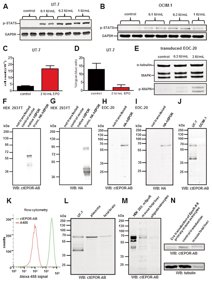
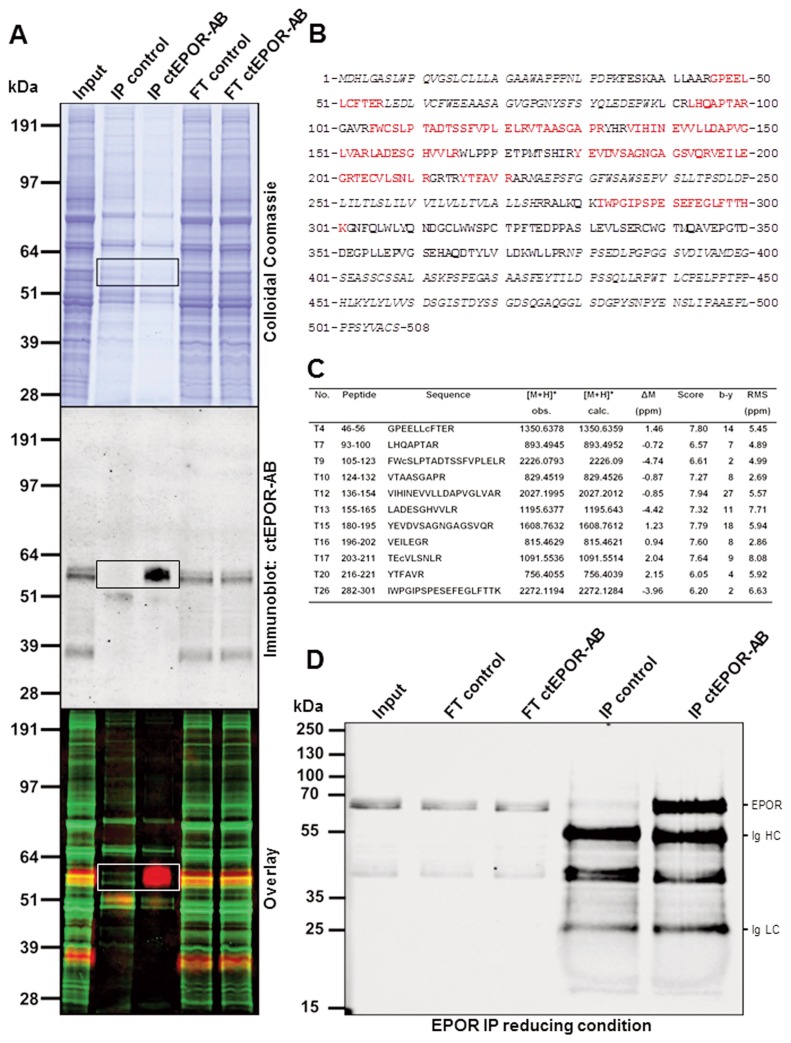
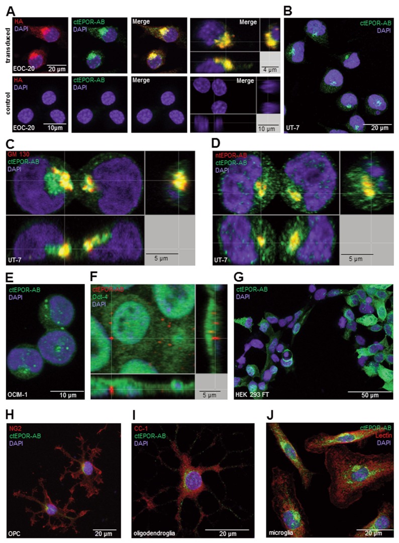
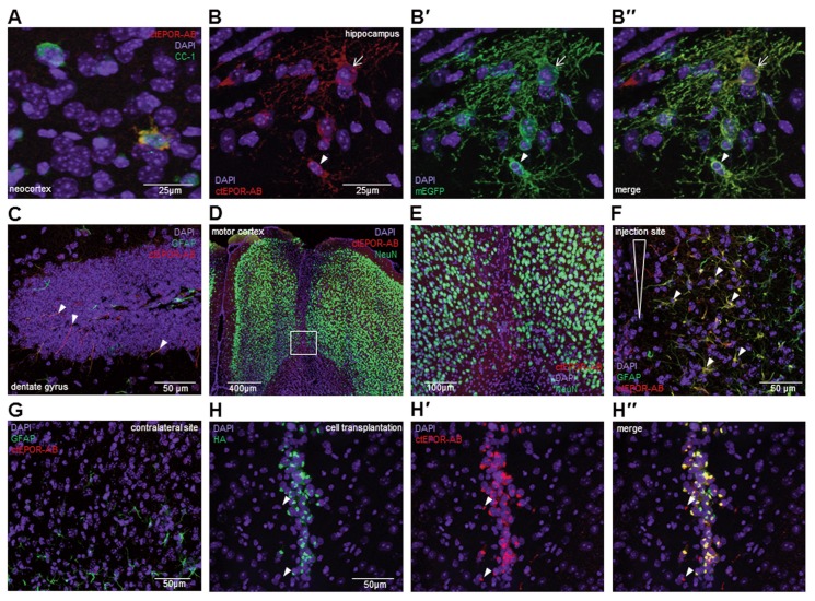
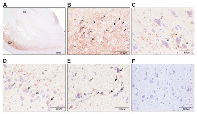
Erythropoietin (EPO) exerts potent neuroprotective, neuroregenerative and procognitive functions. However, unequivocal demonstration of erythropoietin receptor (EPOR) expression in brain cells has remained difficult since previously available anti-EPOR antibodies (EPOR-AB) were unspecific. We report here a new, highly specific, polyclonal rabbit EPOR-AB directed against different epitopes in the cytoplasmic tail of human and murine EPOR and its characterization by mass spectrometric analysis of immuno-precipitated endogenous EPOR, Western blotting, immunostaining and flow cytometry. Among others, we applied genetic strategies including overexpression, Lentivirus-mediated conditional knockout of EpoR and tagged proteins, both on cultured cells and tissue sections, as well as intracortical implantation of EPOR-transduced cells to verify specificity. We show examples of EPOR expression in neurons, oligodendroglia, astrocytes and microglia. Employing this new EPOR-AB with double-labeling strategies, we demonstrate membrane expression of EPOR as well as its localization in intracellular compartments such as the Golgi apparatus. Moreover, we show injury-induced expression of EPOR. In mice, a stereotactically applied stab wound to the motor cortex leads to distinct EpoR expression by reactive GFAP-expressing cells in the lesion vicinity. In a patient suffering from epilepsy, neurons and oligodendrocytes of the hippocampus strongly express EPOR. To conclude, this new analytical tool will allow neuroscientists to pinpoint EPOR expression in cells of the nervous system and to better understand its role in healthy conditions, including brain development, as well as under pathological circumstances, such as upregulation upon distress and injury.


Figures

Figure 1
Figure 1
Functional EPOR validation and EPOR/EpoR detection using ctEPOR-AB in Western blots. (A) Incubation of EPO-dependent UT-7 cells (after 12 h of EPO deprivation) for 15 min with increasing EPO concentrations inducing STAT5 phosphorylation. (B) Incubation of OCIM-1 cells for 15 min with increasing EPO concentrations inducing STAT5 phosphorylation. (C) Cell counts of EPO-dependent UT-7 cultures 72 h after seeding in the presence and absence of EPO (n = 6, mean ± SEM; p < 0.0001). (D) Cell death in EPO-dependent UT-7 cultures 72 h after seeding in presence and absence of EPO (n = 5, mean ± SEM; p < 0.03). (E) Incubation of EOC-20 cells transduced with HA-tagged human EPOR for 10 min with increasing EPO concentrations inducing MAPK phosphorylation. (F) EPOR Western blot using ctEPOR-AB on transfected HEK293 FT cell lysates (truncated HA-EPOR: human EPOR lacking the C-terminus, HA-tag at the N-terminus; control vector: anchored human EPOR without N-terminus and C-terminus). (G) HA Western blot of the same transfected HEK293 FT cell lysates used in (F). (H) EPOR Western blot using ctEPOR-AB on EOC-20 cell lysates transduced with N-terminally HA-tagged human EPOR and respective controls. (I) HA Western blot of the same EOC-20 cell lysates used in (H). (J) EPOR Western blot using ctEPOR-AB on UT-7 and OCIM-1 cell lysates. (K) Flow cytometry of fixed UT-7 cells stained with ctEPOR-AB and Alexa Fluor 488 donkey anti-rabbit secondary AB; as control secondary AB only. (L) EPOR detection in human placenta and human fetal brain using ctEPOR-AB. (M) Detection of murine EpoR in transfected HEK293 FT cells overexpressing murine EpoR, mouse fetal liver and mouse primary oligodendrocytes using ctEPOR-AB. (N) Lentivirus-mediated conditional EpoR knockout in primary EpoR-fl/fl mouse astrocytes; anti-α tubulin as stably expressed comparator.
Figure 2
Figure 2
EPOR IP using ctEPOR-AB and protein identification by mass spectrometry. (A) Colloidal Coomassie staining and immunoblot of the same EPOR IP using ctEPOR-AB from UT-7 protein lysates. The overlay was used to determine the region to be excised from the Coomassie gel for subsequent mass spectrometric protein identification (area indicated by rectangles; abbreviations: FT = flow-through, IP = immunoprecipitation). (B) Amino acid sequence of EPOR (UniProtKB/Swiss-Prot P19235). Peptides identified by mass spectrometry are indicated in red. Note that large parts of the EPOR precursor sequence (indicated in italics) cannot be covered in a standard proteomic experiment with tryptic cleavage as they are either modified (amino acids 1–34, signal peptide; 57–89, N-glycosylation site), attached to the transmembrane domain (224–275) or too large (>5 kDa) to reveal useful information by mass spectrometric sequencing (379–453, 454–508). (C) Table with details on peptide identification. Columns show from left to right: numbering of tryptic peptides; numbering of amino acids according to the sequence in B; peptide sequence (c, carbox-amidomethyl-Cys); observed and calculated mass of the singly protonated peptide; peptide mass deviation in ppm; PLGS score; number of b–y fragment ions; root mean square fragment mass deviation in ppm. (D) Immunoblot of EPOR IP using ctEPOR-AB from UT-7 lysates. In contrast to the IP used for mass spectrometry, EPOR was eluted from the beads in reducing conditions (Laemmli buffer with β-mercaptoethanol; abbreviations: FT = flow-through, IP = immunoprecipitation, Ig HC = immunoglobulin heavy chains, Ig LC = immunoglobulin light chains). The prominent band at around 40 kDa in both IP conditions has to be an immunoglobulin fragment eluted from the beads in the reducing condition only, since it was not eluted without β-mercaptoethanol (see subpanel 2A immunoblot).
Figure 3
Figure 3
Detection of EPOR by immunocytochemistry: (A) EPOR detection using ctEPOR-AB and monoclonal HA-AB on transduced (N-terminally HA-tagged human EPOR, upper row) and control (lower row) EOC-20 cells. (B) EPOR staining with ctEPOR-AB on EPO-dependent UT-7 cells. (C) Double-immunostaining of UT-7 cells with anti-GM130 AB as marker for the Golgi apparatus and ctEPOR-AB. (D) EPOR double staining of UT-7 cells with ntEPOR AB and ctEPOR-AB. (E) EPOR staining with ctEPOR-AB on OCIM-1 cells. (F) Distinct EPOR staining in human Oct-4 + IPS cells using ctEPOR-AB. (G) EpoR staining with ctEPOR-AB of HEK293 FT cells transfected with full-length murine EpoR. Neighboring nontransfected cells show no immunofluorescence. (H) EpoR and NG2 double-staining with ctEPOR-AB in primary mouse oligodendrocyte precursor cells. (I) EpoR and CC-1 double-staining of primary mouse oligodendrocytes with ctEPOR-AB. (J) Detection of EpoR in primary mouse microglia using ctEPOR-AB and lectin as counterstain.
Figure 4
Figure 4
EPOR detection using ctEPOR-AB in healthy and injured mouse brain by immunohistochemistry: (A) EpoR staining in a subpopulation of CC-1 positive mature oligodendrocytes in the neocortex of a 5-wk-old healthy mouse. (B, B′, B″) EpoR staining in the hippocampus of a 5-wk-old NG2-CreERT2:R26-td-tomato-mEGFP mouse. Some oligodendrocyte precursor cells (arrow head) and newly differentiated oligodendrocytes (arrow) express EpoR. Both cell types are endogenously labeled with membrane-tagged EGFP. (C) EpoR staining of GFAP+ cellular processes in the dentate gyrus of a 5-wk-old mouse (arrow heads). (D) Overview of the injection site in the motor cortex of an 8-wk-old mouse injected with medium only (stab wound analogue). The section was stained for neuronal nuclei with NeuN and for EpoR with ctEPOR-AB. (E) Close-up of the white-rectangle region in (D) shows reactive cells with upregulated EpoR expression. (F) Many of the cells at the injection site with upregulated EpoR expression are GFAP+ (arrow heads). (G) Contralateral to the injection site, GFAP+ cells show no EpoR expression at 24 h after lesion. (H, H′, H″) Shown is EPOR and HA double-labeling of injected EOC-20 microglial cells transduced with an HA-tagged human EPOR. In addition, HA-negative cells at the injection site show strong EpoR expression (arrow heads).
Figure 5
Figure 5
EPOR upregulation in neurons and oligodendrocytes of the hippocampal formation of a patient suffering from temporomesial complex-focal epilepsy as demonstrated by immunohistochemistry using ctEPOR-AB. (A) Overview of the surgical sample stained with ctEPOR-AB. (B) Upregulation of EPOR, visualized by brown color of the AEC-chromogen, in some (arrows) but not all (arrow heads) neurons of the CA1 region. (C) EPOR positive neurons in the CA4 region (arrows). (D) Dentate gyrus neurons (arrows) expressing EPOR. (E) EPOR positive oligodendrocytes (arrows) as well as endothelial cells of capillaries (arrow heads) in the adjacent white matter. (F) No staining was observed when omitting the primary antibody.

Similar articles

Cited by

References

    1. Brines M, Cerami A. Emerging biological roles for erythropoietin in the nervous system. Nat Rev Neurosci. 2005;6:484–94. - PubMed
    1. Jelkmann W. Erythropoietin after a century of research: younger than ever. Eur J Haematol. 2007;78:183–205. - PubMed
    1. Jelkmann W. Physiology and pharmacology of erythropoietin. Transfus Med Hemother. 2013;40:302–9. - PMC - PubMed
    1. Arcasoy MO. The non-haematopoietic biological effects of erythropoietin. Br J Haematol. 2008;141:14–31. - PubMed
    1. Sargin D, Friedrichs H, El-Kordi A, Ehrenreich H. Erythropoietin as neuroprotective and neuroregenerative treatment strategy: comprehensive overview of 12 years of preclinical and clinical research. Best Pract Res Clin Anaesthesiol. 2010;24:573–94. - PubMed