Review
. 2015 Nov 11;11(42):5600-10.
doi: 10.1002/smll.201501236. Epub 2015 Sep 9.

Interfacing Inorganic Nanowire Arrays and Living Cells for Cellular Function Analysis

Affiliations
Review

Interfacing Inorganic Nanowire Arrays and Living Cells for Cellular Function Analysis

Minsuk Kwak et al. Small. .

Abstract

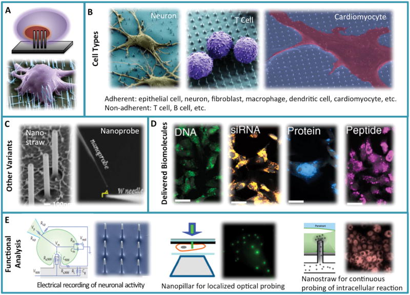
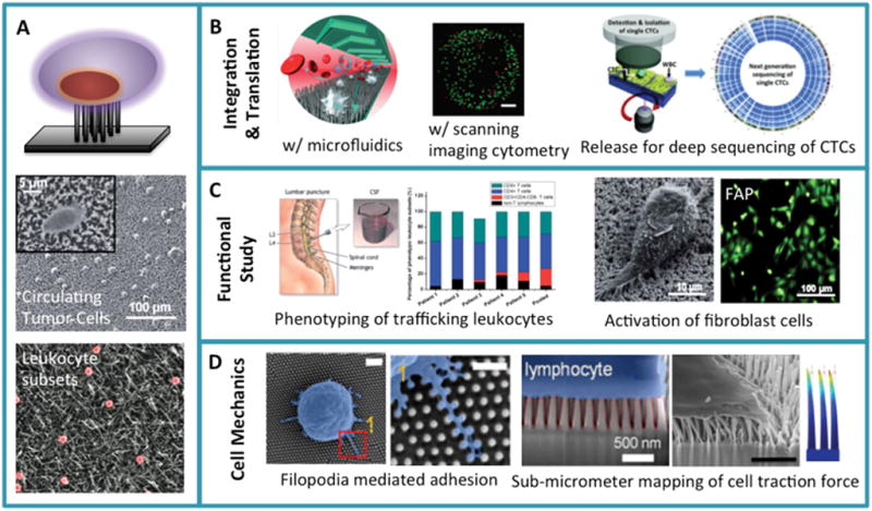
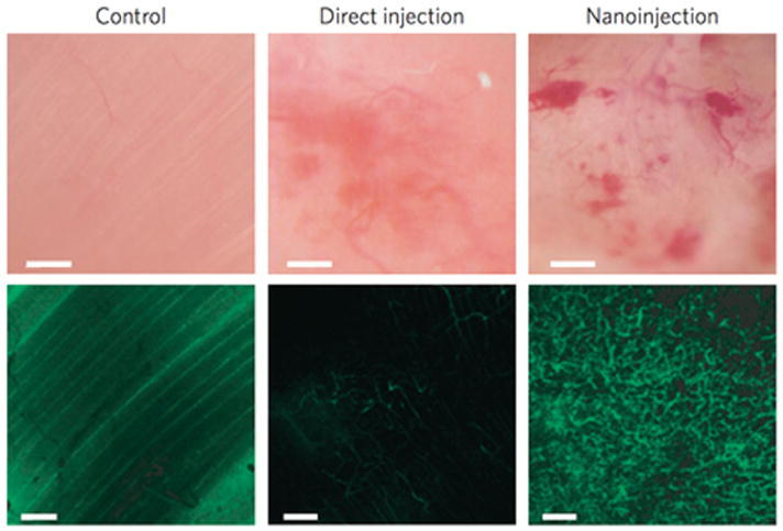
Inorganic nanowires are among the most attractive functional materials, which have emerged in the past two decades. They have demonstrated applications in information technology and energy conversion, but their utility in biological or biomedical research remains relatively under-explored. Although nanowire-based sensors have been frequently reported for biomolecular detection, interfacing nanowire arrays and living mammalian cells for the direct analysis of cellular functions is a very recent endeavor. Cell-penetrating nanowires enabled effective delivery of biomolecules, electrical and optical stimulation and recording of intracellular signals over a long period of time. Non-penetrating, high-density nanowire arrays display rich interactions between the nanostructured substrate and the micro/nanoscale features of cell surfaces. Such interactions enable efficient capture of rare cells including circulating tumor cells and trafficking leukocytes from complex biospecimens. It also serves as a platform for probing cell traction force and neuronal guidance. The most recent advances in the field that exploits nanowire arrays (both penetrating and non-penetrating) to perform rapid analysis of cellular functions potentially for disease diagnosis and monitoring are reviewed.

Keywords: cell-substrate interactions, cellular function analysis; intracellular delivery; nanowire arrays; rare cell analysis.


Figures

Figure 1
Figure 1
Summary - interfacing inorganic nanowire arrays and living cells for a wide range of biological and biomedical applications. In general, this can be classified into two major categories: (1) cell-penetrating nanowire array (red) for biomolecular delivery, intracellular stimulation and probing; (2) non-penetrating nanowire array (blue) for high efficiency capture, separation and molecular phenotyping of rare cells and the biomechanical characterization.
Figure 2
Figure 2
Cell-penetrating nanowire arrays. (A) Schematic illustration of nanowire penetration into living cells and the first demonstration. Reproduced with permission.[5] 2007, American Chemical Society. (B) A range of adherent and non-adherent cells can be interfaced with and penetrated by nanowire arrays. Reproduced with permission.[14, 34] 2010, National Academy of Sciences, USA. (C) Variants of cell penetrating nanowires including nanostraws (Reproduced with permission.[25] 2011, American Chemical Society.) and nanoprobe integrated on a scanning optical tip (Reproduced with permission.[33] 2012, Macmillan Publishers Limited.). (D) A range of biomolecules including DNA, siRNA, protein and peptide can be delivered via the generic nanowire array delivery platform Reproduced with permission.[14] 2010, National Academy of Sciences, USA. (E) Functional study of intracellular signals. Left: cell-penetrating nanoneedle array for electrical stimulation and recording of intracellular action potential. Reproduced with permission.[40] 2012, Macmillan Publishers Limited. Middle: cell-penetrating nanopillars act as subwavelength waveguides and enabled highly localized excitation and fluorescence imaging of single molecule events inside the living cells. Reproduced with permission.[32] 2011, National Academy of Sciences, USA. Right: nanostraws integrated with a microfluidic system permits direct fluid access and molecular delivery into cytosol in a continuous or repeated manner. Reproduced with permission.[46] 2014, Macmillan Publishers Limited.
Figure 3
Figure 3
Non-penetrating nanowire arrays. (A) Schematic illustration of interfacing living cells with a dense array of nanowires that do not penetrate into a cell’s interior. It also shows the two pioneer works demonstrating the use of such nanowire arrays for high efficiency capture of circulating tumor cells (Reproduced with permission.[6] 2009, Wiley-VCH.) and the separation of immune cell subsets (Reproduced with permission.[7] 2010, American Chemical Society.). (B) Integration of nanowire rare cell capture with microfluidics (Reproduced with permission.[67] 2011, Wiley-VCH.), laser scanning cytometry (Reproduced with permission.[73] 2012, American Chemical Society.), or informative molecular characterization (Reproduced with permission.[70] 2013, Wiley-VCH.) represents important endeavors towards the translation to clinical applications. (C) Phenotypic or functional characterization of rare cells using nanowire arrays. Examples include the molecular typing of trafficking leukocytes from cerebrospinal fluid of patients with Alzheimer’s Disease (Reproduced with permission.[82] 2014, The Royal Society of Chemistry.) and the study of fibroblast activation (Reproduced with permission.[88] 2014, The Royal Society of Chemistry.). (D) Study of cell surface microstructure (microvilli, filopodia, etc) interacting with the nanopillar surface (left two panels. Reproduced with permission.[93] 2014, American Chemical Society.) and the measurement of nanopillar deflection to map cell traction force generated by T lymphocytes (third panel. Reproduced with permission.[90] 2013, Springer.) or fibroblasts. The right most panel show the finite element simulation of nanorod deflection upon interfacial traction force.
Figure 4
Figure 4
First demonstration of nanoneedle arrays to deliver proangeiogentic factor and promote in vivo neovascularization. Intravital bright-field (top panels) and confocal (bottom panels) microscopy images of the vasculature of untreated (left) and hVEGF-165-treated muscles with either direct injection (centre) or nanoneedle array-mediated delivery (called nanoinjection, right). The fluorescence signal originates from systemically injected FITC–dextran. Scale bars, bright-field 100 μm; confocal 50 μm. (Reproduced with permission.[102] 2015, Macmillan Publishers Limited.)

Similar articles

Cited by

References

    1. Hochbaum AI, Yang P. Chemical Reviews. 2010;110(1):527. doi: 10.1021/cr900075v. - DOI - PubMed
    1. Li Y, Qian F, Xiang J, Lieber CM. Materials Today. 2006;9(10):18. doi: 10.1016/S1369-7021(06)71650-9. - DOI
    1. Cui Y, Wei Q, Park H, Lieber CM. Science. 2001;293(5533):1289. doi: 10.1126/science.1062711. - DOI - PubMed
    1. Yogeswaran U, Chen S-M. Sensors. 2008;8(1):290. - PMC - PubMed
    1. Kim W, Ng JK, Kunitake ME, Conklin BR, Yang PD. Journal of the American Chemical Society. 2007;129(23):7228. doi: 10.1021/Ja071456k. - DOI - PubMed

Publication types