. 2016 Mar;9(2):527-38.
doi: 10.1038/mi.2015.82. Epub 2015 Sep 9.

β7-Integrin exacerbates experimental DSS-induced colitis in mice by directing inflammatory monocytes into the colon

Affiliations

β7-Integrin exacerbates experimental DSS-induced colitis in mice by directing inflammatory monocytes into the colon

A Schippers et al. Mucosal Immunol. 2016 Mar.

Abstract

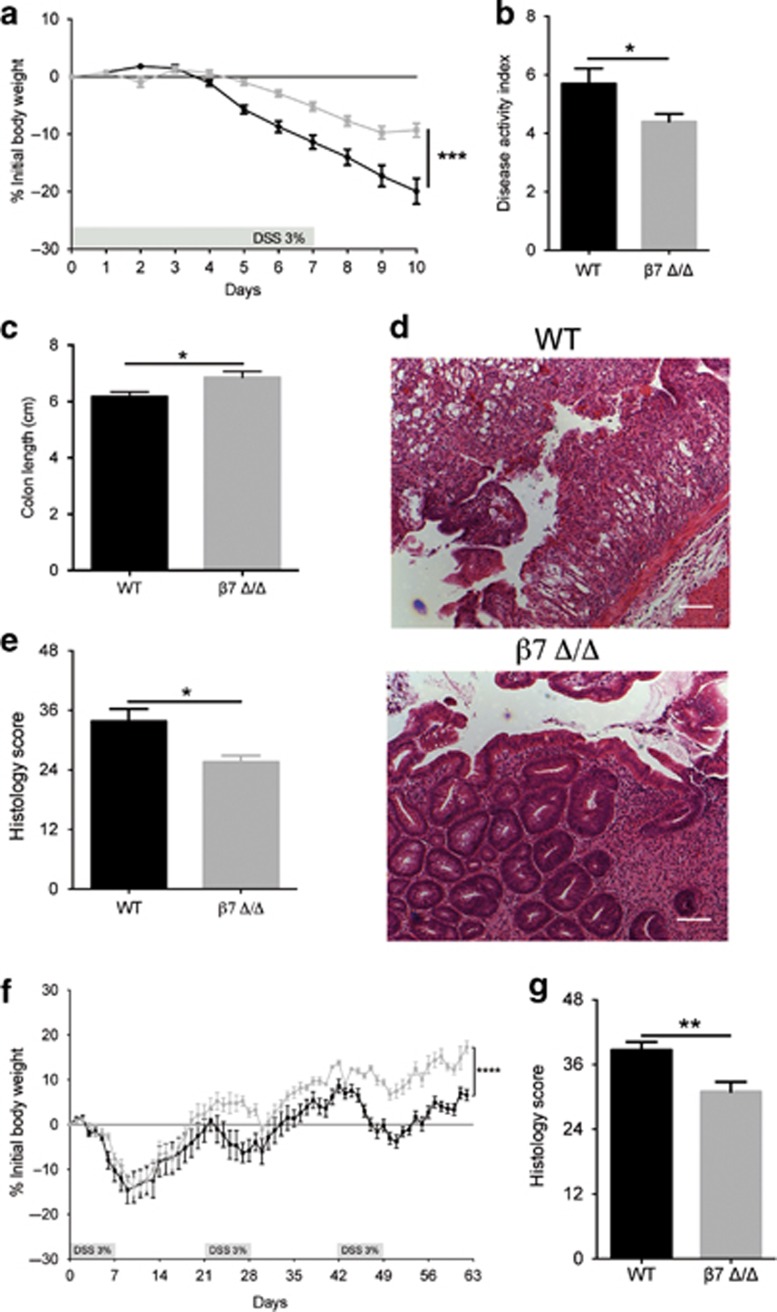
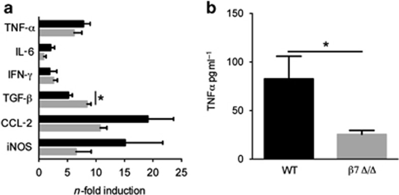
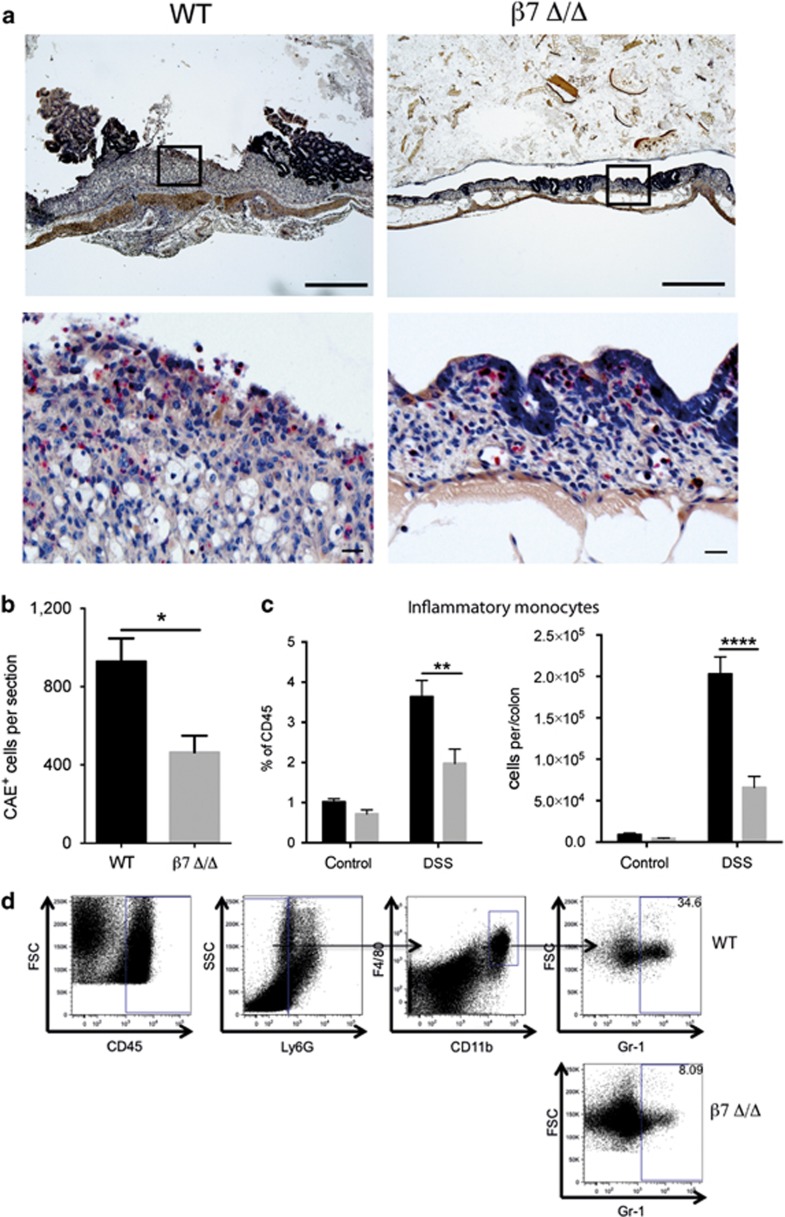
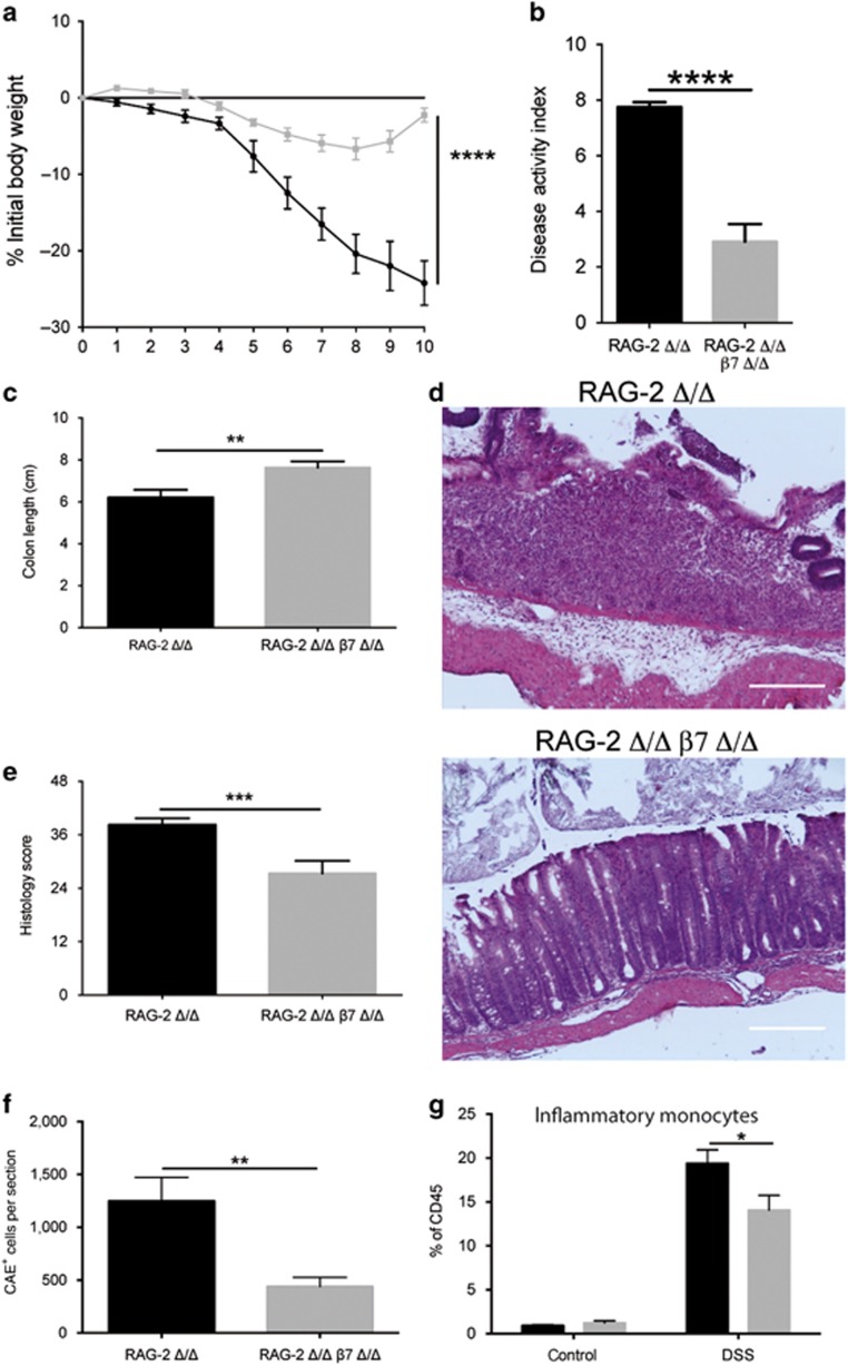
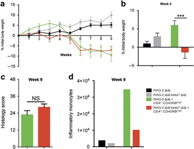
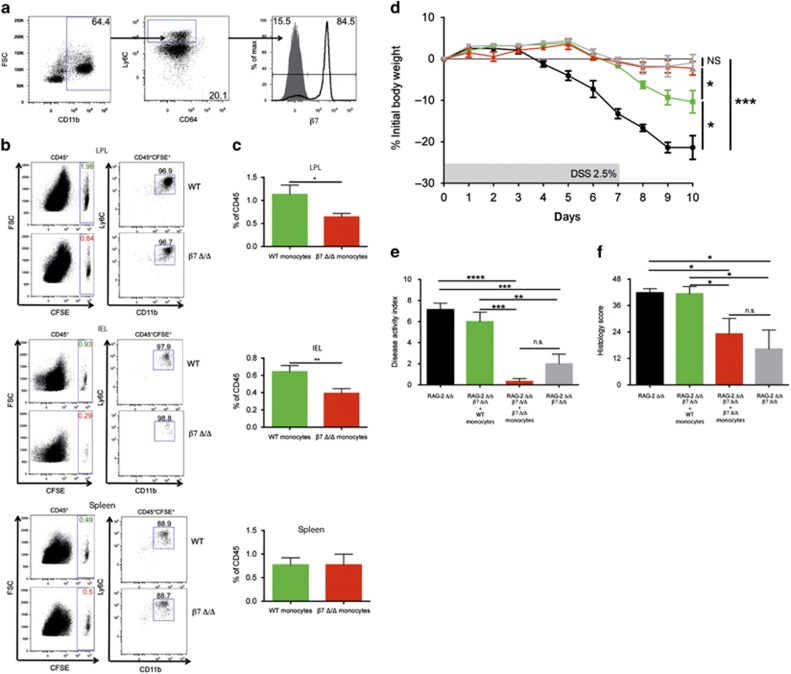
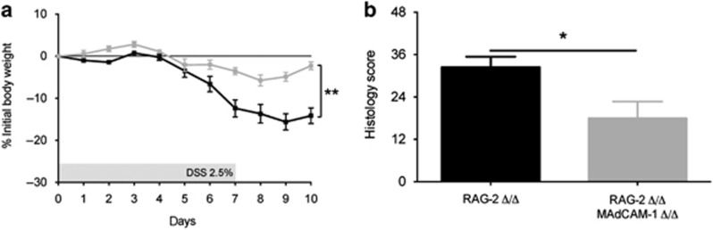
Leukocyte recruitment is pivotal for the initiation and perpetuation of inflammatory bowel disease (IBD) and controlled by the specificity and interactions of chemokines and adhesion molecules. Interactions of the adhesion molecules α4β7-integrin and mucosal addressin cell-adhesion molecule-1 (MAdCAM-1) promote the accumulation of pathogenic T-cell populations in the inflamed intestine. We aimed to elucidate the significance of β7-integrin expression on innate immune cells for the pathogenesis of IBD. We demonstrate that β7-integrin deficiency protects recombination-activating gene-2 (RAG-2)-deficient mice from dextran sodium sulfate (DSS)-induced colitis and coincides with decreased numbers of colonic effector monocytes. We also show that β7-integrin is expressed on most CD11b(+)CD64(low)Ly6C(+) bone marrow progenitors and contributes to colonic recruitment of these proinflammatory monocytes. Importantly, adoptive transfer of CD115(+) wild-type (WT) monocytes partially restored the susceptibility of RAG-2/β7-integrin double-deficient mice to DSS-induced colitis, thereby demonstrating the functional importance of β7-integrin-expressing monocytes for the development of DSS colitis. We also reveal that genetic ablation of MAdCAM-1 ameliorates experimental colitis in RAG-2-deficient mice as well. In summary, we demonstrate a previously unknown role of α4β7-integrin-MAdCAM-1 interactions as drivers of colitis by directing inflammatory monocytes into the colon.


Figures

Figure 1
Figure 1
β7-Integrin deficiency ameliorates acute (ae) and chronic (f and g) dextran sodium sulfate (DSS)-induced colitis. (a) Percentage of body weight loss (measured daily) of wild-type (WT) (black squares, n=12) and β7-integrin-deficient (β7 Δ/Δ) (gray squares, n=12) mice. (b) Disease activity index (DAI) and (c) colon length from DSS-treated WT (black bar, n=12) and β7 Δ/Δ (gray bar, n=12) mice at day 10 of acute DSS colitis. (d) Representative photomicrographs of hematoxylin and eosin (H&E)-stained colon sections from the indicated groups at day 10 of acute DSS colitis at original magnification × 100 (Bar=200 μm). (e) Results of histological scoring of sections from WT (black bar, n=4) and β7 Δ/Δ (gray bar, n=5) mice. The data shown are representative of three independent experiments. (f) Percentage of body weight loss (measured daily) of WT (black squares, n=7) and β7 Δ/Δ (gray squares, n=6) mice. (g) Results of histological scoring of sections from WT (black bar, n=7) and β7 Δ/Δ (gray bar, n=6) mice. The data shown are representative of two independent experiments. Data represent mean±s.e.m. Statistical significance was calculated by two-tailed t-test and is indicated as followings: *P<0.05; **P<0.01, ***P<0.001, ****P<0.0001.
Figure 2
Figure 2
β7-Integrin deficiency results in decreased production of dextran sodium sulfate (DSS)-induced proinflammatory mediators. (a) Levels of the indicated mRNAs were measured in colon tissue at day 10 of acute DSS colitis. For quantification, values are expressed as fold increase over the mean values obtained for healthy control colon tissue. Black bars represent data from wild-type (WT) mice (n=4), gray bars from β7-integrin-deficient (β7Δ/Δ) (n=4) mice, tumor necrosis factor-α (TNF-α), interleukin-6 (IL-6), interferon-γ (IFN-γ), tumor growth factor-β (TGF-β), C–C chemokine ligand-2 (CCL-2), and inducible nitric oxide reductase (iNOS). Data represent mean±s.e.m. (b) TNF-α levels were measured in the serum from WT (n=5) and β7 Δ/Δ mice (n=5) at day 10 of acute DSS colitis. The data shown are representative of two independent experiments. Statistical significance was calculated by two-tailed t-test and is indicated as followings: *P<0.05.
Figure 3
Figure 3
Ameliorated colitis correlates with decreased immigration of myeloid cells in β7-integrin-deficient mice. (a) Representative photomicrographs of chloroacetate esterase (CAE)-stained colon sections from the indicated groups (wild-type (WT), β7-integrin-deficient (β7Δ/Δ) mice) at day 10 of acute dextran sodium sulfate (DSS) colitis at original magnification × 5 (Bar=500 μm); boxed regions are shown below at original magnification × 20 (Bar=20 μm). (b) Quantification of CAE+ cells on sections from WT (black bar, n=4) and β7 Δ/Δ (gray bar, n=5) mice. (c) Flow cytometric quantification of inflammatory monocytes (CD45+Ly6GCD11b+F4/80+GR1+) in colon tissue of untreated and DSS-treated WT (black bar, n=6) and β7 Δ/Δ (gray bar, n=7) mice at day 10 of acute DSS colitis shown as the percent of CD45+ cells (left side) and as absolute number of cells per colon (right side). (d) Representative FACS dot plots illustrating the gating strategy for inflammatory monocytes of DSS-treated mice. The data shown are representative of two independent experiments. Statistical significance is indicated as follows: *P<0.05, **P<0.01, and ****P<0.0001. (b) Two-tailed t-test. (c) Two-way analysis of variance (ANOVA) with Sidak's post-test.
Figure 4
Figure 4
Loss of β7 integrin protects recombination activating gene-2-deficient (RAG-2 Δ/Δ) mice from acute dextran sodium sulfate (DSS)-induced colitis. (a) Percentage of body weight loss (measured daily over the course of DSS treatment), (b) disease activity index (DAI), and (c) Colon length (at day 10 of acute DSS colitis) of RAG-2 Δ/Δ (black squares, black bars, n=12) and RAG-2 Δ/Δ β7 integrin-deficient (β7Δ/Δ) (gray squares, gray bars, n=13) mice. (d) Representative photomicrographs of hematoxylin and eosin (H&E)-stained colon sections from the indicated groups at day 10 of acute DSS colitis at original magnification × 100 (bar represents 100 μm) and (e) results of histological scoring of sections from RAG-2 Δ/Δ (n=7) and RAG-2 Δ/Δ β7 integrin Δ/Δ (n=7) mice. (f) Quantification of CAE+ cells on sections from RAG-2 Δ/Δ (black bar, n=13) and RAG-2 Δ/Δ β7 integrin Δ/Δ (gray bar, n=11) mice at day 10 of acute DSS colitis. (g) Flow cytometric quantification of inflammatory monocytes (CD45+Ly6GCD11b+F4/80+GR1+) in colon tissue of untreated and DSS-treated RAG-2 Δ/Δ (black bars, n=5 (untreated), 4 (DSS-treated)) and RAG-2 Δ/Δ β7 integrin Δ/Δ (gray bars, n=3 (untreated), 4 (DSS-treated)) mice at day 10 of acute DSS colitis. Gating was performed as illustrated in Figure 3. The data shown are representative of three independent experiments. Data represent mean±s.e.m. Statistical significance is indicated as follows: *P<0.05; **P<0.01; ***P<0.001; ****P<0.0001. (ac, e, f) Two-tailed t-test. (g) Two-way analysis of variance (ANOVA) with Sidak's post-test.
Figure 5
Figure 5
β7-Integrin on innate immune cells is of minor importance for the chronic stage of T-cell-mediated colitis. Recombination activating gene-2-deficient (RAG-2 Δ/Δ) and RAG-2 Δ/Δ β7-integrin-deficient (RAG-2 Δ/Δ β7Δ/Δ) mice were adoptively transferred with 0.5 × 10⁶ CD4+CD25-CD45RBhi naïve T cells from wild-type mice and assessed for colitis development. Mice were weighed weekly. At 9 weeks after transfer, the mice were killed and analyzed by flow cytometry and histology. Color code: black=RAG-2 Δ/Δ mice that received no cells (control); gray=RAG-2 Δ/Δ β7Δ/Δ mice that received no cells (control); green=RAG-2 Δ/Δ recipient mice that received CD4+CD25CD45RBhi cells; red=RAG-2 Δ/Δ β7Δ/Δ recipient mice that received CD4+CD25CD45RBhi cells. (a, b) Body weight as a percent of starting weight of RAG-2 Δ/Δ control mice (black, n=5), RAG-2 Δ/Δ β7Δ/Δ control mice (gray, n=5), RAG-2 Δ/Δ recipient mice (green, n=11), and RAG-2 Δ/Δ β7Δ/Δ recipient mice (red, n=11) over the course of 9 weeks (a) and after 3 weeks (b). (c) Results of histological scoring of sections from RAG-2 Δ/Δ recipient mice (green, n=8) and RAG-2 Δ/Δ β7 integrin Δ/Δ recipient mice (red, n=11) mice after 9 weeks of chronic T-cell-mediated colitis. (d) Representative example of a flow cytometric quantification of inflammatory monocytes per small intestine (SI) after week 9 of chronic T-cell-mediated colitis of RAG-2 Δ/Δ control mice (black), RAG-2 Δ/Δ β7Δ/Δ control mice (gray), RAG-2 Δ/Δ recipient mice (green), and RAG-2 Δ/Δ β7Δ/Δ recipient mice (red). Flow cytometry data are from pooled small intestinal samples (n=5 mice each). The data shown are representative of two independent experiments. Data are presented as mean±s.e.m. Statistical significance was calculated by two-way analysis of variance (ANOVA) with Sidak's post-test and is indicated as follows: ***P<0.001.
Figure 6
Figure 6
β7-Integrin-mediated immigration of inflammatory monocytes drives colitis. (a) Flow cytometric detection of β7-integrin surface expression on CD45+CD11b+Ly6ChiCD64low cells isolated from bone marrow of recombination activating gene-2-deficient (RAG-2 Δ/Δ) mice. (b and c) Comparison of the migration efficiency of 5-(and 6) carboxyfluorescein diacetate (CFSE)-labeled CD115-enriched wild-type (WT) or β7-integrin-deficient (β7 Δ/Δ) bone marrow monocytes into the colonic lamina propria lymphocyte (LPL) fraction, intraepithelial lymphocyte (IEL) fraction, and spleen of dextran sodium sulfate (DSS)-treated RAG-2 Δ/Δ mice 16 h after intravenous transfer. (b) Representative FACS plots. (c) Quantification of immigrated CD45+CFSE+ WT (green bars) and β7 Δ/Δ (red bars) monocytes. Data are representative of three independent experiments. (d) Percentage (%) of body weight loss (measured daily over the course of DSS treatment), (e) disease activity index (DAI), and (f) histology score at day 10 of acute DSS colitis of RAG-2 Δ/Δ mice (black squares, black bar, n=7), RAG-2 Δ/Δ-β7Δ/Δ mice (gray squares, gray bars, n=4), RAG-2 Δ/Δ-β7-integrin Δ/Δ mice that had received WT monocytes (green squares, green bars, n=5), and RAG-2 Δ/Δ-β7-integrin Δ/Δ mice that had received β7 Δ/Δ monocytes (red squares, red bars, n=5). Data are representative of three independent experiments. Data represent mean±s.e.m. and significance is indicated as follows: NS=nonsignificant; *P<0.05; **P<0.01; ***P<0.001; and ****P<0001. (c) One-tailed t-test. (df) One-way analysis of variance (ANOVA) with Tukey's post-test.
Figure 7
Figure 7
MAdCAM-1 (mucosal addressin cell-adhesion molecule-1)-deficiency ameliorates acute dextran sodium sulfate (DSS)-induced colitis. Disease severity is expressed in terms of (a) percentage (%) body weight loss (measured daily) and (b) results of histological scoring of sections from DSS-treated recombination activating gene-2-deficient (RAG-2 Δ/Δ) (black squares and bar, n=5 (% initial body weight), 5 (histology score)), and RAG-2/MAdCAM-1 double-deficient (RAG-2 Δ/Δ MAdCAM-1 Δ/Δ), (gray squares and bar, n=5 (% initial body weight), 3 (histology score)) mice at day 10 of the experiment. The data are representative of three independent experiments and represent mean±s.e.m. Statistical significance was calculated by two-tailed t-test and is indicated as follows: *P<0.05 and **P<0.01.

Similar articles

Cited by

References

    1. Springer, T.A. Traffic signals for lymphocyte recirculation and leukocyte emigration: The multistep paradigm. Cell 76, 301–314 (1994). - PubMed
    1. Powrie, F. T cells in inflammatory bowel disease: protective and pathogenic roles. Immunity 3, 171–174 (1995). - PubMed
    1. Wagner, N. et al. Critical role for beta7 integrins in formation of the gut-associated lymphoid tissue. Nature 382, 366–370 (1996). - PubMed
    1. Berlin, C. et al. Alpha 4 beta 7 integrin mediates lymphocyte binding to the mucosal vascular addressin MAdCAM-1. Cell 74, 185–195 (1993). - PubMed
    1. Briskin, M.J., McEvoy, L.M. & Butcher, E.C. MAdCAM-1 has homology to immunoglobulin and mucin-like adhesion receptors and to IgA1. Nature 363, 461–464 (1993). - PubMed

Publication types

MeSH terms