. 2016 Mar;9(2):492-502.
doi: 10.1038/mi.2015.79. Epub 2015 Sep 9.

CD4(+) T-cell survival in the GI tract requires dectin-1 during fungal infection

Affiliations

CD4(+) T-cell survival in the GI tract requires dectin-1 during fungal infection

R A Drummond et al. Mucosal Immunol. 2016 Mar.

Abstract

Dectin-1 is an innate antifungal C-type lectin receptor necessary for protective antifungal immunity. We recently discovered that Dectin-1 is involved in controlling fungal infections of the gastrointestinal (GI) tract, but how this C-type lectin receptor mediates these activities is unknown. Here, we show that Dectin-1 is essential for driving fungal-specific CD4(+) T-cell responses in the GI tract. Loss of Dectin-1 resulted in abrogated dendritic cell responses in the mesenteric lymph nodes (mLNs) and defective T-cell co-stimulation, causing substantial increases in CD4(+) T-cell apoptosis and reductions in the cellularity of GI-associated lymphoid tissues. CD8(+) T-cell responses were unaffected by Dectin-1 deficiency. These functions of Dectin-1 have significant implications for our understanding of intestinal immunity and susceptibility to fungal infections.


Figures

Figure 1
Figure 1
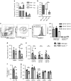
Dectin-1 is required for fungal-specific CD4+ T-cell responses in the GI tract. WT (filled bars/circles) and Dectin-1−/− (clear bars/circles) animals were systemically infected via the tail vein with 2 × 10⁵ CFU C. albicans and analyzed at 72 h post infection for (a) kidney (WT n=25, KO n=25) and (b) small intestine fungal burdens (WT n=17, KO n=21). (c) Bile-acid concentration in the small intestine of infected WT (n=5) and Dectin-1 KO (n=6) mice. (d) The fold change (to naïve controls) in genes regulating bile-acid synthesis (Cyp7a1) or in bile-acid-negative feedback (Fgf15) were measured in 3 day infected livers and intestines (respectively) by quantitative PCR (n=3). (e) WT (n=10) and Dectin-1 KO (n=9) animals maintained on cholestyramine diets starting at 24 h pre-infection until 72 h post infection, when bile acids were measured as in (c), and intestine fungal burdens determined. In other experiments, 3 × 10⁶ OT.II T-cells were transferred into WT and Dectin-1−/− animals, and then infected via the tail vein with 2 × 10⁵ CFU Calb-Ag and analyzed at 72 h post infection. (f) Frequency of OT.II T-cells (CD4+Vα2+CD45.1+) was assessed by flow cytometry in the spleen (n=12), mLNs (n=12), and small intestine (n=8). Example plots are shown for the small intestine and are gated on CD4+ lymphocytes. (g) Total number of OT.II T-cells in the mLN at the indicated time points post infection. (h) Expression of CCR9 (WT n=20, KO n=18) and α4β7 (WT n=16, KO n=14) by CD4+ T-cells in the mLN. (i) The dilution of carboxyfluorescein succinimidyl ester (CFSE) was used to measure proliferation of OT.II cells at day 3 (n=12) and day 6 (WT n=12, KO n=10) post infection in the mLNs. Graph shows percentage of cells that diluted CFSE; see Supplementary Figure S2 for data expressed as the division index. (j) Activation of OT.II cells in the spleen (n=8) and mLN (n=12) was determined by staining for CD69 and comparing with naive controls. (k) Expression of indicated transcription factors by OT.II cells in the mLN was analyzed by intracellular staining and flow cytometry at day 3 post infection (WT n=6, KO n=8). (l) 2 × 10⁶ cells from mLN suspensions at day 3 post infection were restimulated in vitro with OVA323-339 peptide and IL-17 in the supernatant measured by ELISA (WT n=10, KO n=6). Bar charts show pooled data (2–4 experiments); overlaid dot plots show a single representative experiment. TCR, T-cell receptor. *P<0.05.
Figure 2
Figure 2
Dectin-1 is not required for antigen-specific CD8+ T-cell responses. (a) Frequency, (b) division, and (c) activation of OT.I T-cells (CD8+Vα2+CD45.1+) was assessed as in Figure 1 in the indicated tissues (WT n=10, KO n=9) at day 3 post infection. Bar charts show pooled data (two experiments); overlaid dot plots show a single representative experiment. TCR, T-cell receptor.
Figure 3
Figure 3
Dectin-2 is not required for antigen-specific CD4+ T-cell responses in the GI tract. WT (filled bars/circles) and Dectin-2 KO (light gray bars/circles) mice were analyzed as described for Dectin-1 KO mice for (a) intestinal fungal burdens (WT n=13, KO n=12), (b) frequency of OT.II cells (WT n=11, KO n=10), (c) division (WT n=12, KO n=10), (d) activation (WT n=11, KO n=10) and (e, f) polarization of OT.II cells in the mLN (WT n=6, KO n=4) at day 3 post infection. Bar charts show pooled data from three experiments; overlaid dot plot shows a representative experiment. *P<0.05.
Figure 4
Figure 4
Loss of Dectin-1 does not confer global CD4+ T-cell defects. In total, 3 × 10⁶ OT.II T-cells were adoptively transferred into WT (filled bars/circles; n=5) and Dectin-1 KO (clear bars/circles; n=5) mice that were subsequently immunised with 3 μg ovalbumin and 2 μg CpG in the foot pad. Draining popliteal LNs were isolated at 4 days post immunization and OT.II cell (a) frequency, (b) division, and (c) activation analyzed as in Figure 1. (df) WT (n=4) and Dectin-1 KO (n=4) mice were immunised with 20 μg ovalbumin and 20 μg CpG systemically via the tail vein, and OT.II responses analyzed at 4 days post immunization as above. Data are from one experiment. CpG, oligodeoxynucleotide.
Figure 5
Figure 5
Loss of Dectin-1 causes defects in the DC response. (a) WT bone marrow restores the frequency of OT.II T-cells in the Dectin-1−/− mLN (WT→WT n=7, KO→KO n=5, WT→KO n=7) and small intestine (WT→WT n=3, KO→KO n=3, WT→KO n=4), and also leads to a reduction in (b) intestinal fungal burdens (WT→WT n=3, KO→KO n=4, WT→KO n=7). WT→WT denotes WT animals reconstituted with WT bone marrow. WT→KO denotes KO animals reconstituted with WT bone marrow. KO→KO denotes KO animals reconstituted with KO bone marrow. (c) DCs in the mLN were defined as B220CD11c+MHCII+ (left panel), and migratory/resident sub-populations grouped into three sub-populations based on CD103 and CD11b expression (middle panel). Dectin-1 expression was assessed by staining with 2A11 and analysis by flow cytometry (right panel and graph, n=4). (d) The number of total CD11c+MHCII+ cells (infected: WT n=29, KO n=23, naive: WT n=4, KO n=4) and (e) the migratory/resident populations (WT n=19, KO n=16) within the mLN at 72 h post infection in WT and Dectin-1−/− mice. (f) 4 × 10⁴ CD11c+ cells from WT (n=8) and Dectin-1−/− (n=8) mice were purified from the mLN at 72 h post infection and cultured with 2 × 10⁵ naive OT.II T-cells and 10 μg ml−1 OVA323-339 peptide. IL-2 in the supernatants was measured 24 h later and expressed relative to the WT. (g) FACS analysis of the indicated co-stimulation molecules relative to naive controls at 72 h post infection by CD11c+MHCII+ cells in the mLN (n=12). Bar charts show pooled data (chimera/IL-2: two experiments; DC data: five experiments); overlaid dot plots show one representative experiment. *P<0.05.
Figure 6
Figure 6
Dectin-1 is required for CD4+ T-cell survival. (a) The frequency of apoptotic CD4+ T-cells was measured by Annexin-V staining in mLN cell suspensions at the indicated time points post infection (WT n=9, KO n=9 at each time point). (b) CD8+ T-cell apoptosis and necrosis was measured by Annexin-V/7-AAD staining in mLN cell suspensions at 72 h post infection (n=6). (c) TUNEL staining performed on paraformaldehyde-fixed mLNs from infected WT and KO mice 72 h post infection; photo taken at 5 × magnification, insert at 40 × magnification. Blue=DAPI, green=apoptotic cells. (d) The total number of recoverable cells from the mLN (WT n=9, KO n=9 at each time point). (e) H&E staining of mLNs at 72 h post infection compared with naive controls (scale bars are 500 μm). (f) The total number of visible (>1 mm3) Peyer's patches (infected n=9, naive n=8) in WT and Dectin-1−/− mice at 72 h post infection. (g) The total number of recoverable cells from the mLN of infected WT (n=10) and KO (n=9) mice at 3 days post infection while maintained on cholestyramine diets, as described in Figure 1. (h) Schematic of the colitis model where co-housed WT and Dectin-1 KO animals were maintained on water supplemented with penicillin, streptomycin, and fluconazole for 4 days, and then switched to water containing 3.5% DSS and C. tropicalis for 5 days. The number of (i) Peyer's patches and (j) number of DCs (CD11c+MHCII+) isolated from mLNs from WT (n=20) and Dectin-1 KO (n=16) mice at 5 days post DSS exposure was compared with Candida only WT animals (control, n=6). Bar charts show pooled data (2–5 experiments); overlaid dot plots show one representative experiment. *P<0.05.
Figure 7
Figure 7
Transfer of WT DCs restores T-cell responses and mLN cellularity in Dectin-1 KO mice. CD11c+ cells from the mLN of naive WT mice were pre-loaded with OVA or left untreated, and injected intraperitoneally into Dectin-1 KO recipients. OT.II cells were then transferred and Calb-Ag used to infected mice, as for previous experiments. At 72 h post infection, animals were analyzed for (a) total number cells in the mLN, (b) frequency of OT.II cells, (c) CD4+ T-cell apoptosis and (d) necrosis, and (e) intestine fungal burdens. All data are pooled from two independent experiments; overlaid dot plots show one representative experiment. *P<0.05.

Similar articles

Cited by

References

    1. Hardison, S.E. & Brown, G.D. C-type lectin receptors orchestrate antifungal immunity. Nat. Immunol. 13, 817–822 (2012). - PMC - PubMed
    1. Taylor, P.R. et al. Dectin-1 is required for β-glucan recognition and control of fungal infection. Nat. Immunol. 8, 31–38 (2007). - PMC - PubMed
    1. Saijo, S. et al. Dectin-1 is required for host defense against Pneumocystis carinii but not against Candida albicans. Nat. Immunol. 8, 39–46 (2007). - PubMed
    1. Werner, J.L. et al. Requisite role for the Dectin-1 beta-glucan receptor in pulmonary defense against Aspergillus fumigatus. J. Immunol. 182, 4938–4946 (2009). - PMC - PubMed
    1. LeibundGut-Landmann, S. et al. Syk- and CARD9-dependent coupling of innate immunity to the induction of T helper cells that produce interleukin 17. Nat. Immunol. 8, 630–638 (2007). - PubMed

Publication types

MeSH terms