Review
. 2015 Sep;9(3):367-402.
doi: 10.1007/s11682-015-9444-y.

Advanced neuroimaging applied to veterans and service personnel with traumatic brain injury: state of the art and potential benefits

Affiliations
Review

Advanced neuroimaging applied to veterans and service personnel with traumatic brain injury: state of the art and potential benefits

Elisabeth A Wilde et al. Brain Imaging Behav. 2015 Sep.

Abstract

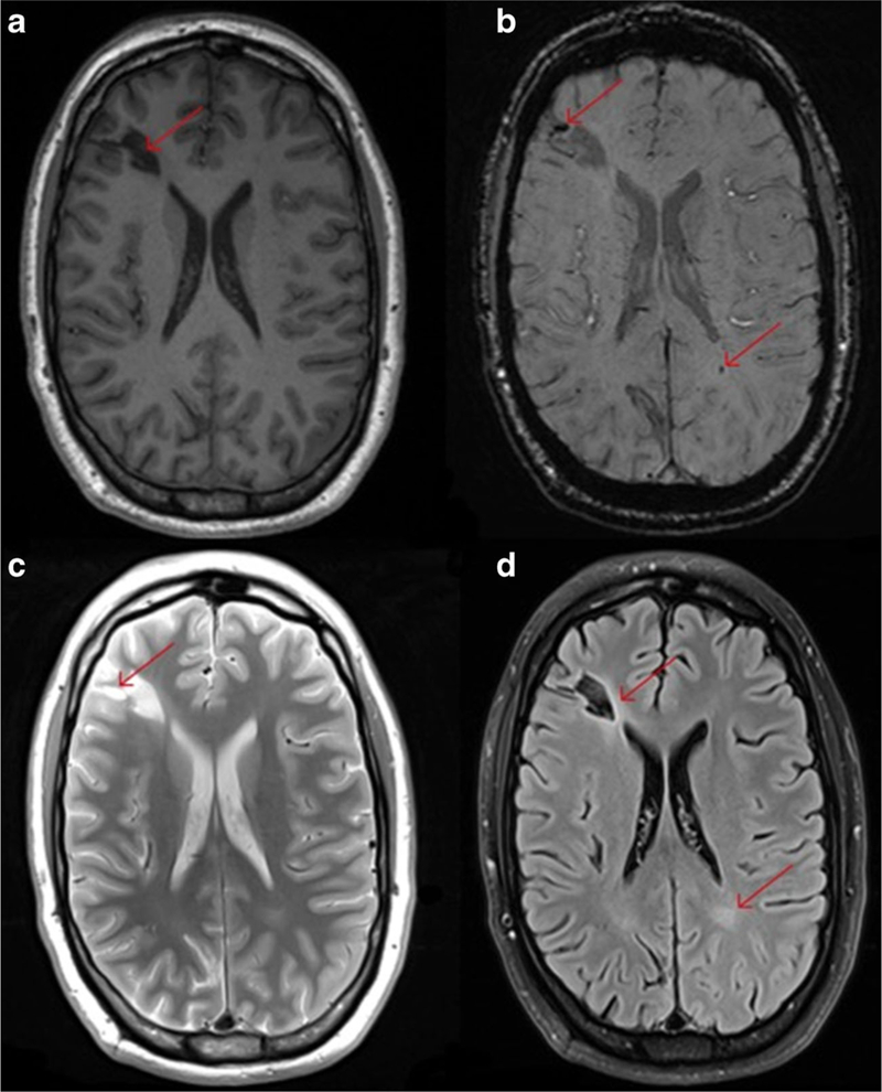
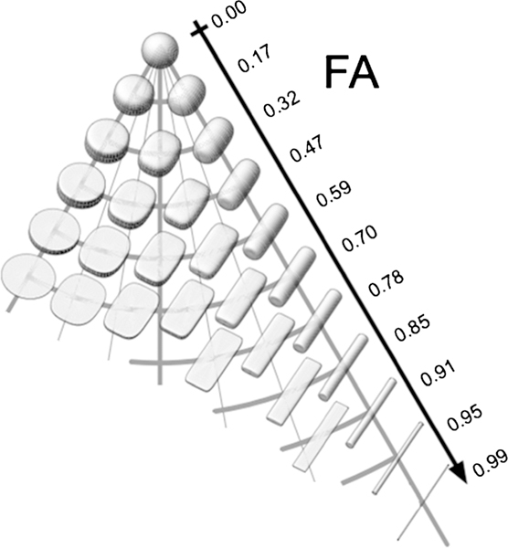
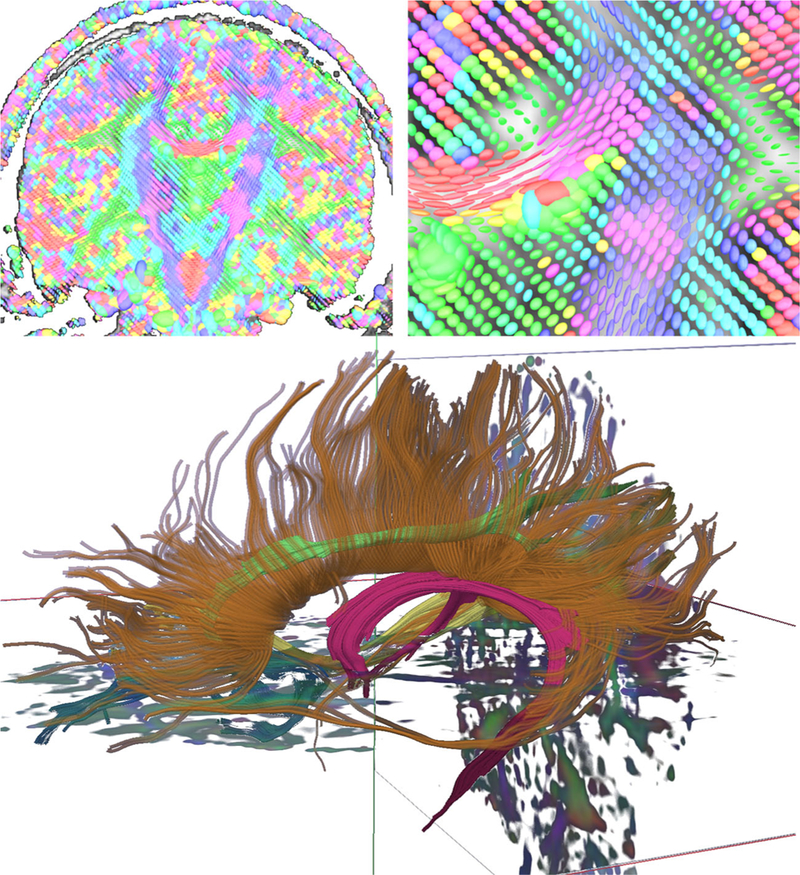
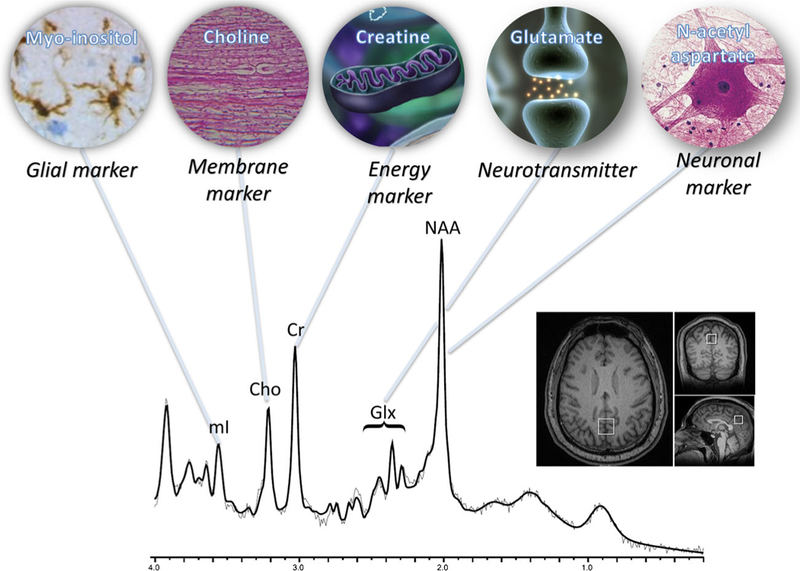
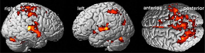
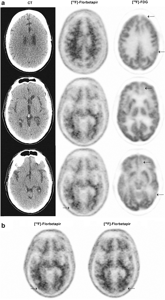
Traumatic brain injury (TBI) remains one of the most prevalent forms of morbidity among Veterans and Service Members, particularly for those engaged in the conflicts in Iraq and Afghanistan. Neuroimaging has been considered a potentially useful diagnostic and prognostic tool across the spectrum of TBI generally, but may have particular importance in military populations where the diagnosis of mild TBI is particularly challenging, given the frequent lack of documentation on the nature of the injuries and mixed etiologies, and highly comorbid with other disorders such as post-traumatic stress disorder, depression, and substance misuse. Imaging has also been employed in attempts to understand better the potential late effects of trauma and to evaluate the effects of promising therapeutic interventions. This review surveys the use of structural and functional neuroimaging techniques utilized in military studies published to date, including the utilization of quantitative fluid attenuated inversion recovery (FLAIR), susceptibility weighted imaging (SWI), volumetric analysis, diffusion tensor imaging (DTI), magnetization transfer imaging (MTI), positron emission tomography (PET), magnetoencephalography (MEG), task-based and resting state functional MRI (fMRI), arterial spin labeling (ASL), and magnetic resonance spectroscopy (MRS). The importance of quality assurance testing in current and future research is also highlighted. Current challenges and limitations of each technique are outlined, and future directions are discussed.

Keywords: Diffusion tensor imaging; Magnetic resonance imaging; Magnetic resonance spectroscopy; Positron emission tomography; Traumatic brain injury; Veteran; fMRI.


Conflict of interest statement

Conflict of interest The authors declare that they have no competing interests.

Figures

Fig. 1
Fig. 1
This figure illustrates the different contrast available when using structural MRI to examine a young OIF/OIF Active Duty Service Member diagnosed with a mild TBI (LOC<30 min; AOC<24 h; PTA <24 h). Utilization of multimodal imaging (even structural imaging) can be informative with each sequence potentially adding additional clinically meaningful information about the specific injury incurred. Panel a shows the T1-weighted image and the large hypointense lesion in the right frontal lobe (red arrow). Panel b shows the SWI image and not only shows the large hypointense lesion but several smaller hemosiderin deposits including one in the white matter in the right parietal occipital region (red arrows). Panel c is the T2-weighted image and shows the bright areas indicating inflammation or CSF accumulation around the larger lesion. Panel d is the FLAIR image and shows enhancement around the larger lesion and an area abnormality in the white matter in the left parietal occipital region (contracoup injury)
Fig. 2
Fig. 2
Visualization of the Fractional Anisotropy of Diffusion Tensors. Note that tensors of different shapes can have the same FA. Adapted from Ennis and Kindlmann (Ennis and Kindlmann 2006)
Fig. 3
Fig. 3
Top Left: A coronal slice of a DTI volume at the level of the posterior internal capsule. The image is composed of many individual tensor estimations, and the orientation of these estimations indicates the presumed direction of the fiber. Consistent with convention, red indicates fibers coursing in a right-left orientation, green represents an anterior-posterior fiber orientation, and blue/purple reflects a superior/inferior orientation. Top right: Magnification of an area near the junction of the corpus callosum and internal capsule showing highly organized anisotropic tensors in the corpus callosum. Bottom: Tractography of the corpus callosum (brown), fornix (magenta), and cingulum bundle (green)
Fig. 4
Fig. 4
Major metabolite resonances and associated biological functions. Each peak is labeled with the abbreviation and inset which describes the role that each metabolite plays as biomarkers for traumatic brain injury. Data acquired using single voxel, PRESS, TR/TE: 2000/30 ms, 2×2×2 cc in posterior cingulate at 3Tas shown in bottom right inset and postprocessed using LCModel. Modified from Lin et al. (2012a)
Fig. 5
Fig. 5
Subjects with blast mTBI who performed a stimulus–response compatibility task demonstrated greater activation than subjects who had not been exposed to blast, a pattern which was augmented after covarying for PTSD and depression symptoms and reaction time
Fig. 6
Fig. 6
59 year old with TBI following a fall demonstrating increased uptake of 18F-florbetapir (arrows) and decreased uptake of 18F-FDG (arrows) within the region of a previous traumatic cerebral contusion to the occiput (Mitsis et al. 2014). Figure used with permission

References

    1. Allen EA, Erhardt EB, Damaraju E, Gruner W, Segall JM, Silva RF, et al. (2011). A baseline for the multivariate comparison of resting-state networks. Frontiers in Systems Neuroscience, 5, 2. doi: 10.3389/fnsys.2011.00002. - DOI - PMC - PubMed
    1. Alsop DC, Detre JA, Golay X, Gunther M, Hendrikse J, Hernandez-Garcia L, et al. (2014). Recommended implementation of arterial spin-labeled perfusion MRI for clinical applications: a consensus of the ISMRM perfusion study group and the European consortium for ASL in dementia. Magnetic Resonance in Medicine. doi:10.1002/mrm.25197. - DOI - PMC - PubMed
    1. Amann M, Sprenger T, Naegelin Y, Reinhardt J, Kuster P, Hirsch JG, et al. (2015). Comparison between balanced steady-state free precession and standard spoiled gradient echo magnetization transfer ratio imaging in multiple sclerosis: methodical and clinical considerations. Neuroimage, 108, 87–94. doi:10.1016/j.neuroimage.2014.12.045. - DOI - PubMed
    1. Aoki Y, Inokuchi R, Gunshin M, Yahagi N, & Suwa H (2012). Diffusion tensor imaging studies of mild traumatic brain injury: a meta-analysis. [Meta-Analysis]. Journal of Neurology, Neurosurgery, and Psychiatry, 83(9), 870–876. doi:10.1136/jnnp-2012-302742. - DOI - PMC - PubMed
    1. Aron AR, Fletcher PC, Bullmore ET, Sahakian BJ, & Robbins TW (2003). Stop-signal inhibition disrupted by damage to right inferior frontal gyrus in humans. [Research Support, Non-U.S. Gov’t]. Nature Neuroscience, 6(2), 115–116. doi:10.1038/nn1003. - DOI - PubMed

Publication types

LinkOut - more resources