. 2015 Dec 2;43(21):10277-91.
doi: 10.1093/nar/gkv896. Epub 2015 Sep 8.

Human HLTF mediates postreplication repair by its HIRAN domain-dependent replication fork remodelling

Affiliations

Human HLTF mediates postreplication repair by its HIRAN domain-dependent replication fork remodelling

Yathish Jagadheesh Achar et al. Nucleic Acids Res. .

Abstract

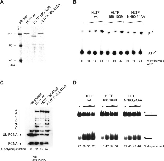
Defects in the ability to respond properly to an unrepaired DNA lesion blocking replication promote genomic instability and cancer. Human HLTF, implicated in error-free replication of damaged DNA and tumour suppression, exhibits a HIRAN domain, a RING domain, and a SWI/SNF domain facilitating DNA-binding, PCNA-polyubiquitin-ligase, and dsDNA-translocase activities, respectively. Here, we investigate the mechanism of HLTF action with emphasis on its HIRAN domain. We found that in cells HLTF promotes the filling-in of gaps left opposite damaged DNA during replication, and this postreplication repair function depends on its HIRAN domain. Our biochemical assays show that HIRAN domain mutant HLTF proteins retain their ubiquitin ligase, ATPase and dsDNA translocase activities but are impaired in binding to a model replication fork. These data and our structural study indicate that the HIRAN domain recruits HLTF to a stalled replication fork, and it also provides the direction for the movement of the dsDNA translocase motor domain for fork reversal. In more general terms, we suggest functional similarities between the HIRAN, the OB, the HARP2, and other domains found in certain motor proteins, which may explain why only a subset of DNA translocases can carry out fork reversal.


Figures

Figure 1.
Figure 1.
The effect of HIRAN deletion on cellular function of HLTF. (A) Domain structure of the HLTF protein. The SNF2-type ATPase-helicase, the C3HC4-type RING finger domain and the HIRAN motif are shown by gray boxes numbered I–VI, a black box, and a striped box, respectively. In HLTF 156–1009, the N-terminal 155 amino acids containing the whole HIRAN domain were deleted, in HLTF NN90,91AA the two asparagines in position 90 and 91 were mutated to alanines. (B) Multiple alignment of the HIRAN domain. Conserved residues are shaded, and asterisks indicate residues mutated to generate HLTF NN90,91AA. (C) The crystal structure of the HIRAN fragment (hHLTF.56–175) with a dinucleotide fragment reveals a six-stranded β-barrel covered with two α-helices. The DNA-binding site of the HIRAN domain is positioned in a cavity region formed by two loops and one β-strand. (D) Deletion or mutation of the HIRAN domain of HLTF sensitises cells to UV irradiation. Complementation of the UV sensitivity of stable shRNA-depleted HLTF knockdown HCT116 cells was tested by expressing the shRNA-resistant form of WT (HLTF WT), RING mutant (HLTF C759S), ATPase mutant (HLTF DE557,558AA), HIRAN deletion mutant (HLTF 156–1009), and HIRAN point mutant (HLTF NN90,91AA) HLTF proteins.
Figure 2.
Figure 2.
HLTF-mediated postreplication repair depends on the HIRAN domain. (A) Experimental setup of alkaline BrdU comet postreplication repair assay. Cells were first pulse-labeled with BrdU for 20 min, washed with PBS and UV-irradiated or mock-treated, followed by chasing by adding 4× dNTP for 6 hours before alkaline single cell electrophoresis. (B) Representative images of the UV-treated cells after comet assay. HLTF was stably depleted in HCT116 cells by shRNA, and for testing complementation the shRNA-resistant form of WT (HLTF WT) or HIRAN mutant HLTF 156–1009 or HLTF NN90,91AA expressing plasmids were transfected transiently. (C) Representation of the percentage of comet tail DNA without and after UV treatment. Standard deviations were calculated from three independent experiments.
Figure 3.
Figure 3.
The effect of HIRAN point mutation or deletion on the enzymatic function of HLTF. (A) Purity of the FLAG-tagged wild-type (HLTF WT), HIRAN deletion (HLTF156–1009), and point mutant (HLTF NN90,91AA) proteins. Purified proteins were run on 8% denaturing polyacrylamide gel and stained with Coomassie blue. (B) Mutation of the HIRAN domain of HLTF does not compromise its ATPase activity. [γ-32P]ATP was incubated with increasing concentrations of wild-type, HIRAN domain-deleted, and HIRAN point mutant HLTF proteins in the presence of 75-mer double-stranded oligonucleotides. Hydrolyzed gamma-phosphate (Pi) was detected by thin layer chromatography. (C) Mutation of the HIRAN domain of HLTF does not affect its ubiquitin ligase function. PCNA was first loaded onto nicked plasmid DNA by RFC, followed by adding ubiquitin, Uba1, Rad6-Rad18, Mms2-Ubc13 and wild-type, HIRAN domain-deleted or HIRAN domain point mutant purified HLTF proteins. Ubiquitylation of PCNA was followed by western blot using anti-PCNA antibody. (D) Mutation of the HIRAN domain of HLTF does not affect its double-stranded DNA translocase activity. Radioactively labeled triple-helix substrate was incubated with increasing amounts of wild-type, HIRAN-deleted or HIRAN domain point mutant HLTF proteins, and the release of single-stranded DNA products was followed by native polyacrylamide gel electrophoresis.
Figure 4.
Figure 4.
The HIRAN domain is necessary for HLTF fork regression activity. (A) Fork regression activity of HLTF on oligonucleotide-based replication fork-like structure. Increasing amounts of wild-type (HLTF WT), HIRAN deletion mutant (HLTF 156–1009), or HIRAN point mutant (HLTF NN9,91AA) HLTF were incubated with a radioactively labeled oligonucleotide-based replication fork-like structure. The release of the parental (75-mer) and daughter duplexes (30-mer) as products of fork regression were followed by electrophoresis on nondenaturing polyacrylamide gel. (B) Schematic representation of the plasmid-based model replication fork substrate used for testing HLTF fork regression activity. Fork regression activity of purified wild-type (HLTF WT), HIRAN-deleted (HLTF 156-1009), and HIRAN point mutant (NN90,91AA) HLTF proteins on radioactively labeled plasmid-based replication fork-like structure. Fork regression was revealed by digesting the products with restriction endonucleases BamHI (B), EcoRI (E) and AflIII (A). (C) Schematic representation and purity of wild-type and mutant HIRAN domain fragments. (D) Restoration of the fork regression activity of the HIRAN-deleted HLTF156-1009 protein by purified HIRAN domain fragment (containing only the 56–168 residues of HLTF). Purified wild-type or point mutant HIRAN domains were mixed with HIRAN-deleted HLTF156-1009 proteins and assayed for fork reversal activity using fluorescently labeled oligonucleotide-based replication fork-like structure. The release of parental duplex (75-mer) was monitored to reveal fork regression activity.
Figure 5.
Figure 5.
The HIRAN domain specifically binds to a replication fork-like structure. (A) Gel-retardation assay to reveal the binding of HLTF to various DNA substrates. Purified wild-type or mutant HIRAN domains were incubated with dsDNA, ssDNA, partial heteroduplex, and replication fork-like oligonucleotide-based DNA substrates before electrophoresis on nondenaturing acrylamide gels. (B) DNA competition assay. Purified HIRAN domain was preincubated with oligonucleotide-based Cy5-labeled replication fork-like substrate, followed by adding fluorescently labeled competitor ssDNA or replication fork substrates in the same increasing concentrations. After electrophoresis, the same gel was visualized first for Cy5 (upper panel) and next for fluorescent signals (lower panel). (C) Quantification of the DNA competition assay shown in (B).
Figure 6.
Figure 6.
Schematic representation of the function of HIRAN. (A) Without the HIRAN domain, HLTF cannot be positioned at the junction point of a replication fork; although it can translocate on the dsDNA region, the junction and the two arms present a barrier for translocation into that direction. These reasons explain why HIRAN-mutated HLTF is able to displace ssDNA from a partial triple helix DNA but is unable to carry out fork reversal. (B) The HIRAN domain of HLTF can provide direct binding for HLTF to the junction point of the fork; serve as a pin for separating the two strands of parental/daughter dsDNA arms; orient the dsDNA translocase activity of HLTF into the right direction toward the junction. These reasons explain the critical role of HIRAN in HLTF-mediated fork reversal enabling its cellular function.

References

    1. Ciccia A., Elledge S.J. The DNA damage response: making it safe to play with knives. Mol. Cell. 2010;40:179–204. - PMC - PubMed
    1. Izhar L., Ziv O., Cohen I.S., Geacintov N.E., Livneh Z. Genomic assay reveals tolerance of DNA damage by both translesion DNA synthesis and homology-dependent repair in mammalian cells. Proc. Natl. Acad. Sci. U.S.A. 2013;110:E1462–E1469. - PMC - PubMed
    1. Zhang H., Lawrence C.W. The error-free component of the RAD6/RAD18 DNA damage tolerance pathway of budding yeast employs sister-strand recombination. Proc. Natl. Acad. Sci. U.S.A. 2005;102:15954–15959. - PMC - PubMed
    1. Yoon J.-H., Prakash S., Prakash L. Requirement of Rad18 protein for replication through DNA lesions in mouse and human cells. Proc. Natl. Acad. Sci. U.S.A. 2012;109:7799–7804. - PMC - PubMed
    1. Prakash S., Johnson R.E., Prakash L. Eukaryotic translesion synthesis DNA polymerases: specificity of structure and function. Annu. Rev. Biochem. 2005;74:317–353. - PubMed

Publication types

MeSH terms