

Novel genetic causes for cerebral visual impairment
- PMID: 26350515
- PMCID: PMC4930090
- DOI: 10.1038/ejhg.2015.186
Novel genetic causes for cerebral visual impairment
Abstract
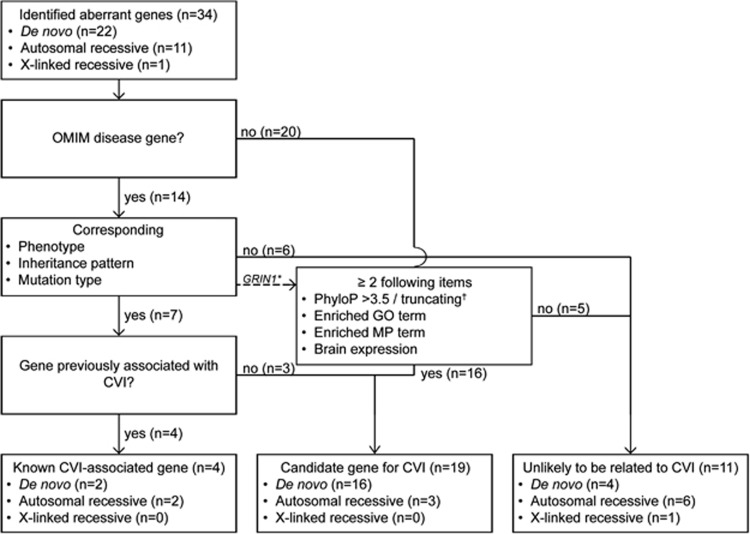
Cerebral visual impairment (CVI) is a major cause of low vision in children due to impairment in projection and/or interpretation of the visual input in the brain. Although acquired causes for CVI are well known, genetic causes underlying CVI are largely unidentified. DNAs of 25 patients with CVI and intellectual disability, but without acquired (eg, perinatal) damage, were investigated by whole-exome sequencing. The data were analyzed for de novo, autosomal-recessive, and X-linked variants, and subsequently classified into known, candidate, or unlikely to be associated with CVI. This classification was based on the Online Mendelian Inheritance in Man database, literature reports, variant characteristics, and functional relevance of the gene. After classification, variants in four genes known to be associated with CVI (AHDC1, NGLY1, NR2F1, PGAP1) in 5 patients (20%) were identified, establishing a conclusive genetic diagnosis for CVI. In addition, in 11 patients (44%) with CVI, variants in one or more candidate genes were identified (ACP6, AMOT, ARHGEF10L, ATP6V1A, DCAF6, DLG4, GABRB2, GRIN1, GRIN2B, KCNQ3, KCTD19, RERE, SLC1A1, SLC25A16, SLC35A2, SOX5, UFSP2, UHMK1, ZFP30). Our findings show that diverse genetic causes underlie CVI, some of which will provide insight into the biology underlying this disease process.
Figures
References
-
- Boonstra N, Limburg H, Tijmes N, van Genderen M, Schuil J, van Nispen R: Changes in causes of low vision between 1988 and 2009 in a Dutch population of children. Acta Ophthalmol 2012; 90: 277–286. - PubMed
-
- Dutton GN, Jacobson LK: Cerebral visual impairment in children. Semin Neonatol 2001; 6: 477–485. - PubMed
-
- Good WV, Jan JE, DeSa L, Barkovich AJ, Groenveld M, Hoyt CS: Cortical visual impairment in children. Surv Ophthalmol 1994; 38: 351–364. - PubMed
-
- Ortibus E, Lagae L, Casteels I, Demaerel P, Stiers P: Assessment of cerebral visual impairment with the L94 visual perceptual battery: clinical value and correlation with MRI findings. Dev Med Child Neurol 2009; 51: 209–217. - PubMed
-
- Saidkasimova S, Bennett DM, Butler S, Dutton GN: Cognitive visual impairment with good visual acuity in children with posterior periventricular white matter injury: a series of 7 cases. J AAPOS 2007; 11: 426–430. - PubMed
Publication types
MeSH terms
Grants and funding
LinkOut - more resources
Full Text Sources
Other Literature Sources
Molecular Biology Databases
Miscellaneous
