Review
. 2015 Dec;34(4):753-64.
doi: 10.1007/s10555-015-9595-8.

EPLIN: a fundamental actin regulator in cancer metastasis?

Affiliations
Review

EPLIN: a fundamental actin regulator in cancer metastasis?

Ross J Collins et al. Cancer Metastasis Rev. 2015 Dec.

Abstract

Treatment of malignant disease is of paramount importance in modern medicine. In 2012, it was estimated that 162,000 people died from cancer in the UK which illustrates a fundamental problem. Traditional treatments for cancer have various drawbacks, and this creates a considerable need for specific, molecular targets to overcome cancer spread. Epithelial protein lost in neoplasm (EPLIN) is an actin-associated molecule which has been implicated in the development and progression of various cancers including breast, prostate, oesophageal and lung where EPLIN expression is frequently lost as the cancer progresses. EPLIN is important in the regulation of actin dynamics and has multiple associations at epithelial cells junctions. Thus, EPLIN loss in cancer may have significant effects on cancer cell migration and invasion, increasing metastatic potential. Overexpression of EPLIN has proved to be an effective tool for manipulating cancerous traits such as reducing cell growth and cell motility and rendering cells less invasive illustrating the therapeutic potential of EPLIN. Here, we review the current state of knowledge of EPLIN, highlighting EPLIN involvement in regulating cytoskeletal dynamics, signalling pathways and implications in cancer and metastasis.

Keywords: Actin; Cancer; EPLIN; Metastasis.


Figures

Fig. 1
Fig. 1
Schematic diagram of the LIMA1 genomic structure and EPLIN structural isoforms. The LIMA1 gene consists of 11 exons and ten introns. EPLINα differs from EPLINβ at the amino terminus where an additional 160 amino acids are present in EPLINβ. Shown below EPLINβ is the 52-amino acid centrally located LIM domain common to both EPLIN isoforms. Adapted from [4]
Fig. 2
Fig. 2
Protein structure of EPLIN LIM domain (PDB ID=2D8Y). Protein structure of the EPLIN centrally located LIM domain. Zinc-binding domains depicted. This domain may aid self-dimerisation. Image generated using UCSF Chimera software
Fig. 3
Fig. 3
ClustalW protein alignment of human, mouse and pig EPLINβ. Areas of amino acids that are conserved across species are highlighted. The region shown is amino side of the EPLINβ protein, where EPLINα originates at amino acid (AA) p. 161. ClustalW generated using BioEdit Biological Sequence Alignment software
Fig. 4
Fig. 4
EPLIN predicted functional partners. EPLIN (LIMA1) has various associations including cadherin and catenin molecules which contribute to cytoskeleton regulation. LIMA1 LIM domain and actin binding 1; CDH1 cadherin 1; CTNNA1 catenin-α1; CDH1 cadherin 1; CTNND1 catenin-δ1; CTNNB1 catenin-β1; UBC ubiquitin C; PTPLAD1 protein tyrosine phosphatase-like A domain-containing 1; ARPC1A actin-related protein 2/3 complex, subunit 1A; ATP6V1B1 ATPase, H+ transporting, lysosomal 56–58 kDa, V1 subunit B1; YWHAH tyrosine 3-monooxygenase/tryptophan 5-monooxygenase activation protein; SVIL supervillin. Image generated and extracted from online STRING database
Fig. 5
Fig. 5
Schematic representation of adherens junctions. The AJ between epithelial cells consists of various protein complexes to orchestrate actin cytoskeletal dynamics. The cadherin–catenin complex is associated with filamentous actin via EPLIN and/or vinculin which binds α-catenin in the cytoplasm. Adapted from [27]
Fig. 6
Fig. 6
Predicted phosphorylation sites in EPLINα. The protein structure of human EPLIN has multiple putative phosphorylation sites at all regions of the protein, including various sites where serine kinases would likely act. Predicted threonine and tyrosine phosphorylation sites not shown. The phosphorylated residue suggested in [20] is indicated. Phosphorylation status predicted using NetPhos 2.0 software
Fig. 7
Fig. 7
EPLIN profile in clinical prostate and breast cancer. Immunohistochemical staining (×20 objective magnification) of normal and cancerous a prostate and b breast clinical samples demonstrating EPLIN localisation and expressional differences. c, d Semi-quantitative analysis of EPLIN staining within prostate clinical cohort demonstrates that lower levels of EPLIN staining are associated with cancerous and higher-grade samples. e Within a clinical breast cancer cohort, lower transcript expression of EPLIN is seen in tumour samples compared to normal breast tissue and was associated with a higher grade (f), a poorer patient prognosis (g) and reduced overall survival rates (h). Figure modified from [10, 12]
Fig. 8
Fig. 8
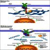
Proposed EPLIN signalling pathways and implications for loss in cancer. When cancer is not present, EPLIN associates with the actin cytoskeleton linking the cadherin–catenin complex to F-actin via interaction with α-catenin. The signal transduction protein, paxillin, interacts with EPLIN in the cytoplasm, and this complex likely stabilises actin dynamics. ERK phosphorylates EPLIN regulating cell motility and migration. When cancer is present and EPLIN is lost, the actin cytoskeleton becomes less organised and this induces membrane ruffling. Paxillin targeting is likely lost reducing focal adhesion between the cadherin–catenin complex and actin. These molecular, cellular and morphological consequences may result in increased metastatic potential including enhanced cell migration and motility. Signalling pathways summarised from [12, 16, 20, 47]. Image generated using Pathway Builder 2.0 software

References

    1. Stewart, B., & Wild, C. (2014). World Cancer Report 2014.
    1. CancerResearchUK (2015). Cancer stats: cancer statistics for the UK. http://www.cancerresearchuk.org/cancer-info/cancerstats/. Accessed 20th November 2014.
    1. Chang DD, Park NH, Denny CT, Nelson SF, Pe M. Characterization of transformation related genes in oral cancer cells. Oncogene. 1998;16(15):1921–1930. doi: 10.1038/sj.onc.1201715. - DOI - PubMed
    1. Maul RS, Chang DD. EPLIN, epithelial protein lost in neoplasm. Oncogene. 1999;18(54):7838–7841. doi: 10.1038/sj.onc.1203206. - DOI - PubMed
    1. Chen S, Maul RS, Kim HR, Chang DD. Characterization of the human EPLIN (Epithelial Protein Lost in Neoplasm) gene reveals distinct promoters for the two EPLIN isoforms. Gene. 2000;248(1–2):69–76. doi: 10.1016/S0378-1119(00)00144-X. - DOI - PubMed

Publication types

LinkOut - more resources