. 2016 Jan;241(2):177-83.
doi: 10.1177/1535370215600548. Epub 2015 Sep 7.

Hypoxia promotes apoptosis of neuronal cells through hypoxia-inducible factor-1α-microRNA-204-B-cell lymphoma-2 pathway

Affiliations

Hypoxia promotes apoptosis of neuronal cells through hypoxia-inducible factor-1α-microRNA-204-B-cell lymphoma-2 pathway

Xiuwen Wang et al. Exp Biol Med (Maywood). 2016 Jan.

Abstract

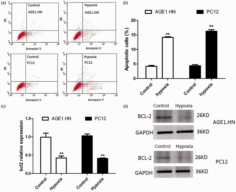
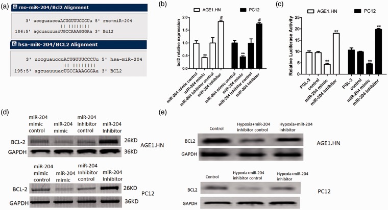
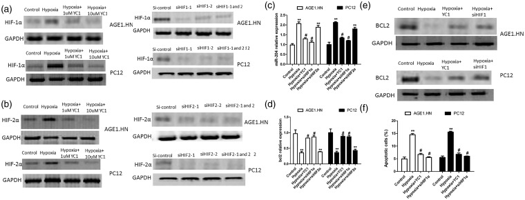
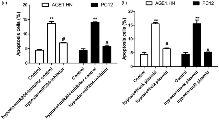
Neuronal cells are highly sensitive to hypoxia and may be subjected to apoptosis when exposed to hypoxia. Several apoptosis-related genes and miRNAs involve in hypoxia-induced apoptosis. This study aimed to examine the role of HIF1α-miR-204-BCL-2 pathway in hypoxia-induced apoptosis in neuronal cells. Annexin V/propidium iodide assay was performed to analyze cell apoptosis in AGE1.HN and PC12 cells under hypoxic or normoxic conditions. The expression of BCL-2 and miR-204 were determined by Western blot and qRT-PCR. The effects of miR-204 overexpression or knockdown on the expression of BCL-2 were evaluated by luciferase assay and Western blot under hypoxic or normoxic conditions. HIF-1α inhibitor YC-1 and siHIF-1α were employed to determine the effect of HIF-1α on the up-regulation of miR-204 and down-regulation of BCL-2 induced by hypoxia. Apoptosis assay showed the presence of apoptosis induced by hypoxia in neuronal cells. Moreover, we found that hypoxia significantly down-regulated the expression of BCL-2, and increased the mRNA level of miR-204 in neuronal cells than that in control. Bioinformatic analysis and luciferase reporter assay demonstrated that miR-204 directly targeted and regulated the expression of BCL-2. Specifically, the expression of BCL-2 was inhibited by miR-204 mimic and enhanced by miR-204 inhibitor. Furthermore, we detected that hypoxia induced cell apoptosis via HIF-1α/miR-204/BCL-2 in neuronal cells. This study demonstrated that HIF-1α-miR-204-BCL-2 pathway contributed to apoptosis of neuronal cells induced by hypoxia, which could potentially be exploited to prevent spinal cord ischemia-reperfusion injury.

Keywords: B-cell lymphoma-2; Spinal cord ischemia–reperfusion injury; hypoxia; hypoxia-inducible factor-1α; microRNA-204; neuronal cells.


Figures

Figure 1
Figure 1
Hypoxia induces apoptosis in neuronal cells and decreases the expression of BCL-2. (a) Exposed to hypoxia or normoxia for 18 h, cell apoptosis was tested by Annexin V/PI flow cytometry in AGE1.HN and PC12 cells. (b) Graphical representation of the data shown in a. (c) qRT-PCR analyses were performed to examine the mRNA expression of BCL-2 in AGE1.HN and PC12 cells. (d) Western blotting was performed to determine BCL-2 protein levels in AGE1.HN and PC12 cells. The data are the mean ± SD. **P < 0.01 vs. control. (A color version of this figure is available in the online journal.)
Figure 2
Figure 2
Hypoxia increases the expression of miR-204 in neuronal cells. (a) The expression of miR-204 in peripheral plasma of patients with spinal cord ischemia–reperfusion. (b) qRT-PCR analyses were performed to examine the expression of miR-204 in AGE1.HN and PC12 cells exposed to hypoxia or normoxia for different duration. The data are the mean ± SD. *P < 0.05 vs. control, **P < 0.01 vs. control
Figure 3
Figure 3
BCL-2 is the target of miR-204. (a) Sequence alignment of miR-204 and 3′-UTR of BCL-2 using mirco-RNA.org. (b) AGE1.HN and PC12 cells were transfected with miR-204 mimic or miR-204 inhibitor or controls under normal oxygen conditions. The expression of BCL-2 was detected with qRT-PCR. (c) Relative luciferase activity in cells transfected with miR-204 mimics (or miR-204 inhibitor) and luciferase expression vector of BCL-2 under normal oxygen conditions. (d) AGE1.HN and PC12 cells were transfected with miR-204 mimic or miR-204 inhibitor or controls under normal oxygen conditions. The protein expression of BCL-2 was detected with Western blotting. (e) AGE1.HN and PC12 cells were transfected with miR-204 inhibitor or controls and then exposed to hypoxia. The protein expression of BCL-2 was detected with Western blotting. The data are the mean ± SD. **P < 0.01 vs. control; #P < 0.05 vs. inhibitor control. (A color version of this figure is available in the online journal.)
Figure 4
Figure 4
Hypoxia increases the expression of miR-204 and reduces the expression of BCL-2 via HIF1-α pathway. (a) AGE1.HN and PC12 cells were treated with 1 µmol/L and 10 µmol/L YC-1 for 6 h and then exposed to hypoxia for 24 h or transfected with siHIF-1α. The protein expression of HIF1α was detected with Western blotting. (b) AGE1.HN and PC12 cells were treated with 1 µmol/L and 10 µmol/L YC-1 for 6 h and then exposed to hypoxia for 24 h or transfected with siHIF-2α. The protein expression of HIF-2α was detected with Western blotting. (c) The mRNA level of miR-204 was detected in cells transfected with YC-1 or siHIF-1α or siHIF-2α and exposed to hypoxia. (d) The mRNA level of BCL-2 was detected in cells transfected with YC-1 or siHIF-1α or siHIF-2α and exposed to hypoxia. (e) The protein level of BCL-2 was detected in AGE1.HN and PC12 cells transfected with YC-1 and siHIF-1α and exposed to hypoxia. (f) The percentage of apoptotic cells was detected by flow cytometry in AGE1.HN and PC12 cells transfected with YC-1 or siHIF-1α. The data are the mean ± SD. **P < 0.01 vs. control; #P < 0.05 vs. hypoxia
Figure 5
Figure 5
Hypoxia induces cell apoptosis via miR-204 and BCL-2. (a) AGE1.HN and PC12 cells were transfected with miR-204 inhibitor or controls and then exposed to hypoxia. Flow cytometry was performed to detect the levels of apoptosis. (b) AGE1.HN and PC12 cells were transfected with BCL-2 plasmid or controls and then exposed to hypoxia. Flow cytometry was performed to detect the levels of apoptosis. The data are the mean ± SD. **P < 0.01 vs. control; #P < 0.05 vs. hypoxia

References

    1. Li Y, Gu J, Liu Y, Long H, Wang G, Yin G, Fan J. iNOS participates in apoptosis of spinal cord neurons via p-BAD dephosphorylation following ischemia/reperfusion (I/R) injury in rat spinal cord. Neurosci Lett 2013; 545: 117–22. - PubMed
    1. Nazli Y, Colak N, Namuslu M, Erdamar H, Haltas H, Alpay MF, Aksoy ON, Akkaya IO, Cakir O. Cilostazol attenuates spinal cord ischemia-reperfusion injury in rabbits. J Cardiothorac Vasc Anesth. Epub ahead of print 2014. - PubMed
    1. Yavuz C, Demirtas S, Guclu O, Karahan O, Caliskan A, Yazici S, Mavitas B. Rosuvastatin may have neuroprotective effect on spinal cord ischemia reperfusion injury. CNS Neurol Disord Drug Targets 2013; 12: 1011–6. - PubMed
    1. Savas S, Delibas N, Savas C, Sutcu R, Cindas A. Pentoxifylline reduces biochemical markers of ischemia-reperfusion induced spinal cord injury in rabbits. Spinal Cord 2002; 40: 224–9. - PubMed
    1. Greijer AE, van der Wall E. The role of hypoxia inducible factor 1 (HIF-1) in hypoxia induced apoptosis. J Clin Pathol 2004; 57: 1009–14. - PMC - PubMed

Substances