

Stem Cells in Teeth and Craniofacial Bones
- PMID: 26350960
- PMCID: PMC4622324
- DOI: 10.1177/0022034515603972
Stem Cells in Teeth and Craniofacial Bones
Abstract
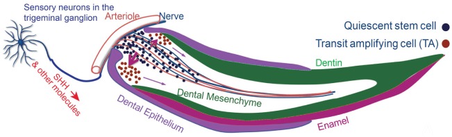
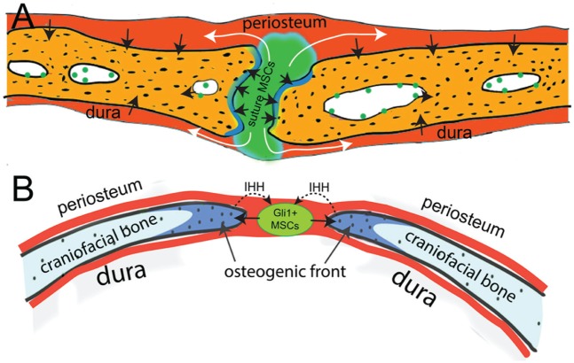
Stem cells are remarkable, and stem cell-based tissue engineering is an emerging field of biomedical science aiming to restore damaged tissue or organs. In dentistry and reconstructive facial surgery, it is of great interest to restore lost teeth or craniofacial bone defects using stem cell-mediated therapy. In the craniofacial region, various stem cell populations have been identified with regeneration potential. In this review, we provide an overview of the current knowledge concerning the various types of tooth- and craniofacial bone-related stem cells and discuss their in vivo identities and regulating mechanisms.
Keywords: Gli1 protein; hedgehogs; mesenchymal stromal cells; neural crest; skull; stem cell niche.
© International & American Associations for Dental Research 2015.
Conflict of interest statement
The authors declare no potential conflicts of interest with respect to the authorship and/or publication of this article.
Figures

References
-
- Akintoye SO, Lam T, Shi S, Brahim J, Collins MT, Robey PG. 2006. Skeletal site-specific characterization of orofacial and iliac crest human bone marrow stromal cells in same individuals. Bone. 38(6):758–768. - PubMed
-
- Aslan H, Zilberman Y, Kandel L, Liebergall M, Oskouian RJ, Gazit D, Gazit Z. 2006. Osteogenic differentiation of noncultured immunoisolated bone marrow-derived CD105+ cells. Stem Cells. 24(7):1728–1737. - PubMed
-
- Awad HA, Butler DL, Boivin GP, Smith FN, Malaviya P, Huibregtse B, Caplan AI. 1999. Autologous mesenchymal stem cell-mediated repair of tendon. Tissue Eng. 5(3):267–277. - PubMed
Publication types
MeSH terms
Grants and funding
LinkOut - more resources
Full Text Sources
Other Literature Sources
Medical
