. 2015 Nov 21;13(43):10716-25.
doi: 10.1039/c5ob01536g.

RGD-conjugated two-photon absorbing near-IR emitting fluorescent probes for tumor vasculature imaging

Affiliations

RGD-conjugated two-photon absorbing near-IR emitting fluorescent probes for tumor vasculature imaging

Xiling Yue et al. Org Biomol Chem. .

Abstract

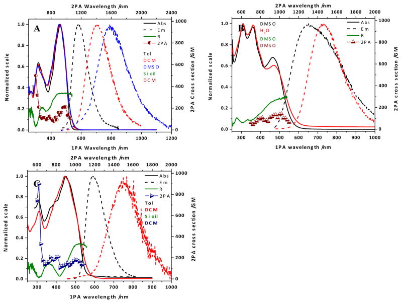
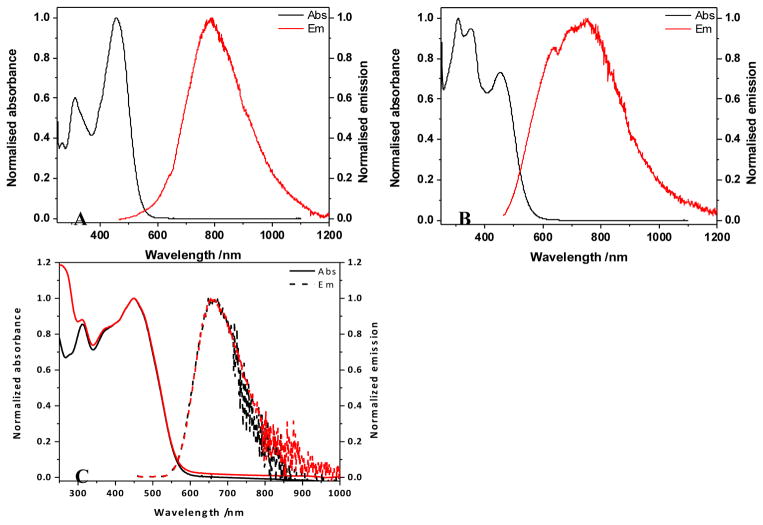
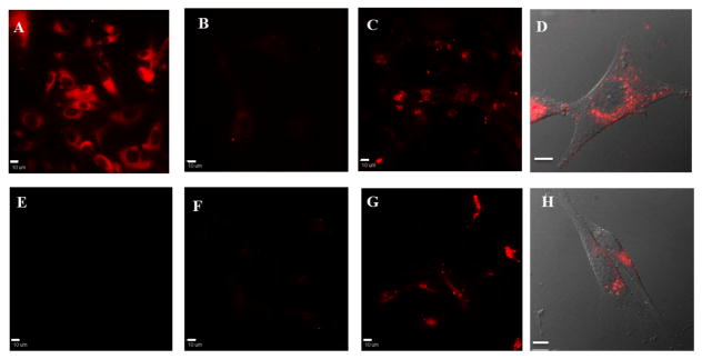
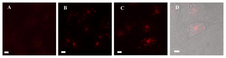
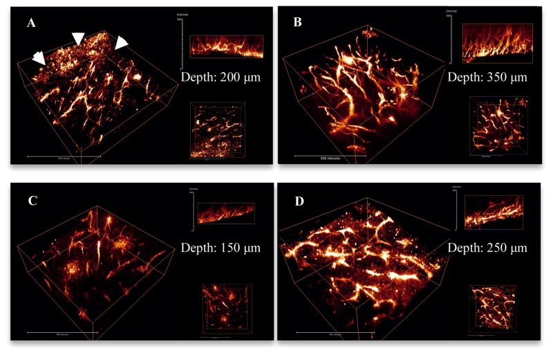
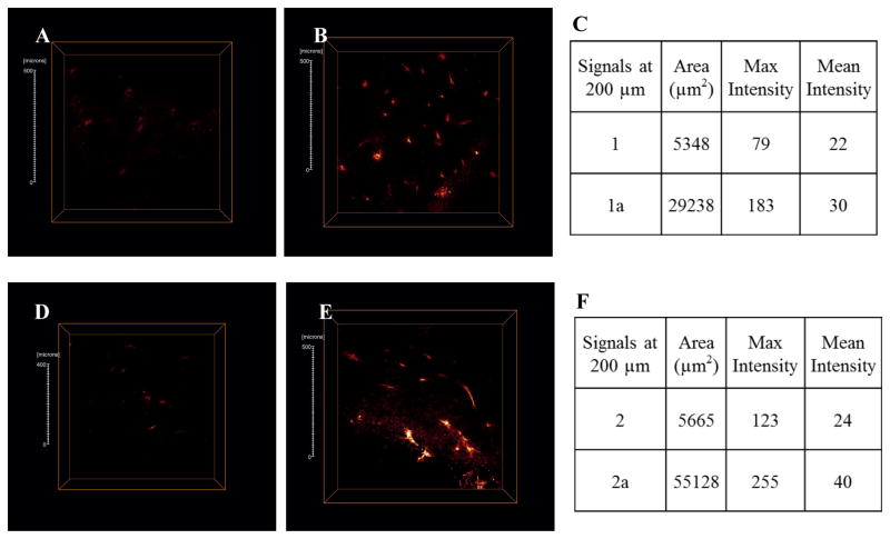
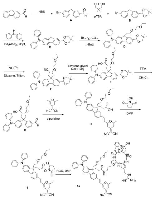
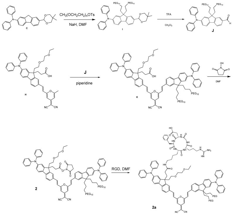
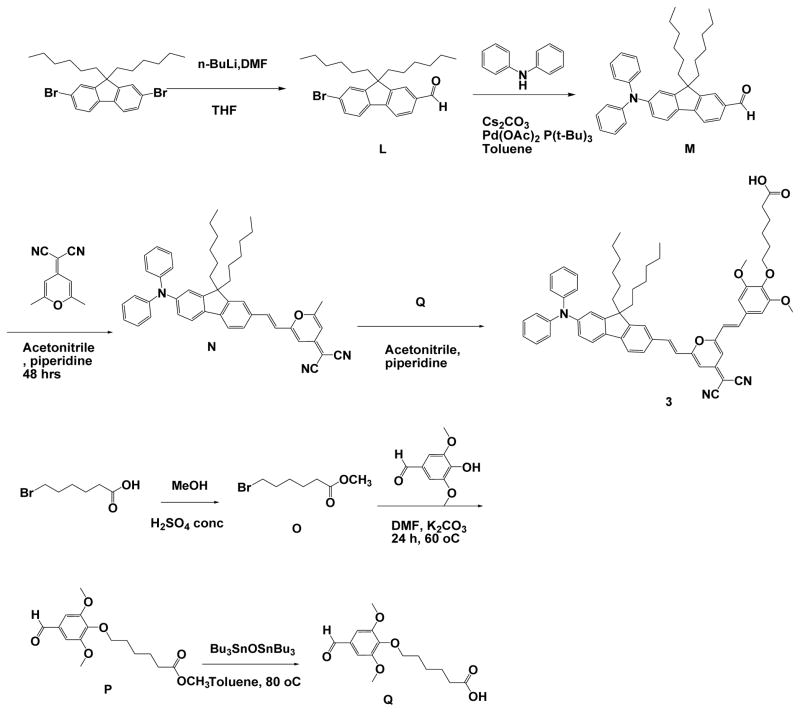
Observation of the activation and inhibition of angiogenesis processes is important in the progression of cancer. Application of targeting peptides, such as a small peptide that contains adjacent L-arginine (R), glycine (G) and L-aspartic acid (D) residues can afford high selectivity and deep penetration in vessel imaging. To facilitate deep tissue vasculature imaging, probes that can be excited via two-photon absorption (2PA) in the near-infrared (NIR) and subsequently emit in the NIR are essential. In this study, the enhancement of tissue image quality with RGD conjugates was investigated with new NIR-emitting pyranyl fluorophore derivatives in two-photon fluorescence microscopy. Linear and nonlinear photophysical properties of the new probes were comprehensively characterized; significantly the probes exhibited good 2PA over a broad spectral range from 700-1100 nm. Cell and tissue images were then acquired and examined, revealing deep penetration and high contrast with the new pyranyl RGD-conjugates up to 350 μm in tumor tissue.


Figures

Figure 1
Figure 1
(A) Absorption (solid lines) and emission (dashed lines) spectra in toluene (black), DCM (red), and DMSO (blue), excitation anisotropy in silicone oil (dark green), and two-photon absorption spectrum in DCM (dark red circles) for 1; (B) absorption (solid lines) and emission (dashed lines) spectra in DMSO (black), and water (red), excitation anisotropy in DMSO (dark green), two-photon absorption spectrum in DMSO (dark red triangles) for 2; (C) absorption (solid lines) an emission (dashed lines) spectra in toluene (black), and DCM (red), excitation anisotropy in silicone oil (dark green), and two-photon absorption spectrum in DCM (dark blue triangles) for 3.
Figure 2
Figure 2
(A) Absorption and emission spectra for 1a in DMSO; (B) Absorption and emission spectra for 2a in DMSO; (C) Absorption (solid lines) and emission (dashed lines) spectra for 3 in organic nanoparticles without, 3a (black), and with, 3b (red) added lecithin.
Figure 3
Figure 3
Size distribution of organic nanoparticles 3a (A) and 3b (B) determined by DLS.
Figure 4
Figure 4
Fluorescence (A–C, E–G) and DIC overlay (D, H) images of U87MG cells after 1 h incubation with 1 (A), 1a (B–D), 2 (E) or 2a (F–H). B and F were pre-incubated with free RGD peptide. Scale bars are 10 μm.
Figure 5
Figure 5
Fluorescence (A–C) and DIC overlay (D) images of HCT116 cells after 1 h incubation with medium (A), 3a (B), or 3b (C, D). Scale bars are 10 μm.
Figure 6
Figure 6
3D reconstruction images show the vasculature in LLC tumor implants that were extracted from mice injected with 1 (A), 1a (B), 2 (C) and 2a (D). Arrows in A indicate non-specific uptake of 1. Each image shows 3D view (left), x-z view (right top) and x-y view (right bottom). Scale bars are 50 μm.
Figure 7
Figure 7
Optical cross-section fluorescence at 200 μm depth for 1 (A), 1a (B), 2 (D) and 2a (E) were analyzed. Scale bars are 50 μm.
Scheme 1
Scheme 1
Synthesis of compounds 1 and 1a.
Scheme 2
Scheme 2
Synthesis of compounds 2 and 2a.
Scheme 3
Scheme 3
Synthesis of compounds 3.

References

    1. Fukumura D, Duda DG, Munn LL, Jain RK. Microcirculation. 2010;17(3):206–225. - PMC - PubMed
    1. Provenzano PP, Eliceiri KW, Keely PJ. Clin Exp Metastasis. 2009;26(4):357–370. - PubMed
    2. Tozer GM, Ameer-Beg SM, Baker J, Barber PR, Hill SA, Hodgkiss RJ, Locke R, Prise VE, Wilson I, Vojnovic B. Adv Drug Delivery Rev. 2005;57(1):135–152. - PubMed
    3. Beerling E, Ritsma L, Vrisekoop N, Derksen PWB, van Rheenen J. J Cell Sci. 2011;124(3):299–310. - PMC - PubMed
    1. Nishida N, Yano H, Nishida T, Kamura T, Kojiro M. Vasc Health Risk Manage. 2006;2(3):213–9. - PMC - PubMed
    1. Avraamides CJ, Garmy-Susini B, Varner JA. Nat Rev Cancer. 2008;8(8):604–617. - PMC - PubMed
    1. Brooks PC, Clark RA, Cheresh DA. Science. 1994;264(5158):569–71. - PubMed
    2. Friedlander M, Brooks PC, Shaffer RW, Kincaid CM, Varner JA, Cheresh DA. Science. 1995;270(5241):1500–2. - PubMed

Publication types

MeSH terms

LinkOut - more resources