Review
. 2015 Dec;57(12):1181-202.
doi: 10.1007/s00234-015-1571-z. Epub 2015 Sep 9.

A neuroradiologist's guide to arterial spin labeling MRI in clinical practice

Affiliations
Review

A neuroradiologist's guide to arterial spin labeling MRI in clinical practice

M Grade et al. Neuroradiology. 2015 Dec.

Abstract

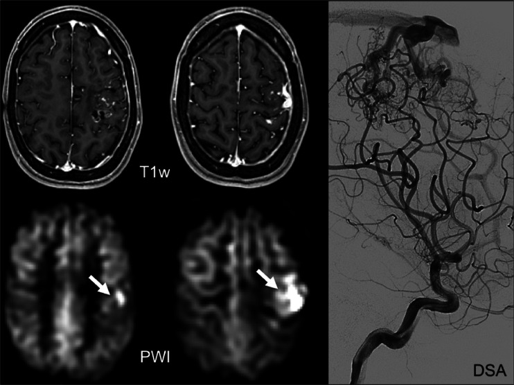
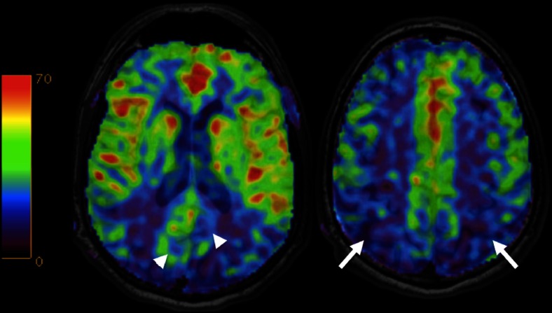
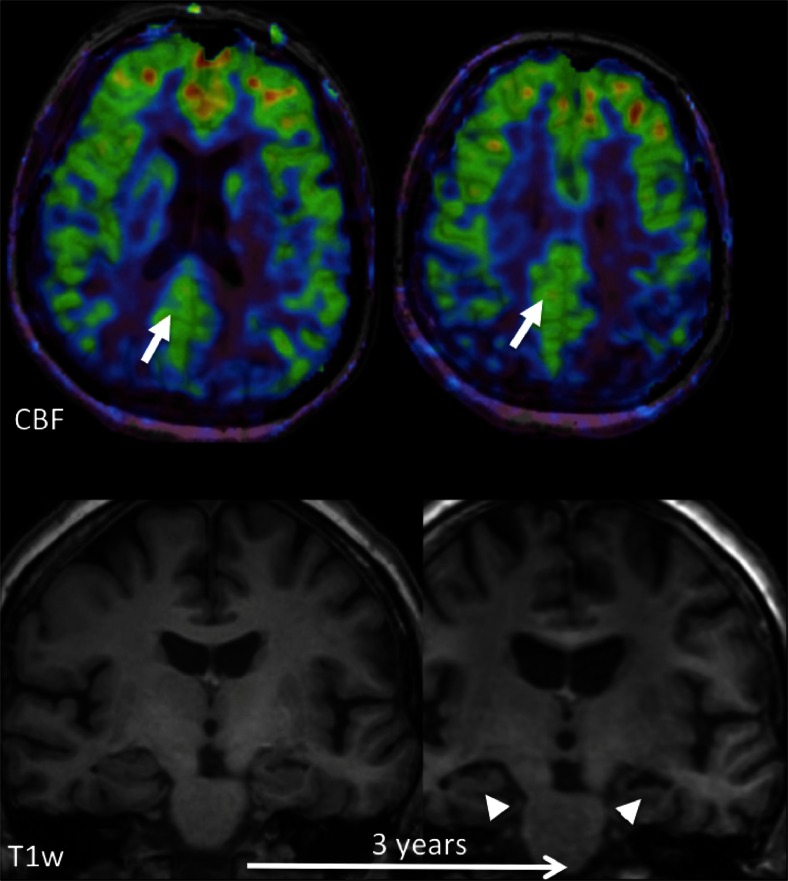
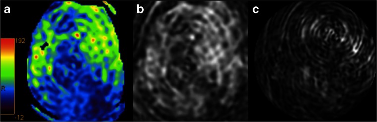
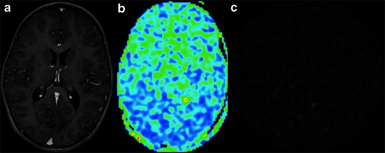
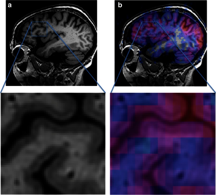
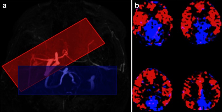
Arterial spin labeling (ASL) is a non-invasive MRI technique to measure cerebral blood flow (CBF). This review provides a practical guide and overview of the clinical applications of ASL of the brain, as well its potential pitfalls. The technical and physiological background is also addressed. At present, main areas of interest are cerebrovascular disease, dementia and neuro-oncology. In cerebrovascular disease, ASL is of particular interest owing to its quantitative nature and its capability to determine cerebral arterial territories. In acute stroke, the source of the collateral blood supply in the penumbra may be visualised. In chronic cerebrovascular disease, the extent and severity of compromised cerebral perfusion can be visualised, which may be used to guide therapeutic or preventative intervention. ASL has potential for the detection and follow-up of arteriovenous malformations. In the workup of dementia patients, ASL is proposed as a diagnostic alternative to PET. It can easily be added to the routinely performed structural MRI examination. In patients with established Alzheimer's disease and frontotemporal dementia, hypoperfusion patterns are seen that are similar to hypometabolism patterns seen with PET. Studies on ASL in brain tumour imaging indicate a high correlation between areas of increased CBF as measured with ASL and increased cerebral blood volume as measured with dynamic susceptibility contrast-enhanced perfusion imaging. Major advantages of ASL for brain tumour imaging are the fact that CBF measurements are not influenced by breakdown of the blood-brain barrier, as well as its quantitative nature, facilitating multicentre and longitudinal studies.

Keywords: Arterial spin labeling; Brain tumour; Dementia; Perfusion; Stroke.


Figures

Fig. 1
Fig. 1
Capillary bed where water and nutrient exchange with the brain parenchyma takes place
Fig. 2
Fig. 2
Schematic of the three periods in any ASL sequence: (I) labeling/control period, (II) post-labeling delay (PLD), to allow for the magnetisation to reach and exchange with the tissue, and (III)) imaging period, during which the images of the brain are acquired. a Labeling acquisition. b Control acquisition. The difference between the two acquisitions only lies in the type of radiofrequency pulse and gradient applied during the first part of the sequence (yes or no labeling)
Fig. 3
Fig. 3
Labeling slice prescription (in blue) in sagittal and coronal views for (p)CASL. Feeding arteries are shown in purple
Fig. 4
Fig. 4
Labeling slice prescription (in blue) in sagittal and coronal views for PASL. Feeding arteries are shown in purple
Fig. 5
Fig. 5
Cerebral blood flow at different ages, for grey matter, white matter, and their ratio. Reprinted with permission from [37]
Fig. 6
Fig. 6
Incorrect (left) and correct (right) positioning of the labeling plane. The labeling plane needs to be placed perpendicular to the feeding arteries and sources of susceptibility artefact (such as air in the sinuses) should be avoided
Fig. 7
Fig. 7
Labeling failure in the left internal carotid artery. There is absence of signal on the ASL perfusion-weighted images (PWI) and corresponding cerebral blood flow (CBF) maps in the arterial territory of the left internal carotid artery. Note the large susceptibility artefact in the area of the left internal carotid area on the T2* weighted (T2*w) image, as the likely cause of the labeling failure. Diffusion weighted imaging (DWI) is normal, supporting interpretation of this finding as an artefact
Fig. 8
Fig. 8
Acute ischaemia in the left occipital lobe, with diffusion restriction on the diffusion-weighted image (DWI) and apparent diffusion coefficient (ADC) map, and high signal intensity on the T2 weighted (T2w) images. The colour-coded cerebral blood flow (CBF) map shows hypoperfusion in the ischaemic region, with the arrow indicating the residual vascular signal in the arteries feeding the ischaemic tissue (arterial transfer artefact: ATA)
Fig. 9
Fig. 9
Post-contrast T1 weighted (T1w: top row) and perfusion-weighted images (PWI: bottom row) obtained with ASL of an arteriovenous malformation as evidenced by digital subtraction angiography (right column: DSA). The arrows indicate high signal in the draining veins
Fig. 10
Fig. 10
Colour-coded cerebral blood flow maps acquired with ASL overlaid on structural T1w images show hypoperfusion in the precuneus and posterior cingulate cortex (arrowheads) and posterior parietal cortex (arrows) bilaterally consistent with Alzheimer’s disease
Fig. 11
Fig. 11
Early perfusion changes in Alzheimer’s disease (AD). Top row: colour-coded cerebral blood flow (CBF) maps acquired with ASL overlaid on structural T1w images at baseline; bottom row: coronal reconstructions at the level of the hippocampus at baseline and after 3 years. At baseline, hippocampal volume is normal, but hypoperfusion in the posterior cingulate cortex/precuneus (arrows) already indicates AD. Note that the hypoperfusion may easily be missed, as there is no clear perfusion deficit. Perfusion in this area however should be much higher than the rest of the cortex, while here it is similar to the rest of the cortex. This is abnormal. After 3 years, structural changes consistent with AD, i.e., hippocampal and global atrophy, also become visible
Fig. 12
Fig. 12
Colour-coded CBF map (a) with severe motion artefacts. The artefacts can easily be appreciated on the source images (b, c) as linear and spiral patterns
Fig. 13
Fig. 13
ASL acquisition—inadvertently—after contrast administration (a). While the post-processing software will provide a ‘CBF’ map (b), the source images (c) clearly show random noise without any signal
Fig. 14
Fig. 14
Zoomed in sagittal views of high-resolution T1-weighted images without (a) and with (b) CBF map overlay. Coloured ASL voxels include grey matter, white matter, and cerebrospinal fluid simultaneously, illustrating the partial volume effect. Partial volume effects can be dealt with by using a post-processing technique called partial volume correction [181]
Fig. 15
Fig. 15
Positioning of the selective labeling planes for territorial ASL based on an MR angiogram of the circle of Willis for the right internal carotid artery (ICA, red box) and the basilar artery (blue box). The labeling plane for the left ICA is not shown, as this vessel was occluded and no signal was obtained. b Colour territorial perfusion maps showing the distribution of brain tissue perfused by the intracranial arteries labeled in (a). The anterior and middle cerebral artery territories in both hemispheres are supplied by the right ICA in this patient with left ICA occlusion (in red). The medial occipital and parietal lobes are supplied by the basilar artery (in blue)

References

    1. Wintermark M, Sesay M, Barbier E, et al. Comparative overview of brain perfusion imaging techniques. Stroke. 2005;36(9):e83–e99. doi: 10.1161/01.STR.0000177884.72657.8b. - DOI - PubMed
    1. Williams DS, Detre JA, Leigh JS, Koretsky AP. Magnetic resonance imaging of perfusion using spin inversion of arterial water. Proc Natl Acad Sci U S A. 1992;89(1):212–216. doi: 10.1073/pnas.89.1.212. - DOI - PMC - PubMed
    1. Sadowski EA, Bennett LK, Chan MR, et al. Nephrogenic systemic fibrosis: risk factors and incidence estimation. Radiology. 2007;243(1):148–157. doi: 10.1148/radiol.2431062144. - DOI - PubMed
    1. Wang J, Licht DJ, Jahng G-H, et al. Pediatric perfusion imaging using pulsed arterial spin labeling. J Magn Reson Imaging. 2003;18(4):404–413. doi: 10.1002/jmri.10372. - DOI - PubMed
    1. Wolf RL, Detre JA. Clinical neuroimaging using arterial spin-labeled perfusion magnetic resonance imaging. Neurotherapeutics. 2007;4(3):346–359. doi: 10.1016/j.nurt.2007.04.005. - DOI - PMC - PubMed

Publication types