

Metachronous Bilateral Testicular Leydig-Like Tumors Leading to the Diagnosis of Congenital Adrenal Hyperplasia (Adrenogenital Syndrome)
- PMID: 26351608
- PMCID: PMC4553183
- DOI: 10.1155/2015/459318
Metachronous Bilateral Testicular Leydig-Like Tumors Leading to the Diagnosis of Congenital Adrenal Hyperplasia (Adrenogenital Syndrome)
Abstract
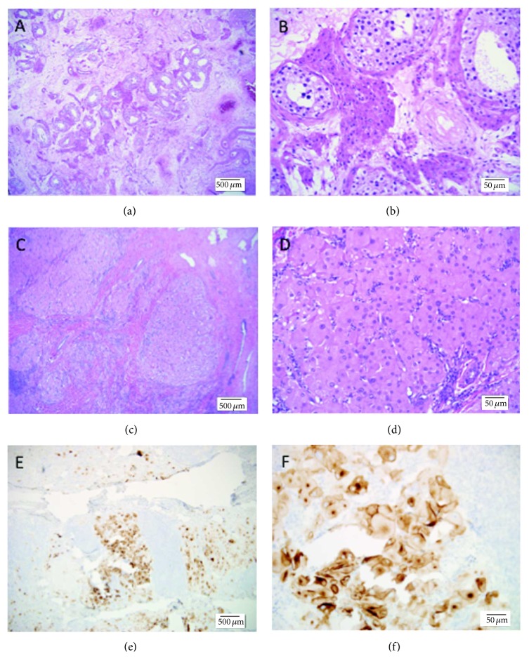
A 33-year-old male with a history of left testis Leydig cell tumor (LCT), 3-month status after left radical orchiectomy, presented with a rapidly enlarging (0.6 cm to 3.7 cm) right testicular mass. He underwent a right radical orchiectomy, sections interpreted as showing a similar Leydig cell-like oncocytic proliferation, with a differential diagnosis including metachronous bilateral LCT and metachronous bilateral testicular tumors associated with congenital adrenal hyperplasia (a.k.a. "testicular adrenal rest tumors" (TARTs) and "testicular tumors of the adrenogenital syndrome" (TTAGS)). Additional workup demonstrated a markedly elevated serum adrenocorticotropic hormone (ACTH) and elevated adrenal precursor steroid levels. He was diagnosed with congenital adrenal hyperplasia, 3β-hydroxysteroid dehydrogenase deficiency (3BHSD) type, and started on treatment. Metachronous bilateral testicular masses in adults should prompt consideration of adult presentation of CAH. Since all untreated CAH patients are expected to have elevated serum ACTH, formal exclusion of CAH prior to surgical resection of a testicular Leydig-like proliferation could be accomplished by screening for elevated serum ACTH.
Figures
Similar articles
-
Extensive Bilateral Adrenal Rest Testicular Tumors in a Patient With 3β-Hydroxysteroid Dehydrogenase Type 2 Deficiency.J Endocr Soc. 2018 May 1;2(6):513-517. doi: 10.1210/js.2018-00082. eCollection 2018 Jun 1. J Endocr Soc. 2018. PMID: 29850650 Free PMC article.
-
Congenital adrenal hyperplasia presented with bilateral testicular tumor: A case report.Caspian J Intern Med. 2021;12(Suppl 2):S431-S434. doi: 10.22088/cjim.12.0.431. Caspian J Intern Med. 2021. PMID: 34760099 Free PMC article.
-
Tumors of the adrenogenital syndrome: an aggressive conservative approach.J Urol. 1998 Nov;160(5):1838-41. J Urol. 1998. PMID: 9783971
-
Bilateral testicular masses and adrenal insufficiency: is congenital adrenal hyperplasia the only possible diagnosis? First two cases of TARTS described in Addison-only X-linked adrenoleukodystrophy and a brief review of literature.J Endocrinol Invest. 2021 Mar;44(3):391-402. doi: 10.1007/s40618-020-01362-x. Epub 2020 Jul 20. J Endocrinol Invest. 2021. PMID: 32691371 Review.
-
Leydig cell tumour and giant adrenal myelolipoma associated with adrenogenital syndrome: a case report with a review of the literature.Urologia. 2016 Jan-Mar;83(1):43-8. doi: 10.5301/uro.5000162. Epub 2015 Dec 4. Urologia. 2016. PMID: 26660266 Review.
Cited by
-
The legacy of the F344 rat as a cancer bioassay model (a retrospective summary of three common F344 rat neoplasms).Crit Rev Toxicol. 2016 Sep;46(8):641-75. doi: 10.1080/10408444.2016.1174669. Epub 2016 Jun 9. Crit Rev Toxicol. 2016. PMID: 27278595 Free PMC article. Review.
-
GATA transcription factors in testicular adrenal rest tumours.Endocr Connect. 2017 Nov;6(8):866-875. doi: 10.1530/EC-17-0215. Epub 2017 Oct 16. Endocr Connect. 2017. PMID: 29038332 Free PMC article.
-
Approach of Heterogeneous Spectrum Involving 3beta-Hydroxysteroid Dehydrogenase 2 Deficiency.Diagnostics (Basel). 2022 Sep 7;12(9):2168. doi: 10.3390/diagnostics12092168. Diagnostics (Basel). 2022. PMID: 36140569 Free PMC article. Review.
-
Management of testicular Leydig cell tumor: A case report.Medicine (Baltimore). 2018 Jun;97(25):e11158. doi: 10.1097/MD.0000000000011158. Medicine (Baltimore). 2018. PMID: 29924022 Free PMC article.
-
Testicular leydig cell tumor revealed by hydrocele.Urol Case Rep. 2020 Dec 5;35:101520. doi: 10.1016/j.eucr.2020.101520. eCollection 2021 Mar. Urol Case Rep. 2020. PMID: 33318945 Free PMC article.
References
-
- Eble J., Sauter G., Epstein J., et al. World Health Organization Classification of Tumours. Pathology and Genetics of Tumours of the Urinary System and Male Genital Organs. Paris, France: IARC Press; 2004.
-
- Wein A., Kavoussi L. R., Novick A. C., et al. Campbell-Walsh Urology. 10th. chapter 31 2010. Neoplasm of the testis.
-
- Ulbright T. M., Young R. H. Tumors of the Testis and Adjacent Structures. 18th. American Registry of Pathology; 2013. (AFIP Atlas of Tumor Pathology Series 4, Fascicle).
-
- Al-Ahmadie H. Pediatric Pathology. Alphen aan den Rijn, The Netherlands: Wolters Kluwer; 2011. Testicular tumors.
LinkOut - more resources
Full Text Sources
Other Literature Sources
