. 2015 Sep 22;112(38):11876-80.
doi: 10.1073/pnas.1509929112. Epub 2015 Sep 8.

Rapid and transient palmitoylation of the tyrosine kinase Lck mediates Fas signaling

Affiliations

Rapid and transient palmitoylation of the tyrosine kinase Lck mediates Fas signaling

Askar M Akimzhanov et al. Proc Natl Acad Sci U S A. .

Abstract

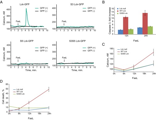
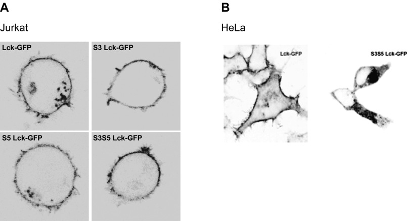
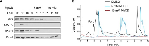
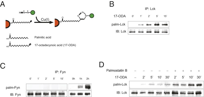
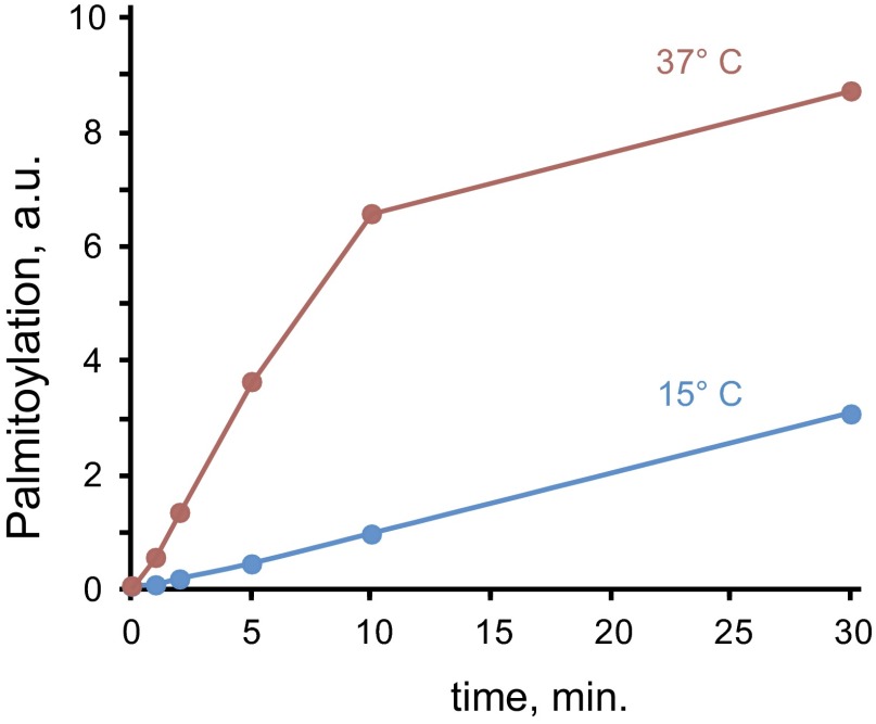
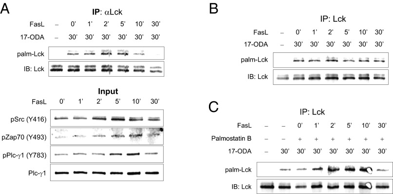
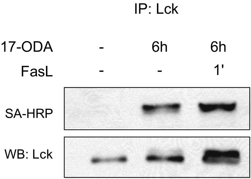
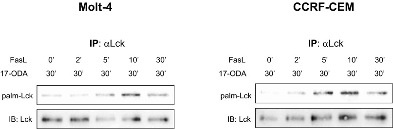
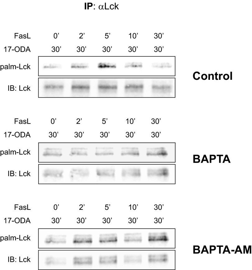
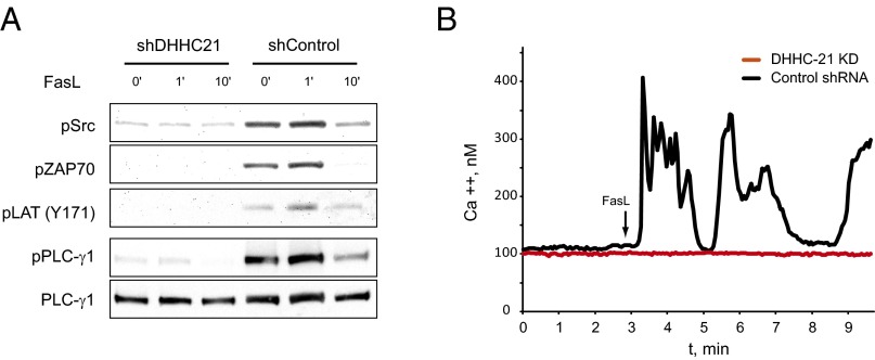
Palmitoylation is the posttranslational modification of proteins with a 16-carbon fatty acid chain through a labile thioester bond. The reversibility of protein palmitoylation and its profound effect on protein function suggest that this modification could play an important role as an intracellular signaling mechanism. Evidence that palmitoylation of proteins occurs with the kinetics required for signal transduction is not clear, however. Here we show that engagement of the Fas receptor by its ligand leads to an extremely rapid and transient increase in palmitoylation levels of the tyrosine kinase Lck. Lck palmitoylation kinetics are consistent with the activation of downstream signaling proteins, such as Zap70 and PLC-γ1. Inhibiting Lck palmitoylation not only disrupts proximal Fas signaling events, but also renders cells resistant to Fas-mediated apoptosis. Knockdown of the palmitoyl acyl transferase DHHC21 eliminates activation of Lck and downstream signaling after Fas receptor stimulation. Our findings demonstrate highly dynamic Lck palmitoylation kinetics that are essential for signaling downstream of the Fas receptor.

Keywords: Fas; Lck; apoptosis; calcium; protein palmitoylation.


Conflict of interest statement

The authors declare no conflict of interest.

Figures

Fig. 1.
Fig. 1.
Palmitoylation of Lck is required for Fas-mediated apoptosis. (A) Fura-2 calcium imaging of Lck-deficient JCam1.6 Jurkat cells transiently transfected with Lck-GFP, S3 Lck-GFP, S5 Lck-GFP, and S3S5 Lck-GFP expression vectors. Black line, Lck-GFP negative; green line, Lck-GFP positive. (B) Caspase 3 activity in JCam1.6 cells stably transfected with WT and S3S5 Lck-GFP expression vectors at 12 and 24 h after Fas ligand stimulation, relative to untreated cells. (C) Time-dependent increases in cytosolic calcium concentration in JCam1.6 cells stably transfected with WT and S3S5 Lck-GFP expression vectors after Fas ligand treatment. (D) Cell death (propidium iodide-positive cells as a percentage of the total) in JCam1.6 cells stably transfected with WT and S3S5 Lck-GFP expression vectors at 12 and 24 h after Fas ligand stimulation. Data in B–D are presented as mean ± SEM from three separate determinations.
Fig. S1.
Fig. S1.
Intracellular localization of palmitoylated Lck. Confocal images of (A) JCam1.6 (Lck null) Jurkat cells and (B) HeLa cells transiently transfected with Lck-GFP, S3 Lck-GFP, S5 Lck-GFP, and S3S5 Lck-GFP expression vectors.
Fig. S2.
Fig. S2.
MβCD inhibits Fas-mediated PLC-γ1 activation and calcium release. (A) Jurkat cells preincubated with 5 mM and 10 mM MβCD for 30 min and treated with Fas ligand for 0, 1, and 10 min. Total cell lysates were analyzed by Western blot analysis. (B) Jurkat cells pretreated for 5 min with MβCD. Intracellular calcium release was determined with Fura-2 imaging.
Fig. 2.
Fig. 2.
Rapid turnover of Lck palmitate in unstimulated cells. (A) Schematic of 17-ODA metabolic labeling and detection of palmitoylated proteins using the click chemistry reaction. (B) Lck palmitate turnover kinetics. Jurkat cells were incubated with 1 μM 17-ODA for the indicated times. Palm-Lck, palmitoylated Lck detected by infrared imaging of labeled 17-ODA; IB, immunoblot of total Lck. (C) Fyn palmitate turnover kinetics. (Left) No labeling was noted after 15 min of 17-ODA incubation at room temperature. (Right) Labeling became evident after 1 h of incubation at 37 °C. (D) Lck palmitate turnover kinetics in the presence of APT1 inhibitor palmostatin B. Jurkat cells were pretreated with 10 μM palmostatin B or DMSO for 30 min before the addition of 1 μM 17-ODA.
Fig. S3.
Fig. S3.
Temperature dependence of Lck palmitoylation. Palmitoylation was determined as in Fig. 2 at 37 °C or 15 °C, and quantified as the percentage of total Lck. Shown is a representative experiment of three separate determinations.
Fig. 3.
Fig. 3.
Rapid and transient Fas-mediated palmitoylation of Lck. (A) Palmitoylation of Lck in the presence of Fas ligand (Upper) and input representing 5% of total protein extracts (Lower). Jurkat cells were incubated with 1 µM 17-ODA or DMSO at room temperature for 30 min. Fas ligand was added during incubation with 17-ODA for the indicated times. (B) Fas-mediated palmitoylation of Lck in J.gamma1 (PLC-γ1 null) Jurkat cells. (C) Fas-mediated palmitoylation of Lck in the presence of APT1 inhibitor palmostatin B. Jurkat cells were pretreated with 10 µM palmostatin B or DMSO for 30 min before the addition of 1 µM 17-ODA and Fas ligand.
Fig. S4.
Fig. S4.
Metabolic labeling of Jurkat cells for 6 h with 17-ODA masks Fas ligand-dependent palmitoylation of Lck. In contrast to the pulse-labeling protocol, extended incubations with 17-ODA saturates the Lck pool and masks FasL-dependent increases in Lck palmitoylation. Palmitoylated Lck was detected by conjugation of 17-ODA with biotin-azide and detection with streptavidin conjugated to horseradish peroxidase (SA-HRP).
Fig. S5.
Fig. S5.
Fas-mediated palmitoylation of Lck in human leukemic T-cell lines. Molt-4 (Left) and CCRF-CEM (Right) cells were incubated with 1 µM 17-ODA at room temperature for 30 min. Fas ligand and 17-ODA were added during incubation for the indicated times. These results are essentially identical to those obtained in Jurkat cells (Fig. 3A).
Fig. S6.
Fig. S6.
Effect of extracellular (BAPTA) and intracellular (BAPTA-AM) calcium chelators on Fas-mediated Lck palmitoylation. Control Jurkat cells (Top) or Jurkat cells preincubated with 5 mM BAPTA (Middle) or 10 µM BAPTA-AM (Bottom) for 30 min at room temperature were stimulated with Fas ligand in the presence of 17-ODA. Both BAPTA and BAPTA-AM inhibited FasL-dependent palmitoylation.
Fig. 4.
Fig. 4.
Palmitoyl acyltransferase DHHC21 mediates activation of the Fas signaling pathway. (A) Activation of the Fas signaling pathway in Jurkat cells stimulated with Fas ligand in cells transduced with DHHC21-specific (shDHHC21) or control (shControl) shRNA. (B) Fas-dependent calcium release in Jurkat cells expressing DHHC21-specific (shDHHC21) or control (shControl) shRNA. Shown are single-cell responses representative of hundreds of determinations.

References

    1. Linder ME, Deschenes RJ. Palmitoylation: Policing protein stability and traffic. Nat Rev Mol Cell Biol. 2007;8(1):74–84. - PubMed
    1. Baekkeskov S, Kanaani J. Palmitoylation cycles and regulation of protein function (review) Mol Membr Biol. 2009;26(1):42–54. - PubMed
    1. Ladygina N, Martin BR, Altman A. Dynamic palmitoylation and the role of DHHC proteins in T cell activation and anergy. Adv Immunol. 2011;109:1–44. - PMC - PubMed
    1. Draper JM, Smith CD. Palmitoyl acyltransferase assays and inhibitors (review) Mol Membr Biol. 2009;26(1):5–13. - PMC - PubMed
    1. Ohno Y, Kihara A, Sano T, Igarashi Y. Intracellular localization and tissue-specific distribution of human and yeast DHHC cysteine-rich domain-containing proteins. Biochim Biophys Acta. 2006;1761(4):474–483. - PubMed

Publication types

MeSH terms

Substances

LinkOut - more resources