. 2015 Sep 22;112(38):11959-64.
doi: 10.1073/pnas.1500525112. Epub 2015 Sep 8.

Isoflurane inhibits synaptic vesicle exocytosis through reduced Ca2+ influx, not Ca2+-exocytosis coupling

Affiliations

Isoflurane inhibits synaptic vesicle exocytosis through reduced Ca2+ influx, not Ca2+-exocytosis coupling

Joel P Baumgart et al. Proc Natl Acad Sci U S A. .

Abstract

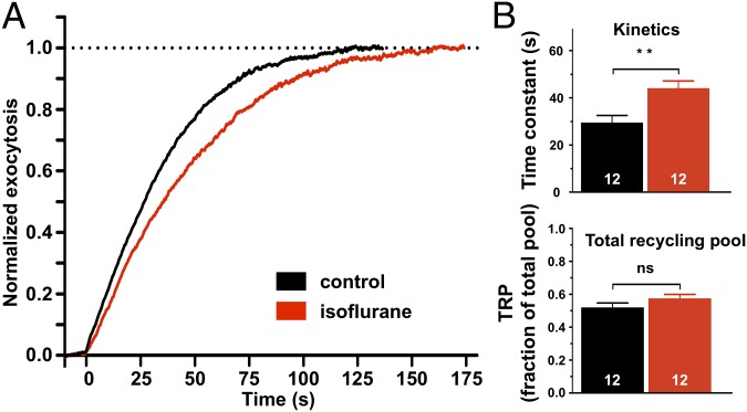
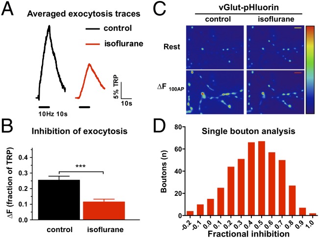
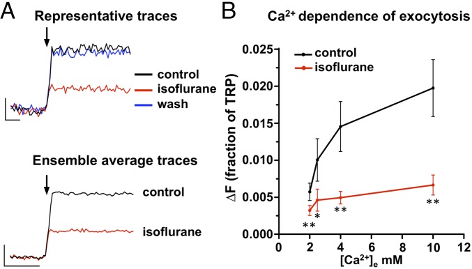
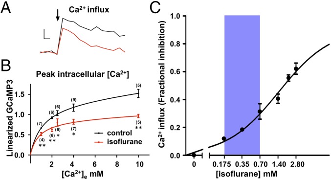
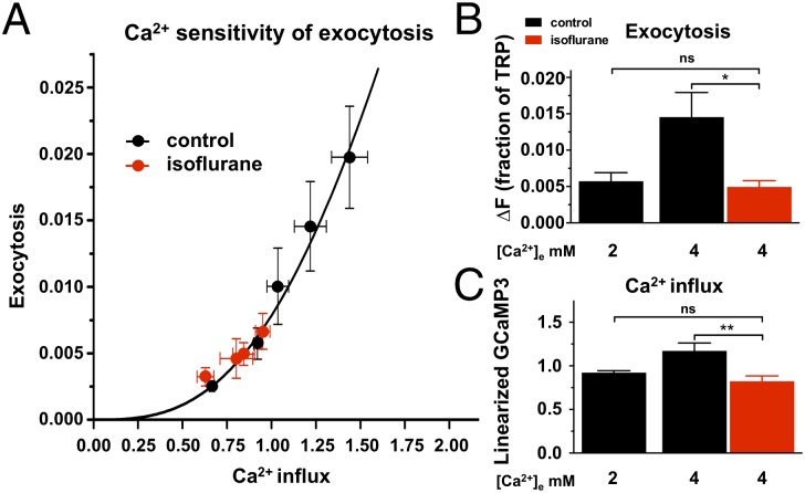
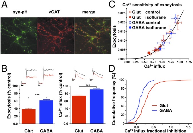
Identifying presynaptic mechanisms of general anesthetics is critical to understanding their effects on synaptic transmission. We show that the volatile anesthetic isoflurane inhibits synaptic vesicle (SV) exocytosis at nerve terminals in dissociated rat hippocampal neurons through inhibition of presynaptic Ca(2+) influx without significantly altering the Ca(2+) sensitivity of SV exocytosis. A clinically relevant concentration of isoflurane (0.7 mM) inhibited changes in [Ca(2+)]i driven by single action potentials (APs) by 25 ± 3%, which in turn led to 62 ± 3% inhibition of single AP-triggered exocytosis at 4 mM extracellular Ca(2+) ([Ca(2+)]e). Lowering external Ca(2+) to match the isoflurane-induced reduction in Ca(2+) entry led to an equivalent reduction in exocytosis. These data thus indicate that anesthetic inhibition of neurotransmitter release from small SVs occurs primarily through reduced axon terminal Ca(2+) entry without significant direct effects on Ca(2+)-exocytosis coupling or on the SV fusion machinery. Isoflurane inhibition of exocytosis and Ca(2+) influx was greater in glutamatergic compared with GABAergic nerve terminals, consistent with selective inhibition of excitatory synaptic transmission. Such alteration in the balance of excitatory to inhibitory transmission could mediate reduced neuronal interactions and network-selective effects observed in the anesthetized central nervous system.

Keywords: GCaMP3; live cell imaging; mechanisms of anesthesia; pHlourin; presynaptic.


Conflict of interest statement

The authors declare no conflict of interest.

Figures

Fig. 1.
Fig. 1.
Effects of isoflurane on synaptic vesicle recycling pool size and exocytosis kinetics. (A) Synaptic vesicle exocytosis kinetics were determined by fitting vGlut-pHluorin fluorescence traces during prolonged action potential (AP) stimulation in the presence of bafilomycin A1 (10 Hz for 150 s; 1,500 APs) for control or 0.7 ± 0.06 mM isoflurane [twice the minimum alveolar concentration (MAC); equivalent to EC50 for immobilization in rat] in the presence of 2 mM extracellular Ca2+ ([Ca2+]e). (B) (Upper) Effect of isoflurane on mean exocytosis time constant. Traces were fit with a single exponential function to determine the time constant for exocytosis of the recycling pool. **P < 0.01 by two-tailed unpaired t test. (Lower) Effect of isoflurane on total recycling pool (TRP) size as a fraction of total pool size (determined using 50 mM NH4Cl), P = 0.21 by two-tailed unpaired t test. n = 12.
Fig. 2.
Fig. 2.
Isoflurane inhibits synaptic vesicle exocytosis evoked by a brief action potential train. (A) Ensemble average traces of a vGlut–pHluorin transfected neuron stimulated at 10 Hz for 10 s (100 APs). Fluorescence intensities were normalized to the peak of a subsequent bafilomycin A1 response (defining the total recycling pool, TRP). (B) Mean values of vGlut–pHluorin response amplitudes for control or 0.7 ± 0.06 mM isoflurane-treated neurons stimulated with 100 APs in the presence of 2 mM extracellular Ca2+ ([Ca2+]e). Average 100 AP response (ΔF100AP) was inhibited 55% by isoflurane, ***P < 0.001 by paired t-test, n = 8. (C) Representative field of vGlut–pHluorin fluorescence images at rest and difference image for 100 AP stimulation (ΔF100AP) for control or isoflurane-treated neurons. (Scale bars: 5 μm.) (D) Histogram of fractional block of vGlut–pHluorin responses in single boutons stimulated with 100 AP. Measurements from 497 boutons collected in eight experiments.
Fig. 3.
Fig. 3.
Reversibility of isoflurane inhibition and Ca2+ dependence of synaptic vesicle exocytosis. (A) (Upper) Representative vGlut–pHlourin fluorescence responses elicited by a single AP at 4 mM extracellular Ca2+ ([Ca2+]e) for control (black), 0.7 mM isoflurane (red), and wash (blue) conditions. Arrow indicates single AP stimulus (n = 8 trials each, ∼25 boutons analyzed per neuron). (Lower) Ensemble average traces of vGlut-pHlourin responses relative to total releasable pool (TRP) size to single AP stimulation at 4 mM extracellular Ca2+ ([Ca2+]e) (10 trial average, ∼25 boutons per neuron). (Scale bar: 0.5% TRP, 100 ms.) (B) Isoflurane inhibition of single AP-stimulated release is not overcome by elevated extracellular Ca2+ ([Ca2+]e). Average relative peak (∆F) evoked by a single AP stimulus expressed as a fraction of the TRP (n = 7–11). *P < 0.05, **P < 0.01 compared with respective control by paired t test. Mean isoflurane concentration 0.70 ± 0.06 mM.
Fig. 4.
Fig. 4.
Isoflurane reduces action potential-stimulated increases in presynaptic Ca2+ influx. (A) Control (black) and 0.70 ± 0.06 mM isoflurane (red) ensemble average traces of single AP-evoked [Ca2+] influx determined by linearized responses synaptophysin–GCaMP3 at 4 mM extracellular Ca2+ ([Ca2+]e). Arrow indicates single AP stimulus. (Scale bar: 30% ∆F/F0, 100 ms.) (B) Inhibition by 0.70 ± 0.06 mM isoflurane of average peak ∆F/F0 evoked by single AP stimulus as a function of [Ca2+]e. Data are fitted to a single site binding model using a Levenberg–Marquardt algorithm optimization method (control: EC50 = 1.3 ± 0.1 mM, Emax = 1.6 ± 0.5, R2 = 99.8; isoflurane: EC50= 0.8 ± 0.4 mM, Emax = 0.9 ± 0.2, R2 = 92.3); both P < 0.05 by two-sample sum-of-squares F test comparison against the null hypothesis that Emax and EC50 values are identical. (C) Inhibition of Ca2+ influx as a function of log [isoflurane] using synaptopysin-GCaMP3 at 4 mM [Ca2+]e (n = 27). Shaded area indicates the clinical concentration range of isoflurane for general anesthesia in rat (0.18–0.7 mM; 0.5–2 × MAC). Data are fitted to a generalized Hill model by least-squares analysis (Imax = 0.92 ± 0.17; IC50 = 1.4 ± 1.0 mM). Inhibition data were all significantly different from paired control values (*P < 0.05 in two-tailed paired t test).
Fig. 5.
Fig. 5.
Effect of isoflurane on synaptic vesicle exocytosis as a function of Ca2+ influx. (A) Exocytosis determined using synaptophysin-pHlourin (ΔF/F) in response to a single AP stimulus relative to total releasable pool (TRP) at 1.5, 2, 2.5, 4, 10 mM [Ca2+]e is plotted against Ca2+ influx determined using synaptophysin-GCaMP3 (ΔF) under similar conditions in the absence or presence of 0.70 ± 0.06 mM isoflurane to reveal the Ca2+ sensitivity of exocytosis (n = 34 for syn-pH experiments, n = 28–33 for syn-GCaMP3 experiments). Data are fitted to a generalized Hill model [exocytosis = [Ca2+]in/(Kn+[Ca2+]in), n = 2.9 ± 0.5, K = 2.8 ± 0.2]. (B) Reduction of [Ca2+]e from 4 mM to 2 mM mimicked the reduction in exocytosis produced by 0.70 ± 0.06 mM isoflurane at 4 mM [Ca2+]e. *P < 0.05 by one-way ANOVA with Tukey’s post hoc test. (C) Reduction of [Ca2+]e from 4 mM to 2 mM mimicked the reduction in syn-GCaMP3 signal produced by 0.70 ± 0.06 mM isoflurane at 4 mM [Ca2+]e. **P < 0.01 by one-way ANOVA with Tukey’s post hoc test.
Fig. 6.
Fig. 6.
Differential effects of isoflurane on synaptic vesicle exocytosis in glutamatergic and GABAergic neurons is proportional to reduction Ca2+ influx. (A) Representative live-cell image of syn-pH-positive boutons (green, Left), GABAergic boutons immunolabeled using vGAT–Oyster 550 (red, Center), and merge shown as yellow (Right), with surrounding vGAT-positive boutons (red, Right) from untransfected neurons. (Scale bar: 20 μm.) (B Left) The effect of 0.70 ± 0.06 mM isoflurane on single AP exocytosis is greater in glutamatergic (vGAT-negative; 62 ± 3% block, n = 11) than in GABAergic (vGAT-positive; 38 ± 3% block, n = 5) boutons at 4 mM extracellular [Ca2+]e, ***P < 0.001 by unpaired t test. (Insets) Exemplar single AP stimulus (arrow) syn-pH fluorescence traces before (black) and after (red) isoflurane in glutamatergic (Left) and GABAergic (Right) boutons. (Scale bars: 0.5% TRP, 100 ms.) (B, Right) Effect of isoflurane on presynaptic Ca2+ influx is greater in glutamatergic (26 ± 2% block, n = 7) than in GABAergic (9.2 ± 2% block, n = 7) boutons at 4 mM extracellular [Ca2+]e, ***P < 0.001 by unpaired t test. (Insets) Exemplar single AP stimulus (arrow) syn-GCaMP3 fluorescence traces before (black) and after (red) isoflurane in glutamatergic and GABAergic boutons. (Scale bars: 30% ∆F/F0, 200 ms.) (C) Effect of 0.70 ± 0.06 mM isoflurane on exocytosis as a function of Ca2+ influx in glutamatergic and GABAergic boutons. Exocytosis determined using syn-pH in response to single AP relative to TRP is plotted against relative increases in Ca2+ influx determined using syn-GCaMP3 over a range of [Ca2+]e from 1.5 to 10 mM under similar conditions to yield the Ca2+ sensitivity of exocytosis (n = 26 for exocytosis, n = 24 for Ca2+ influx). The curve indicates the data from Fig. 5A. (D) Cumulative frequency distribution of 0.70 ± 0.06 mM isoflurane inhibition of peak [Ca2+]i for GABAergic and glutamatergic boutons. Data from 272 boutons collected in 12 experiments.

References

    1. Hemmings HC, Jr, et al. Emerging molecular mechanisms of general anesthetic action. Trends Pharmacol Sci. 2005;26(10):503–510. - PubMed
    1. Alkire MT, Hudetz AG, Tononi G. Consciousness and anesthesia. Science. 2008;322(5903):876–880. - PMC - PubMed
    1. Lewis LD, et al. Rapid fragmentation of neuronal networks at the onset of propofol-induced unconsciousness. Proc Natl Acad Sci USA. 2012;109(49):E3377–E3386. - PMC - PubMed
    1. Blain-Moraes S, et al. Neurophysiological correlates of sevoflurane-induced unconsciousness. Anesthesiology. 2015;122(2):307–316. - PMC - PubMed
    1. Schlame M, Hemmings HC., Jr Inhibition by volatile anesthetics of endogenous glutamate release from synaptosomes by a presynaptic mechanism. Anesthesiology. 1995;82(6):1406–1416. - PubMed

Publication types