. 2015 Sep 29;112(39):E5427-33.
doi: 10.1073/pnas.1516217112. Epub 2015 Sep 8.

Transcellular spreading of huntingtin aggregates in the Drosophila brain

Affiliations

Transcellular spreading of huntingtin aggregates in the Drosophila brain

Daniel T Babcock et al. Proc Natl Acad Sci U S A. .

Abstract

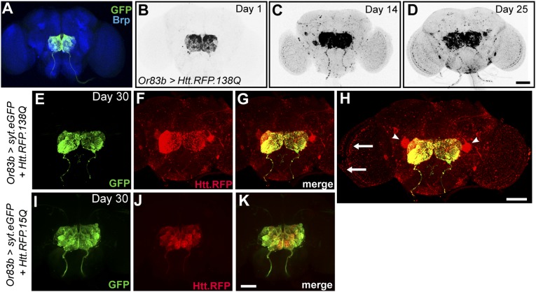
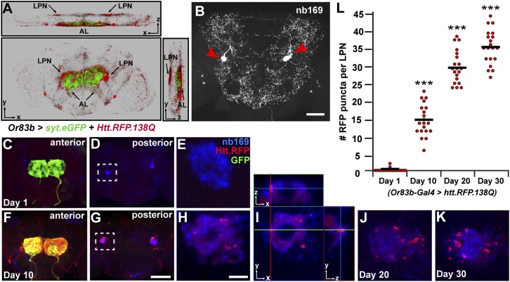
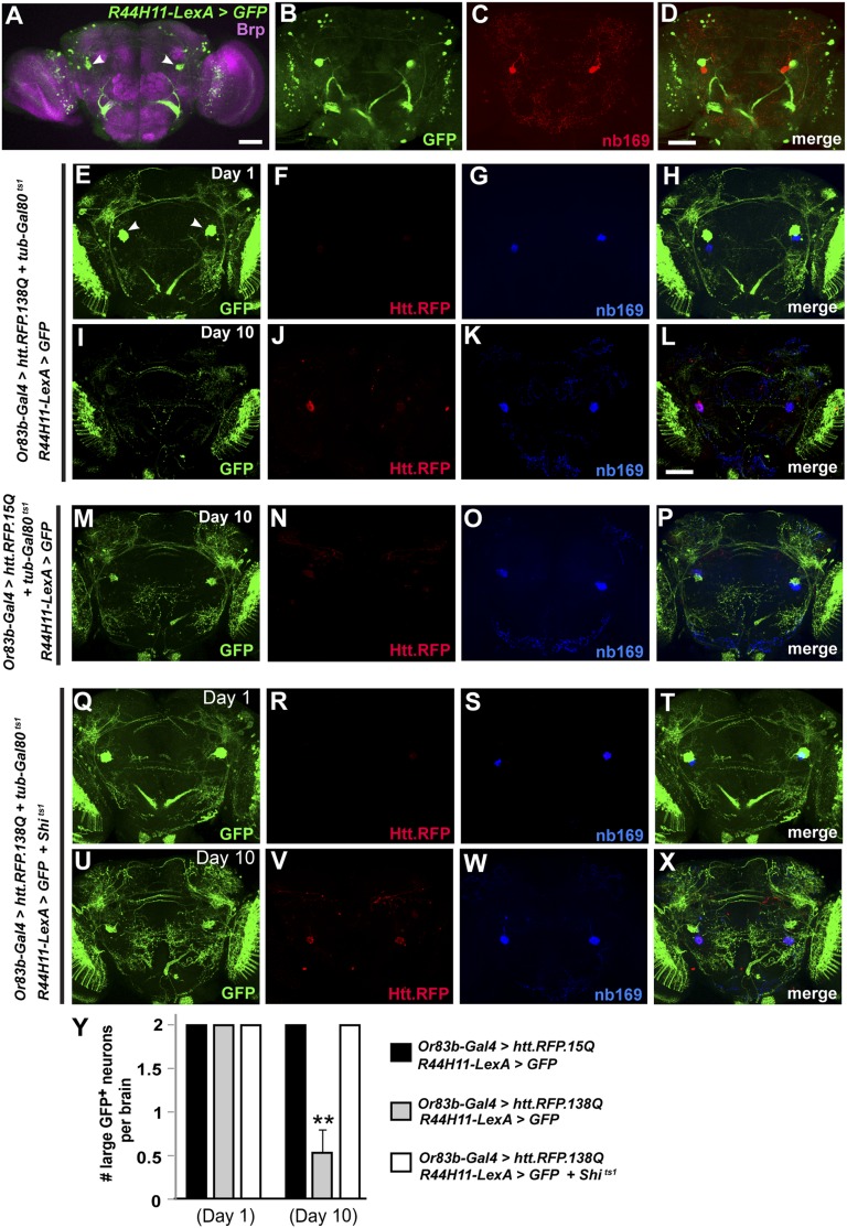
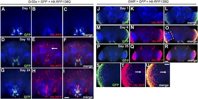
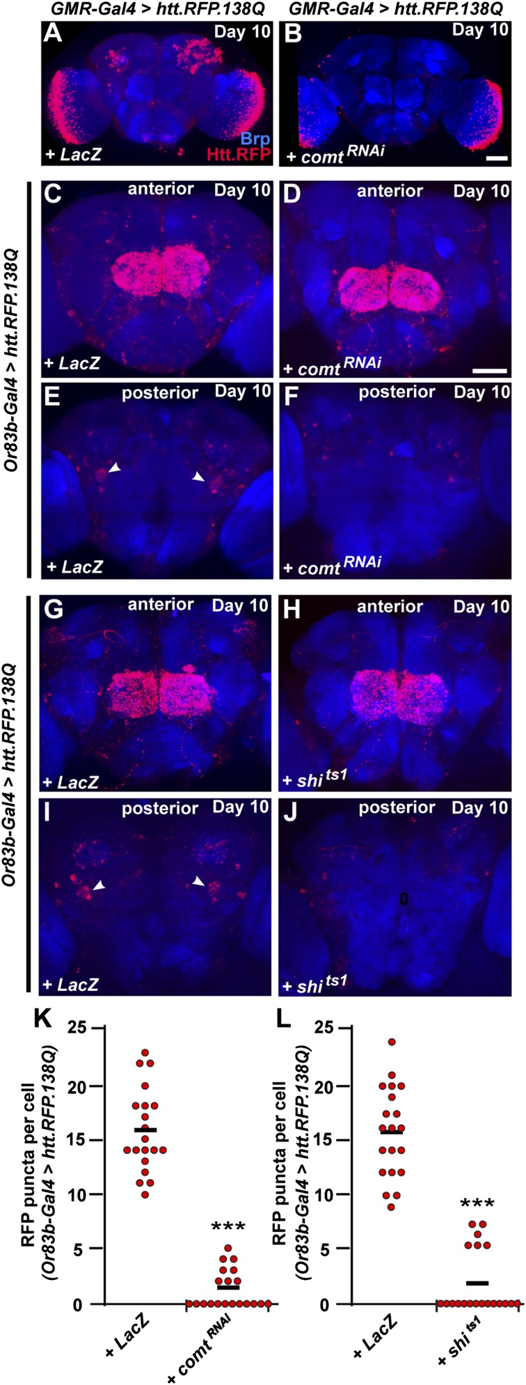
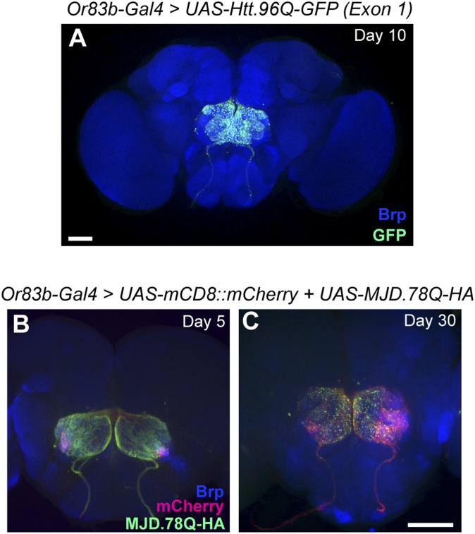
A key feature of many neurodegenerative diseases is the accumulation and subsequent aggregation of misfolded proteins. Recent studies have highlighted the transcellular propagation of protein aggregates in several major neurodegenerative diseases, although the precise mechanisms underlying this spreading and how it relates to disease pathology remain unclear. Here we use a polyglutamine-expanded form of human huntingtin (Htt) with a fluorescent tag to monitor the spreading of aggregates in the Drosophila brain in a model of Huntington's disease. Upon expression of this construct in a defined subset of neurons, we demonstrate that protein aggregates accumulate at synaptic terminals and progressively spread throughout the brain. These aggregates are internalized and accumulate within other neurons. We show that Htt aggregates cause non-cell-autonomous pathology, including loss of vulnerable neurons that can be prevented by inhibiting endocytosis in these neurons. Finally we show that the release of aggregates requires N-ethylmalemide-sensitive fusion protein 1, demonstrating that active release and uptake of Htt aggregates are important elements of spreading and disease progression.

Keywords: Huntington's disease; disease model; expanded triplet repeat; neurodegeneration; transmission.


Conflict of interest statement

The authors declare no conflict of interest.

Figures

Fig. 1.
Fig. 1.
Htt aggregates spread throughout the Drosophila brain. (A) Expression pattern of or83b-Gal4 in the Drosophila brain labeling the antennal lobe. Neuropil is labeled by anti-Brp (blue) (BD). Aggregates of Htt.RFP.138Q expressed in ORNs become more widely distributed throughout the brain as a function of age. (EG) PolyQ-expanded Htt aggregates (red) spread far beyond ORN terminals marked by syt.eGFP (green). (H) Expanded view of G to illustrate Htt aggregates within large posterior neurons (arrowheads) and in the optic lobe (arrows). (IK) Nonpathogenic Htt.RFP is confined to synaptic terminals in the antennal lobe. (Scale bar in D, 50 μm, also applies to AC; scale bar in H, 50 μm; and scale bar in K, 50 μm, also applies to EG, I, and J.)
Fig. 2.
Fig. 2.
Htt aggregates are taken up by large posterior neurons. (A) Three-dimensional projection of the distribution of Htt.RFP aggregates on day 30, as shown from frontal (Bottom), top (Upper), and side (Right panel) views. AL, antennal lobe; LPN, large posterior neurons. (B) Central brain stained with monoclonal antibody nb169. Red arrowheads mark a pair of large posterior neurons in the posterior protocerebrum. (CE) On day 1, Htt.RFP aggregates (red) have started to accumulate within ORN terminals (green) on the anterior side of the brain (C), but not within large posterior cells (blue) (D and E). (FH) By day 10, Htt.RFP aggregates colocalize with GFP in ORN terminals (F) and are now present in posterior cells as well (G and H). E and H are enlarged areas marked by boxes within D and G, respectively. (I) Orthogonal view of a single optical slice from H, demonstrating that Htt.RFP aggregates are localized within large posterior cells. (J and K) Accumulation of Htt.RFP aggregates in large posterior neurons on day 20 (J) and day 30 (K). (L) The number of Htt.RFP aggregates within individual large posterior neurons is quantified at various time points. ***P < 0.001 using Student’s t test. Black bars represent mean values for each condition. (Scale bar in B, 50 μm; scale bar in G, 50 μm, also applies to C, D, and F; and scale bar in H, 5 μm, also applies to E, J, and K.)
Fig. 3.
Fig. 3.
Blocking endocytosis with shibire mutant protects against neurodegeneration. (A) Expression pattern of R44H11-LexA in the adult central brain. Pair of large posterior neurons are marked by white arrowheads. Neuropil is labeled by anti-Brp (pink). (BD) Comparison of large cells labeled by R44H11-LexA (B) and nb169 (C) showing that these labels mark distinct cells (D). (EH) At day 1 following expression of Htt.RFP.138Q in ORNs, few HTT aggregates are observed and both R44H11-LexA > GFP-positive cells and nb169-positive cells are present. (IL) At day 10, spreading of Htt aggregates is noticeable and the GFP-positive cells are no longer detectable, although the nb169-positive cells are still present. (MP) Expression of Htt.RFP.15Q as a control, showing the presence of GFP-positive cells at day 10. (QX) Identical brain images as in EL, but with endocytosis now blocked in R44H11-LexA > GFP-positive cells by coexpression of LexAop-Shits1. Note that at day 10 (UX) under these conditions GFP-positive cells are still present. (Y) Average number of large posterior GFP-positive neurons labeled using R44H11-LexA in all conditions at day 1 and day 10. **P < 0.01 using Student’s t test. (Scale bar in A, 50 μm; scale bar in D, 50 μm, also applies to B and C; scale bar in L, 50 μm, also applies to EK and MX.)
Fig. 4.
Fig. 4.
Expression of Htt.RFP.138Q using different Gal4 drivers results in distinct patterns of aggregate spreading. (AI) Expression pattern of Gr32a-Gal4 (green) and distribution of Htt.RFP aggregates (red) in the adult brain at day 1 (AC), day 10 (DF), and day 24 (GI). Arrow in E points to an axonal projection containing aggregates. Neuropil is labeled by anti-Brp (blue). (JR) Expression pattern of GMR-Gal4 (green) in the adult brain and optic lobes and distribution of Htt.RFP aggregates (red) at day 1 (JL), day 6 (MO), and day 25 (PR). (SU) Enlarged view of the optic lobe from O. Arrow in T and U point to a cell body in the central brain that has taken up aggregates. (Scale bar in I, 50 μm, also applies to AH; scale bar in R, 50 μm, also applies to JQ; and scale bar in U, 20 μm, also applies to S and T.)
Fig. 5.
Fig. 5.
Spreading of Htt aggregates requires exocytosis. (A) Spreading pattern of Htt aggregates (red) into the central brain by day 10 when expressed in photoreceptors using GMR-Gal4. (B) Spreading pattern of Htt aggregates when UAS-comtRNAi is coexpressed to inhibit SNARE-mediated fusion. UAS-LacZ is coexpressed in A to standardize the number of transgenes expressed. (C and D) Anterior view of spreading pattern of Htt aggregates when expressed in ORNs using Or83b-Gal4 along with UAS-LacZ (C) or UAS-comtRNAi (D). (E and F) Posterior view of spreading pattern from C and D, respectively. Arrowheads in E mark large posterior cells with accumulated Htt aggregates in control, but not UAS-comtRNAi brains. (G and H) Anterior view of spreading pattern of Htt aggregates when expressed in ORNs using Or83b-Gal4 along with UAS-LacZ (G) or UAS-Shits1 (H). (I and J) Posterior view of spreading pattern from G and H, respectively. Arrowheads in I mark large posterior cells with accumulated Htt aggregates in control, but not UAS-Shits1 brains. Neuropil is labeled by anti-Brp (blue). (Scale bar in B, 50 μm, also applies to A; scale bar in D, 50 μm, also applies to C and EJ.) (K and L) The number of Htt.RFP aggregates found within large posterior neurons at day 10 in controls compared with UAS-comtRNAi (K) or UAS-Shits1 (L) brains. ***P < 0.001 using Student’s t test. Black bars represent mean values for each condition.
Fig. S1.
Fig. S1.
Not all polyQ aggregates exhibit spreading in Drosophila brain. (A) Expression of polyQ-expanded GFP-tagged Htt exon 1 using or83b-Gal4. GFP-tagged aggregates do not spread beyond ORN terminals at day 10. (B and C) Expression of HA-tagged UAS-MJDtr-Q78 (green) together with UAS-mCD8-mCherry (red) to mark synaptic terminals in ORNs. HA-tagged aggregates do not spread beyond ORN terminals at day 5 (B) or day 30 (C). Neuropil is labeled by anti-Brp (blue). (Scale bar in A, 50 μm; scale bar in C, 50 μm, also applies to B.)

Similar articles

Cited by

References

    1. Braak H, Braak E. Neuropathological stageing of Alzheimer-related changes. Acta Neuropathol. 1991;82(4):239–259. - PubMed
    1. Braak H, et al. Staging of brain pathology related to sporadic Parkinson’s disease. Neurobiol Aging. 2003;24(2):197–211. - PubMed
    1. Braak H, Rüb U, Gai WP, Del Tredici K. Idiopathic Parkinson’s disease: Possible routes by which vulnerable neuronal types may be subject to neuroinvasion by an unknown pathogen. J Neural Transm. 2003;110(5):517–536. - PubMed
    1. Ravits J, Laurie P, Fan Y, Moore DH. Implications of ALS focality: Rostral-caudal distribution of lower motor neuron loss postmortem. Neurology. 2007;68(19):1576–1582. - PubMed
    1. Ravits J, Paul P, Jorg C. Focality of upper and lower motor neuron degeneration at the clinical onset of ALS. Neurology. 2007;68(19):1571–1575. - PubMed

Publication types

MeSH terms