. 2015 Sep 22;112(38):11783-8.
doi: 10.1073/pnas.1507309112. Epub 2015 Sep 8.

Piezo1 regulates mechanotransductive release of ATP from human RBCs

Affiliations

Piezo1 regulates mechanotransductive release of ATP from human RBCs

Eyup Cinar et al. Proc Natl Acad Sci U S A. .

Abstract

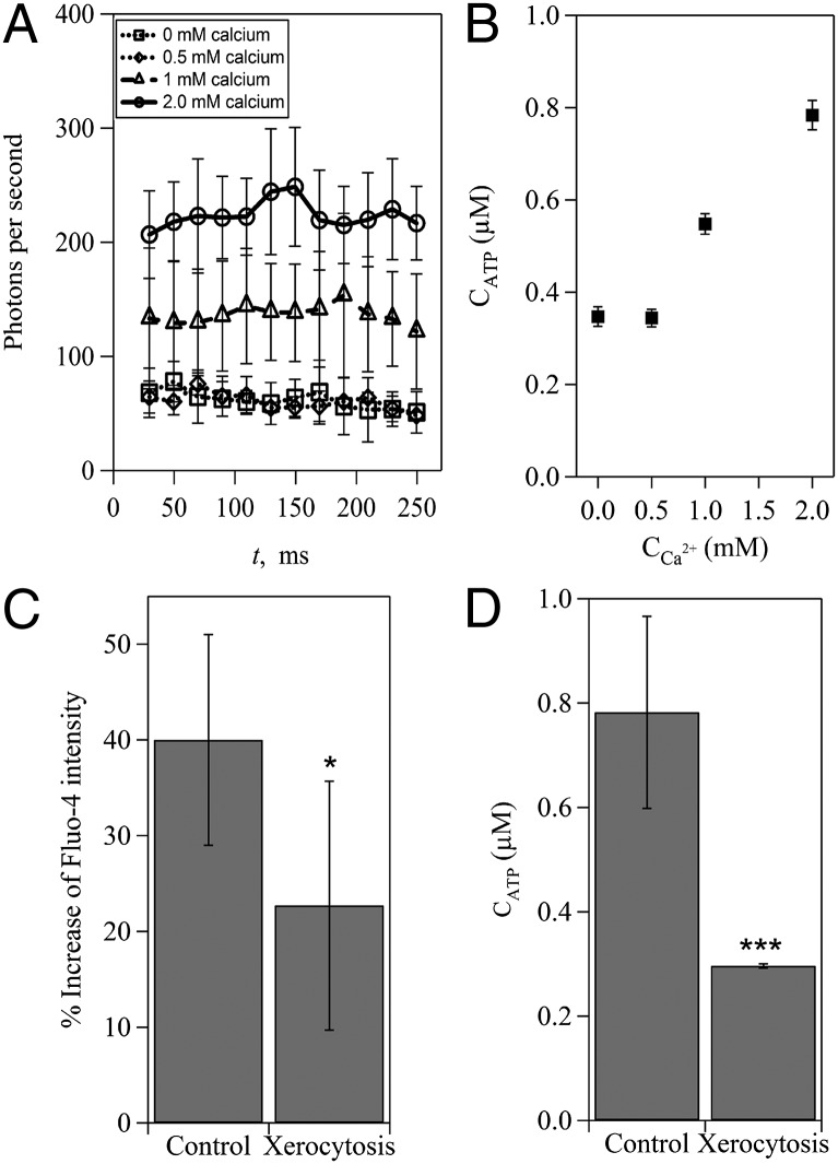
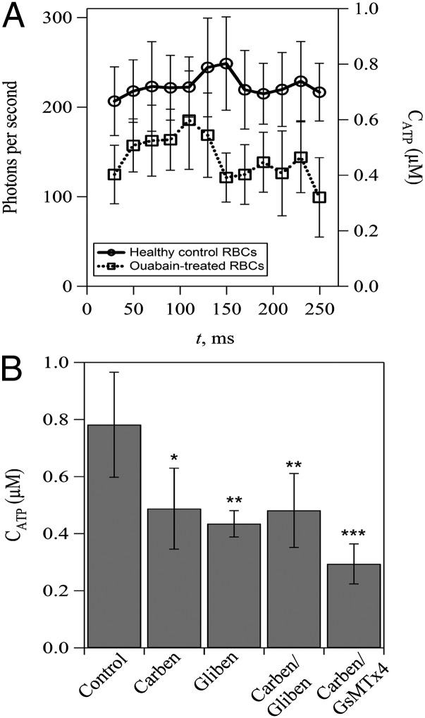
Piezo proteins (Piezo1 and Piezo2) are recently identified mechanically activated cation channels in eukaryotic cells and associated with physiological responses to touch, pressure, and stretch. In particular, human RBCs express Piezo1 on their membranes, and mutations of Piezo1 have been linked to hereditary xerocytosis. To date, however, physiological functions of Piezo1 on normal RBCs remain poorly understood. Here, we show that Piezo1 regulates mechanotransductive release of ATP from human RBCs by controlling the shear-induced calcium (Ca(2+)) influx. We find that, in human RBCs treated with Piezo1 inhibitors or having mutant Piezo1 channels, the amounts of shear-induced ATP release and Ca(2+) influx decrease significantly. Remarkably, a critical extracellular Ca(2+) concentration is required to trigger significant ATP release, but membrane-associated ATP pools in RBCs also contribute to the release of ATP. Our results show how Piezo1 channels are likely to function in normal RBCs and suggest a previously unidentified mechanotransductive pathway in ATP release. Thus, we anticipate that the study will impact broadly on the research of red cells, cellular mechanosensing, and clinical studies related to red cell disorders and vascular disease.

Keywords: ATP release; Pizeo1; RBCs; calcium flux; mechanosensing.


Conflict of interest statement

The authors declare no conflict of interest.

Figures

Fig. 1.
Fig. 1.
Inhibition of Piezo1 impairs shear-induced ATP release from human RBCs. (A) Schematic of the microfluidic setup for measuring shear-induced ATP release from RBCs (not to scale). Note that x = 0 indicates the onset of increased shear. (B) Representative measurements of the photon emission rate resulting from the reaction between luciferase/luciferin and ATP for healthy control and GsMTx4-treated RBCs; t = 0 ms corresponds to the position of x = 0 in A. The approximate average ATP concentration converted from the calibration curve (Fig. S1) is shown on the right axis. Note that GsMTx4 is a peptide that has been used to inhibit mechanically activated Piezo1 channels. The error bars are reported as the SDs of the mean (n = 11 and 4 for control and treated RBCs, respectively). (C) Average concentration of released ATP from control and Piezo1 inhibitor-treated RBCs. **P < 0.01. (D) Superimposed series of time-lapse images showing the deformation of an individual RBC passing through a short constriction (lc = 100 µm; wc = 20 µm). (Scale bar: 20 µm.) (E) Normalized change of RBC length (measured in the flow direction) for cells passing through the short constriction shown in D (δL/L=LstretchedLoriginal/Loriginal). Data were averaged from more than 30 cells for each sample. NS, not significant; RR, ruthenium red.
Fig. S1.
Fig. S1.
Calibration curve of photons per second as a function of ATP concentration in microfluidic channels.
Fig. 2.
Fig. 2.
Inhibition of Piezo1 reduces shear-induced Ca2+ influx in human RBCs. (A) Fluorescence images of Fluo-4–loaded control and Piezo1 inhibitor-treated RBCs stretched by shear in a microfluidic device. Calculated average shear stress is ∼3.4 Pa. (Scale bar: 20 µm.) (B) Average fluorescence intensity of control and Piezo1 inhibitor-treated single RBCs. The error bars are reported as the SDs of the mean (n = 3). ***P < 0.001. (C) Average fluorescence intensity of control and Piezo1 inhibitor-treated RBCs (10% vol/vol) flowing before and after the constriction channel. The error bars are reported as the SDs of the mean (n = 8 and 3 for normal RBCs and treated RBCs, respectively). NS, not significant; RR, ruthenium red. *P < 0.05.
Fig. S2.
Fig. S2.
(A) Shear-induced Ca2+ influx (Fluo-4 intensity) and (B) ATP release (photons per second) vs. time. (C) Normalized increase of Ca2+ influx and ATP release vs. time. Ia is the intensity after the constriction, and Ib is the average intensity before the constriction; t = 0 ms indicates the onset of increased shear. Note that, to emphasize the trend in Ca entry and ATP release, the error bar (±0.05) is not shown in C.
Fig. 3.
Fig. 3.
Ca2+ influx regulates shear-induced ATP release from human RBCs. (A) Measurements of the photon emission rate caused by shear-induced ATP release in solutions with different concentrations of Ca2+. (B) Dependence of shear-induced ATP release on extracellular Ca2+ concentrations. (C) Ca2+ influx-induced increase of Fluo-4 intensity and (D) shear-induced ATP release from healthy control RBCs and RBCs from patients with xerocytosis. The error bars are reported as the SDs of the mean (n = 11 and 4 for control RBCs and RBCs from patients with xerocytosis, respectively). *P < 0.05; ***P < 0.001.
Fig. S3.
Fig. S3.
Effect of extracellular Ca2+ concentration on shear-induced Ca2+ influx in RBCs. The error bars are reported as the SDs of the mean (n = 6, 4, 3, and 8 for 0, 0.5, 1, and 2 mM, respectively).
Fig. 4.
Fig. 4.
Effects of membrane-associated ATP pools and potential ATP-releasing channels on shear-induced ATP release from human RBCs. (A) Effect of ouabain treatment on shear-induced ATP release. Note that ouabain treatment is used to prevent bulk ATP from entering the membrane-associated ATP pools in RBCs. (B) Effect of inhibition of CFTR and/or pannexin-1 and Piezo1 channels on shear-induced ATP release. Carbenoxolone (Carben) and glibenclamide (Gliben) are used to inhibit pannexin-1 and CFTR, respectively. GsMTx4 is used to inhibit Piezo1 channels. The error bars are reported as the SDs of the mean (n = 4 for Carben and Carben/Gliben-treated RBCs; n = 3 for Gliben-treated RBCs; and n = 4 for Carben/GsMTx4-treated RBCs). *P < 0.05; **P < 0.01; ***P < 0.001.
Fig. S4.
Fig. S4.
Schematic of the proposed mechanotransductive release of ATP from RBCs. (A) Shear-induced stretch of RBCs activates Piezo1 channels and induces an increased Ca2+ influx, which (B) activates Ca2+ pumps to remove extra Ca2+ rapidly. (C) Meanwhile, increased Ca2+ influx activates pannexin-1 channels (Px1) directly and triggers ATP release from the membrane-associated ATP pools. (D) It is also possible, although controversial, that increased Ca2+ influx depolymerizes actin filaments (brown spheres), which activates CFTR and induces the release of ATP. Regardless of the precise mechanism, our results show clearly that a critical concentration of intracellular Ca2+ caused by shear-induced Ca2+ influx is required for the mechanotransductive release of ATP.

References

    1. Ho J, Sibbald WJ, Chin-Yee IH. Effects of storage on efficacy of red cell transfusion: When is it not safe? Crit Care Med. 2003;31(12 Suppl):S687–S697. - PubMed
    1. Schmid-Schöenbein H, Wells R. Fluid drop-like transition of erythrocytes under shear. Science. 1969;165(3890):288–291. - PubMed
    1. Vague P, Juhan I. Red cell deformability, platelet aggregation, and insulin action. Diabetes. 1983;32(Suppl 2):88–91. - PubMed
    1. van der Heyde HC, Nolan J, Combes V, Gramaglia I, Grau GE. A unified hypothesis for the genesis of cerebral malaria: Sequestration, inflammation and hemostasis leading to microcirculatory dysfunction. Trends Parasitol. 2006;22(11):503–508. - PubMed
    1. Fischer DJ, Torrence NJ, Sprung RJ, Spence DM. Determination of erythrocyte deformability and its correlation to cellular ATP release using microbore tubing with diameters that approximate resistance vessels in vivo. Analyst (Lond) 2003;128(9):1163–1168. - PubMed

Publication types