

Association between Perivascular Spaces and Progression of White Matter Hyperintensities in Lacunar Stroke Patients
- PMID: 26352265
- PMCID: PMC4564273
- DOI: 10.1371/journal.pone.0137323
Association between Perivascular Spaces and Progression of White Matter Hyperintensities in Lacunar Stroke Patients
Abstract
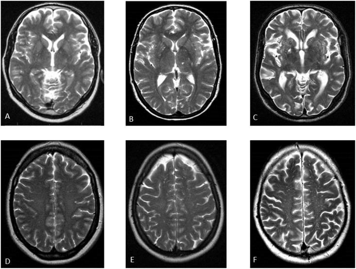
Objectives: Perivascular spaces are associated with MRI markers of cerebral small vessel disease, including white matter hyperintensities. Although perivascular spaces are considered to be an early MRI marker of cerebral small vessel disease, it is unknown whether they are associated with further progression of MRI markers, especially white matter hyperintensities. We determined the association between perivascular spaces and progression of white matter hyperintensities after 2-year follow-up in lacunar stroke patients.
Methods: In 118 lacunar stroke patients we obtained brain MRI and 24-hour ambulatory blood pressure measurements at baseline, and a follow-up brain MRI 2 years later. We visually graded perivascular spaces and white matter hyperintensities at baseline. Progression of white matter hyperintensities was assessed using a visual white matter hyperintensity change scale. Associations with white matter hyperintensity progression were tested with binary logistic regression analysis.
Results: Extensive basal ganglia perivascular spaces were associated with progression of white matter hyperintensities (OR 4.29; 95% CI: 1.28-14.32; p<0.05), after adjustment for age, gender, 24-hour blood pressure and vascular risk factors. This association lost significance after additional adjustment for baseline white matter hyperintensities. Centrum semiovale perivascular spaces were not associated with progression of white matter hyperintensities.
Conclusions: Our study shows that extensive basal ganglia perivascular spaces are associated with progression of white matter hyperintensities in cerebral small vessel disease. However, this association was not independent of baseline white matter hyperintensities. Therefore, presence of white matter hyperintensities at baseline remains an important determinant of further progression of white matter hyperintensities in cerebral small vessel disease.
Conflict of interest statement
Figures
References
Publication types
MeSH terms
LinkOut - more resources
Full Text Sources
Other Literature Sources
Medical
