. 2015 Sep 17;525(7569):333-8.
doi: 10.1038/nature15257. Epub 2015 Sep 9.

Labelling and optical erasure of synaptic memory traces in the motor cortex

Affiliations

Labelling and optical erasure of synaptic memory traces in the motor cortex

Akiko Hayashi-Takagi et al. Nature. .

Abstract

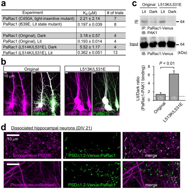
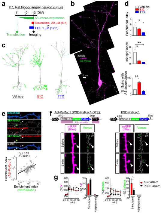
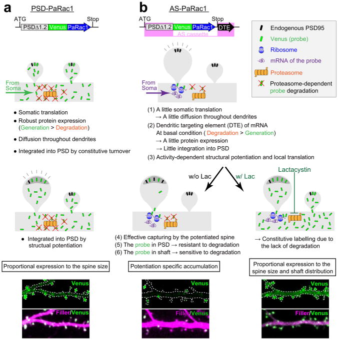
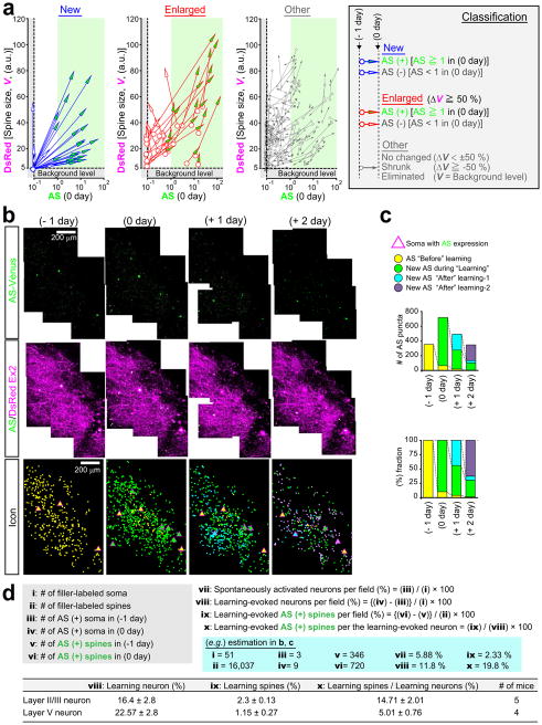
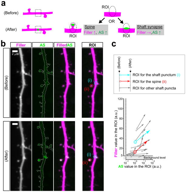
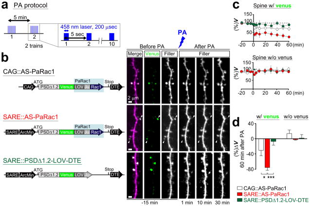
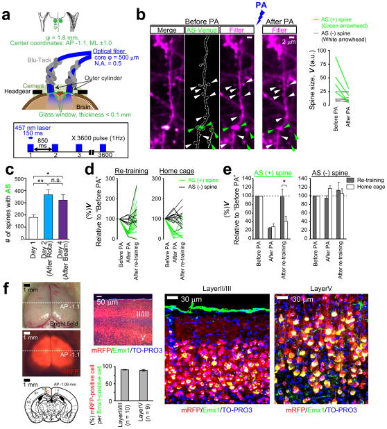
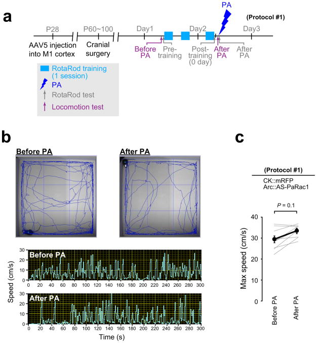
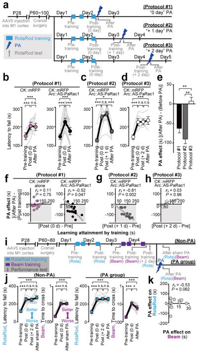
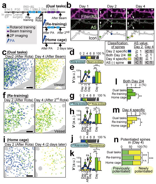
Dendritic spines are the major loci of synaptic plasticity and are considered as possible structural correlates of memory. Nonetheless, systematic manipulation of specific subsets of spines in the cortex has been unattainable, and thus, the link between spines and memory has been correlational. We developed a novel synaptic optoprobe, AS-PaRac1 (activated synapse targeting photoactivatable Rac1), that can label recently potentiated spines specifically, and induce the selective shrinkage of AS-PaRac1-containing spines. In vivo imaging of AS-PaRac1 revealed that a motor learning task induced substantial synaptic remodelling in a small subset of neurons. The acquired motor learning was disrupted by the optical shrinkage of the potentiated spines, whereas it was not affected by the identical manipulation of spines evoked by a distinct motor task in the same cortical region. Taken together, our results demonstrate that a newly acquired motor skill depends on the formation of a task-specific dense synaptic ensemble.


Conflict of interest statement

Conflict of interest statement. The authors declare that they have no competing financial interests.

Figures

Extended Data Figure 1
Extended Data Figure 1. Optimisation of the PaRac1 for the synaptic application
a, Isothermal titration calorimetry (ITC) experiments showing that the introduction of L514K and L531E mutations into the original PaRac1 construct reduced binding with the CRIB domain of PAK1 in the dark. The light-insensitive form of LOV2 (C450A) and the I539E mutant, which mimics the unfolded ‘lit state’, were used as negative and positive controls, respectively. b, Leaky activity of PaRac1 in the dark. In hippocampal neuronal cultures transfected with the original PaRac1, we observed a bearded appearance of the soma with a numerous ectopic dendrites, while neurons transfected with PaRac1 (L514K/L531E) were indistinguishable from normal neurons. c, Assessment of the affinity of PaRac1 to the endogenous PAK1 using a pull-down assay. HEK293 cells, which were transfected with PaRac1-Venus, were divided into two groups: lit and dark. The cells in the lit group were radiated with light with a white fluorescent lamp before cell lysis, and continuous light illumination was present during subsequent immunoprecipitation until the final wash step of protein precipitants. Conversely, cells in the dark group were lit with a yellow fluorescence lamp, which excludes light wavelengths below 500 nm. Co-immunoprecipitation with PAK1 revealed that PaRac1 (L514K/L531E) barely bound with PAK1 in the dark (The number of trial is depicted in the bar graph, **P < 0.01 using the Mann-Whitney U test). d, Targeting of PaRac1 to the postsynaptic density. PSDΔ1.2-PaRac1 [DTE (−)] was transfected into dissociated cortical neurons at 21 days in vitro (DIV). Two days after transfection, cells were fixed with 4% PFA, followed by permeabilization for the subsequent immunostaining procedure. Axons and endogenous PSD-95 were visualized using the anti-phospho-neurofilament and anti-PSD-95 antibodies, respectively, revealing that PSDΔ1.2-PaRac1 co-localised with the endogenous PSD-95. Note that PSDΔ1.2-PaRac1 did not co-localise with the axonal marker.
Extended Data Figure 2
Extended Data Figure 2. The distribution of AS-PaRac1 is regulated by neuronal activity, and is dependent on the dendritic targeting element (DTE)
a, Experimental design. b, Representative image of a cultured hippocampal neuron. c, Bicuculline (BIC) or tetrodotoxin (TTX) was added to the culture media at the designated time points. Images were captured at a high magnification and were tiled to visualize the entire cell. Green circles represent the AS-PaRac1 puncta. d, Quantification of AS-PaRac1 distribution (n = 6/each, *P < 0.05, **P < 0.01 using Kruskal-Wallis test followed by post-hoc Dennett’s test). e, Concomitant accumulation of AS-PaRac1 and SEP-GluA1 in spines. Neurons were co-transfected with mTq (mTurquoise, filler), SEP-GluA1, and AS-PaRac1-mRFP, and the constructs were expressed for 36 h. Potentiated spines during 36 h were shown by SEP-GluA1 fluorescence (arrowheads). Spearman rank correlation revealed a significant correlation between the spine enrichment indices of SEP-GluA1 and AS-PaRac1 (each circle represents one spine, 235 spines, 29 dendrites). f, Schematic of the constructs and representative images of single spine potentiations by glutamate uncaging in the presence of FSK (arrowheads). Rat hippocampal slice cultures were biolistically transfected with either AS-PaRac1 or PSD-PaRac1 [DTE (−)] followed by the uncaging experiments at DIV 13 (equivalent to postnatal day 20). g, Time course of the spine head volume (V) and accumulation of Venus upon uncaging. The mean changes in spine size and Venus accumulation in the stimulated or neighbouring spines are depicted 60 min after uncaging. For quantification, we used pooled data from independent identically designed experiments. The data set for AS-PaRac1 was identical with the FSK-treated group of Fig. 1c–e. Scale bar, 1 μm. *P < 0.05 using the Mann-Whitney U test (n = 6 or 11 dendrites for PSD-PaRac1 or AS-PaRac1, respectively).
Extended Data Figure 3
Extended Data Figure 3. Putative cellular mechanisms of the specific concentration of AS-PaRac1 in potentiated spines
a, Uniform labelling of spines with the PSD-PaRac1 construct that lacks DTE of Arc 3′ UTR (Fig. 1a, construct B). PSD-PaRac1 is translated in the soma that is abundantly equipped with translational machineries. Therefore, the somatic protein expression of the probe is high (data not shown), which would outnumber the degradation, and the resulting proteins are transported throughout dendrites. The overflowing probes integrate into the postsynaptic density (PSD) during the constitutive turnover of PSD molecules. Therefore, probe expression is proportional to the spine size. b, Selective labelling of potentiated spines with AS-PaRac1 (Fig. 1a, construct C). The following six mechanisms endow the potentiation-specific labelling with AS-PaRac1. (1) A little somatic translation: the moderate gene expression of AS-PaRac1, by which the translation of AS-PaRac1 protein is limited in the soma (see Extended Data Fig. 2b), and therefore, the non-specific overflow of this probe from the soma into the dendrites is minimal. (2) Dendritic targeting element (DTE): the essential domains of AS-PaRac1 are the N-terminal PSD-95 (PSDΔ1.2) and the 3′ UTR of Arc mRNA (DTE). DTE has a pivotal role in the dendritic targeting of mRNAs,. One of the most well-known DTE is present in the Arc mRNA, which is targeted to stimulated dendritic segments in an activity-dependent manner. The transport of mRNA out of soma also contributes to the limited translation of the probe in the soma described in (1). In the absence of activation, the limited amount of translational machineries and presence of degradation components in the dendrites maintains the locally translated probe at a low level, which results in a low rate of AS-PaRac1 integration into the PSD during the constitutive turnover of PSD proteins. (3) Local protein synthesis: persistent structural plasticity of the spine depends on the activity-dependent dendritic synthesis of proteins, and the translation of Arc mRNA is controlled by activity levels. (4) Effective capturing of PSD proteins in the structurally potentiated spines: the potentiated spine, which rapidly requires new copies of PSD proteins, captures diffusing PSD proteins more efficiently,. (5) Increased stability of AS-PaRac1 in the PSD: it is likely that the stability of the PSD-integrated AS-PaRac1 increase, as does the typical PSD scaffold proteins. The ubiquitination might be underling mechanism of the increased stability, because the ubiquitination site of AS-PaRac1 resides in the N-terminal domain of PSD-95, the domain of which is aggregated to form head-to-head multimerization in the postsynaptic scaffold. Thus, once AS-PaRac1 is integrated into the PSD, the ubiquitination site may be concealed, and AS-PaRac1 becomes relatively stable. (6) Sensitivity of unbound AS-PaRac1 against the proteasomal degradation: contrary to the PSD-integrated AS-PaRac1, unbound AS-PaRac1 is sensitive to degradation because the ubiquitination site is not concealed. This scenario is supported by the administration of lactacystin (right panel), which inhibits proteasomes and thus completely disrupts the uneven distribution of AS-PaRac1. Similar mechanisms are relevant for newly formed spines, because spine formation is associated with spine enlargement.
Extended Data Figure 4
Extended Data Figure 4. Raw data of the quantification and synaptic mapping shown in Fig. 2
a, Quantification of spine size (based on DsRed fluorescence) and AS-PaRac1 fluorescence after learning are depicted separately based on the classification of spines. The definitions of ‘New spine’, ‘Enlarged spine’ and others are described on the right. Each arrow indicates the trajectory of a spine; beginning and end points represent the absolute values before and after the rotarod task, respectively. b, XY images were captured from the dura to a depth of 300 μm with a step-size of 1.0 μm, and were stacked by the summation of fluorescence values at each pixel. Z-stacked images of 10 overlapping fields were aligned to generate the combined images. AS-PaRac1 and AS-PaRac1/DsRed merged images are shown. AS-PaRac1 that was present before learning (−1 day, yellow), appeared shortly after learning (learning period, 0 day, green), 1 day (after −1, +1 day, blue), or 2 days after learning (after −2, +2 day, purple) are depicted to show the spatiotemporal distribution of AS-PaRac1 triggered in each period. c, Time course of the number and fraction of AS-PaRac1-positive spines in each period. d, Calculation of the learning-evoked spine/neuron ratio (%). Example of the calculation is based on the raw data shown in b and c. The table indicates the comparison between neurons in layer II/III (in utero EP at E14.5) and layer V (in utero EP at E13).
Extended Data Figure 5
Extended Data Figure 5. Assessment of AS-PaRac1 puncta on the dendritic shaft
a, The two possible synapse types that AS-PaRac1 puncta may represent on the dendritic shaft. XY images were captured to encompass the entire Z-range of the dendrite of interest with a step-size of 0.5 μm, and images were stacked by the summation of fluorescence values at each pixel. The fluorescence of both the filler and AS-PaRac1 would increase, if the AS-PaRac1 punctum emerged on the dendritic spine that undergoes structural potentiation. In contrast, fluorescence of the filler would not increase, if AS-PaRac1 was in the shaft synapse. b, Example of the dendrites before and after the emergence of AS-PaRac1. AS-PaRac1 puncta on the shaft and on the dendritic spine are indicated with (i) and (ii), respectively. The ROI used for the calculation of fluorescence in each punctum is shown. c, Quantification of the fluorescence of the filler and AS-PaRac1 upon the emergence of AS-PaRac1 puncta. Each arrow indicates the trajectory of each ROI; beginning and end points represent the absolute values before and after the emergence of AS-PaRac1, respectively. The ROI at (i) exhibited a concomitant fluorescence increase in both the filler and AS-PaRac1, similar to AS-PaRac1 in a typical dendritic spine (ii). All examined AS-PaRac1 puncta on the dendritic shaft exhibited positive correlations, suggesting that the majority of AS-PaRac1 puncta emerge on the dendritic spine during the structural changes of the spine.
Extended Data Figure 6
Extended Data Figure 6. Rac1-dependent shrinkage of dendritic spines induced by low-frequency PA
a, The protocol of PA. PA was performed in the region that encompasses the branch of interest. b, Neurons in the hippocampal slice culture (DIV 11) were biolistically transfected with DNA constructs shown in the schematic image on the left. Representative dendritic images upon PA are shown on the right. Robust shrinkage (arrowheads) was observed in the spines transfected with AS-PaRac1 driven by the SARE-Arc promoter. Despite their adjacent location to the AS-PaRac1-positive spines, AS-PaRac1-negative spines were not affected by the PA. c, Time course of the spine head volume (V) of Venus-positive (upper panel) and negative spines (lower panel). White, red, and blue circles represent CAG::AS-PaRac1, SARE::AS-PaRac1, and SARE::PSDΔ1.2-LOV-DTE, respectively (n = 12 cells/each). d, The mean relative change in spine head size in Venus-positive and negative spines 60 min after PA. Scale bar, 2 μm. *P < 0.05 and ***P < 0.001 according to the Kruskal-Wallis test followed by the post-hoc Scheffé’s test.
Extended Data Figure 7
Extended Data Figure 7. Spine shrinkage in broad areas of the bilateral motor cortices induced by blue laser illumination
a, Schematic of the bilateral cranial windows, optical fibres, and the PA protocol. b, Representative images of spine shrinkage in the M1 cortex upon PA in vivo. AS-PaRac1-positive spines (green arrowheads) shrank, while the AS-PaRac1-negative ones (white arrowheads) did not. Quantification of spine size is shown on the right. c, The mean number of AS-PaRac1 puncta per fields was calculated in mice shown in the Fig. 4i. d, e, Spine structure and AS-PaRac1 were imaged in mice, which were subjected to the re-training and homecage protocols shown in Fig. 5. The majority of AS-PaRac1-positive spines displayed PA-induced shrinkage and subsequent recovery. *P < 0.05 according to the Mann-Whitney U test. f, The success of AAV5 vector injection into the bilateral M1 cortex was confirmed by the presence of mRFP fluorescence after behavioural tests. High efficacy of virus infection in layer II/III and V pyramidal neurons was demonstrated with Emx1 immunostaining, which labels pyramidal neurons. The mice without bilateral mRFP signal in the M1 cortex were excluded from the data analysis.
Extended Data Figure 8
Extended Data Figure 8. No effect of PA on the locomotor activity of mice
a, Experimental schedule. The running speed of AS-PaRac1-injected mice in protocol #1 (Fig. 4a) was measured with a video-tracking system. To minimize the effect of circadian rhythm on locomotion, mice were tested at the same time of the day before and after PA. b, Representative traces of locomotion and temporal sequences of running speed are depicted. c, Statistical analysis shows that PA has only a negligible effect on running speed.
Extended Data Figure 9
Extended Data Figure 9. Detailed illustration of the rotarod and beam tasks in the Fig. 4
a, Experimental flowchart. b, Detailed schedule of the rotarod training/test, locomotion test, and PA. c, To shorten the training time, air puffs were applied to the hind limbs as aversive stimuli to maintain the forward-looking position of mice on the rod, which improved the performance, especially at higher speeds. d, Schematic illustration of the beam test. The test was preceded by a 6-h-long training session that lasted for 2 days.
Figure 1
Figure 1. Potentiation-dependent accumulation of AS-PaRac1 to the dendritic spines in hippocampal slice cultures
a, Mapping for essential domains for the discrete distribution of the probe (arrowheads). b, Representative images of single spine potentiations by glutamate uncaging (arrowheads) in the presence or absence of forskolin (FSK) and anisomycin. c, d, Time courses of spine head volume (c) and AS-PaRac1 accumulation (d). The mean change 60 min after uncaging in the stimulated or neighbouring spines. e, The effect of lactacystin on the discrete accumulation of AS-PaRac1 (arrowheads). Scale bar, 2 μm. Detailed information of statistical methods/results are described in the Extended Data Table.
Figure 2
Figure 2. Spatiotemporal dynamics of AS-PaRac1 labelling in vivo during the rotarod task
a, Schematic of the Arc promoter-driven AS-PaRac1. b, Experimental design. c, Images of spine formation (arrows) and spine enlargement (arrowheads). Green circles, AS-PaRac1; magenta circles, spines that initially acquired AP-PaRac1, but lost it afterward, but the structural change were persistent. d, Fraction of structural change of spines. e–g, Quantification of spine size and AS-PaRac1 (e). Relationship between AS-PaRac1 and ΔV after (0 day, f) and before (−1 day, g) learning. h, Percentage of AS-PaRac1-containing spines (AS-PaRac1 ≥ 1 [a.u.], green shaded area in f). i, Mapping of AS-PaRac1. j, Retention of AS-PaRac1 (green) or structural potentiation (magenta). k, Trajectory of spine size and AS-PaRac1 intensities of the structurally potentiated spines. Scale bar, 2 μm for c; 200 μm for b and i..
Figure 3
Figure 3. Selective shrinkage of AS-PaRac1-containing spines upon photoactivation (PA)
a, Illustration of PA. b, Images of the hind limb regions of cortices. c, Spine size following PA. Dark green circles are eliminated spines. d, The effect of the cortical depth on PA-induced spine shrinkage. e–j, Neurons were co-transfected with GCaMP6s, AS-PaRac1-mTurquoise, and mRFP. Changes in the GCaMP/mRFP ratio (ΔR) in synapse (g) and soma (h) were traced. i,j, Relationships between ΔV and ΔAmplitude (i), or ΔFrequency (j) upon PA. Circles 1, 2, and S correspond to spine #1, #2, and the soma in g and h. Scale bar, 5 μm for b; 50 μm for e, 2 μm for f.
Figure 4
Figure 4. Erasure of acquired learning by the PA of spines labelled with AS-PaRac1
a, Experimental design (see Extended Data Figure 9). b,c,d, Mice were allocated to protocols #1 (b), #2 (c), or #3 (d). An average of three trials of each mouse was used as the task performance (grey line). e, The critical period of PA to erase acquired skills. fh, Relationship between the effect of PA and learning attainments. i, Experimental design. j, Performance trajectory of each skill. k, No correlation between PA effect on acquired rotarod performance and that of beam task..
Figure 5
Figure 5. Visualization of synaptic ensembles for distinct learning tasks
a, Experimental design. Arc::AS-PaRac1 and CAG::mRFP (filler) was transduced by in utero electroporation. b, Images of dendrites upon learning. AS-PaRac1 puncta are colour-coded based on its appearance and duration. Identical colour-codes are used in c–n. c,f,i, Wide view mapping of AS-PaRac1. d,g,j, The fraction of each spine type. e,h,k, The trajectory of spine size (V). l,m, Differential spine potentiation in each condition. n, The proportions of newly potentiated spines. Scale bar, 2 μm for b; 50 μm for c, f and i..

Comment in

  • Neuroscience: Forgetfulness illuminated.
    Lu J, Zuo Y. Lu J, et al. Nature. 2015 Sep 17;525(7569):324-5. doi: 10.1038/nature15211. Epub 2015 Sep 9. Nature. 2015. PMID: 26352474 No abstract available.
  • NEUROSCIENCE: Targeting spines.
    Gundersen BB. Gundersen BB. Nat Methods. 2015 Nov;12(11):1006-7. doi: 10.1038/nmeth.3644. Nat Methods. 2015. PMID: 26824105 No abstract available.

References

    1. Bernstein JG, Boyden ES. Optogenetic tools for analyzing the neural circuits of behavior. Trends Cogn Sci. 2011;15:592–600. - PMC - PubMed
    1. Tye KM, Deisseroth K. Optogenetic investigation of neural circuits underlying brain disease in animal models. Nature Rev Neurosci. 2012;13:251–266. - PMC - PubMed
    1. Liu X, et al. Optogenetic stimulation of a hippocampal engram activates fear memory recall. Nature. 2012;484:381–385. - PMC - PubMed
    1. Holtmaat A, Svoboda K. Experience-dependent structural synaptic plasticity in the mammalian brain. Nature Rev Neurosci. 2009;10:647–658. - PubMed
    1. Kasai H, et al. Structural dynamics of dendritic spines in memory and cognition. Trends Neurosci. 2010;33:121–129. - PubMed

Publication types

MeSH terms

Substances