Clinical Trial
. 2015 Oct 29;373(18):1733-47.
doi: 10.1056/NEJMoa1506583. Epub 2015 Sep 9.

Targeting Mutant BRAF in Relapsed or Refractory Hairy-Cell Leukemia

Affiliations
Clinical Trial

Targeting Mutant BRAF in Relapsed or Refractory Hairy-Cell Leukemia

Enrico Tiacci et al. N Engl J Med. .

Abstract

Background: BRAF V600E is the genetic lesion underlying hairy-cell leukemia. We assessed the safety and activity of the oral BRAF inhibitor vemurafenib in patients with hairy-cell leukemia that had relapsed after treatment with a purine analogue or who had disease that was refractory to purine analogues.

Methods: We conducted two phase 2, single-group, multicenter studies of vemurafenib (at a dose of 960 mg twice daily)--one in Italy and one in the United States. The therapy was administered for a median of 16 weeks in the Italian study and 18 weeks in the U.S. study. Primary end points were the complete response rate (in the Italian trial) and the overall response rate (in the U.S. trial). Enrollment was completed (28 patients) in the Italian trial in April 2013 and is still open (26 of 36 planned patients) in the U.S. trial.

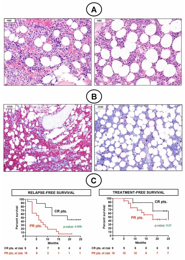
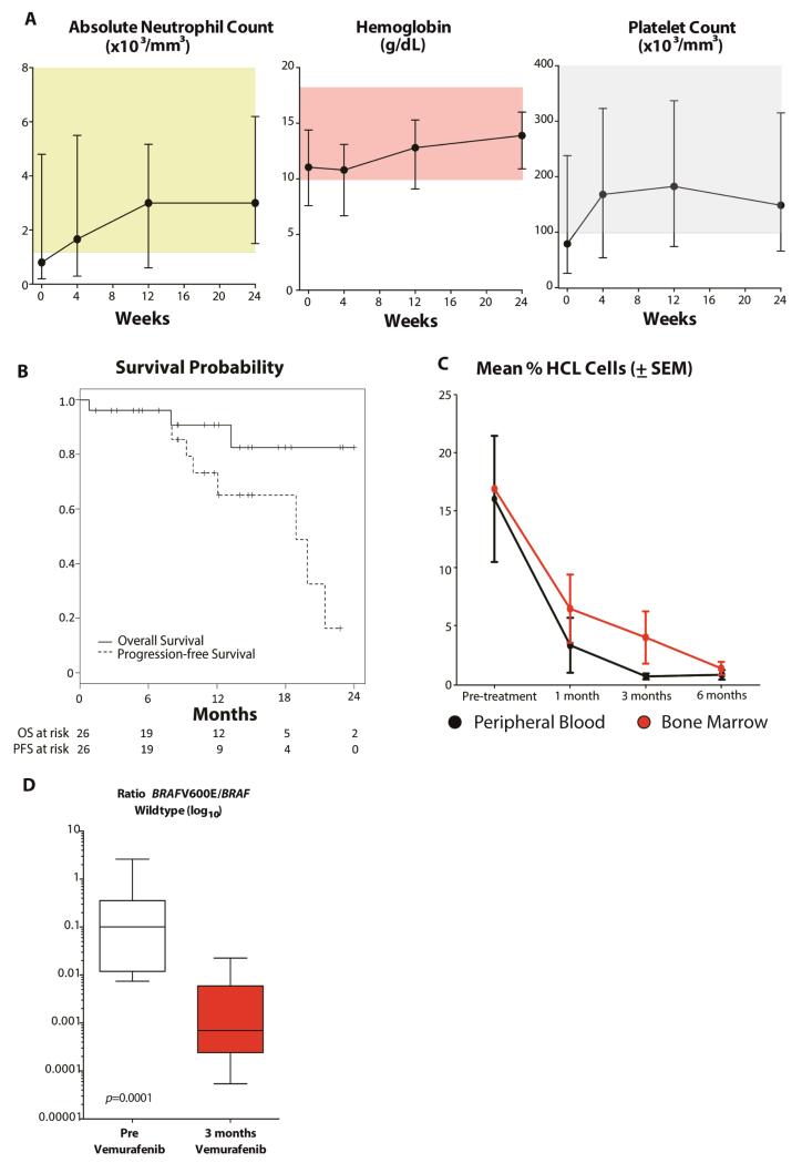
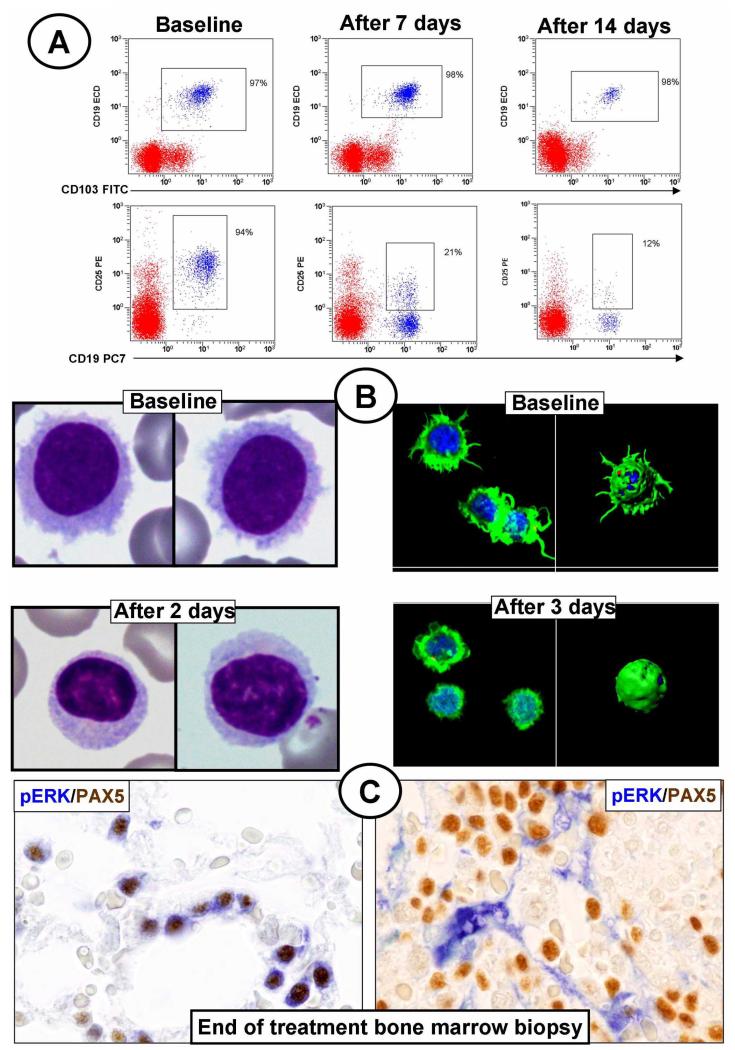
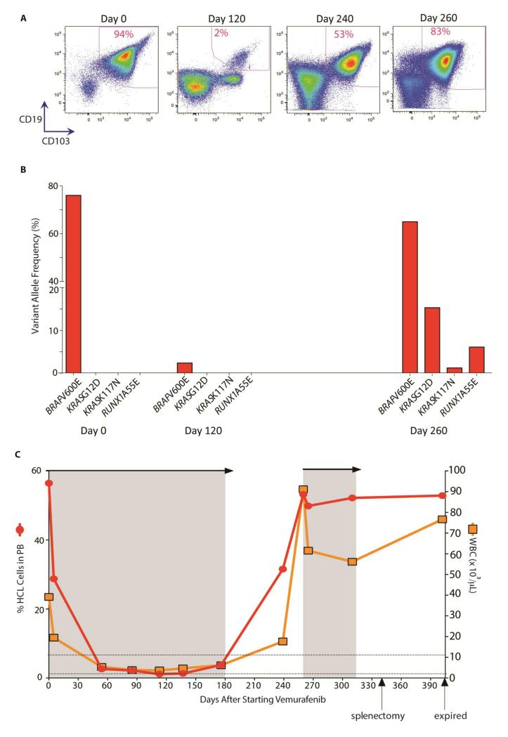
Results: The overall response rates were 96% (25 of 26 patients who could be evaluated) after a median of 8 weeks in the Italian study and 100% (24 of 24) after a median of 12 weeks in the U.S. study. The rates of complete response were 35% (9 of 26 patients) and 42% (10 of 24) in the two trials, respectively. In the Italian trial, after a median follow-up of 23 months, the median relapse-free survival was 19 months among patients with a complete response and 6 months among those with a partial response; the median treatment-free survival was 25 months and 18 months, respectively. In the U.S. trial, at 1 year, the progression-free survival rate was 73% and the overall survival rate was 91%. Drug-related adverse events were usually of grade 1 or 2, and the events most frequently leading to dose reductions were rash and arthralgia or arthritis. Secondary cutaneous tumors (treated with simple excision) developed in 7 of 50 patients. The frequent persistence of phosphorylated ERK-positive leukemic cells in bone marrow at the end of treatment suggests bypass reactivation of MEK and ERK as a resistance mechanism.

Conclusions: A short oral course of vemurafenib was highly effective in patients with relapsed or refractory hairy-cell leukemia. (Funded by the Associazione Italiana per la Ricerca sul Cancro and others; EudraCT number, 2011-005487-13; ClinicalTrials.gov number NCT01711632.).


Figures

Figure 1
Figure 1. Response depth and duration in the 25 patients of the Italian trial responding to vemurafenib
(A) Hematoxylin-eosin staining of a BM biopsy from a patient showing infiltration by leukemic HCL cells with wide, clear cytoplasm recognizable by morphology at baseline (left panel), but not after 8 weeks of vemurafenib (right panel). (B) CD20 immunostaining of the same BM biopsy showing ~75% leukemic infiltration at baseline (left panel), and only few (≤10%) residual scattered hairy cells after 8 weeks of vemurafenib (right panel). (C) Relapse-free (left) and treatment-free survival (right) of patients obtaining a CR (black line) or PR (red line).
Figure 2
Figure 2. Effect of vemurafenib on peripheral blood counts, survival, flow cytometric enumeration of hairy cells, and BRAFV600E allele burden
(A) Median value of the absolute neutrophil count, hemoglobin and platelet cell counts of the 24 evaluable patients over time after initiating vemurafenib treatment. Error bars denote the upper and lower limits. (B) The probability of progression-free survival and overall survival for all 26 patients. Tick marks indicate censored data. (C) Mean percentage of HCL cells in peripheral blood and bone marrow mononuclear cells over time of patients from this study (± standard error of mean (SEM)). Mean percentage of HCL cells decreased with each period of vemurafenib therapy shown (p<0.05 comparing percentage of HCL cells for each time point on therapy relative to pretreatment values). (D) Ratio of the concentration of BRAFV600E:BRAF wildtype alleles (log10) in DNA from peripheral blood mononuclear cells pretreatment with vemurafenib and following 3 months of vemurafenib. The ratios are displayed as box-and-whisker plots with the median value as the middle bar, the ends of the boxes as upper and lower quartile values, and ends of whiskers as highest and lowest values.
Figure 3
Figure 3. Phenotypic and molecular changes of HCL cells during treatment with vemurafenib in the Italian trial
(A) Flow cytometry dot plots of a patient’s BM aspirate gated on CD45 cells and showing at baseline (left panels) CD25 expression by 94% of leukemic cells (CD19+/CD103+, upper row; CD19+/CD25+, bottom row). A progressively loss of CD25 expression (but not of CD19 or CD103 expression) is seen over 1 and 2 weeks of treatment with vemurafenib (down to 21% and 12%, respectively; middle and right panels, respectively). The remaining cells (red events) are normal CD45+ hematopoietic cells. (B) Left panels. May-Grümwald-Giemsa staining of a patient’s peripheral blood smear featuring leukemic cells rich in hairy projections at baseline (top), but not after 2 days of vemurafenib treatment (bottom). Right panels. Confocal fluorescence microscopy analysis of blood leukemic cells purified from the same patient shows prominent surface projections (stained in green by phalloidin) at baseline (top), but not after 3 days of vemurafenib treatment (bottom); these changes are clearly evident both in electronically magnified two-dimensional and three-dimensional reconstructed images of representative cells; (C) Double immunostaining of BM biopsy taken the day after the end of treatment and double stained for PAX5 (a B-cell marker; in brown) and phospho-ERK (pERK; in blue). In some responding patients (one exemplified in the left panel) persistence of pERK+ leukemic hairy cells is observed; conversely, in other patients (one exemplified in the right panel) residual hairy cells do not detectably express phospho-ERK, with stromal cells strongly positive for phospho-ERK as internal positive control.
Figure 4
Figure 4. Serial genomic analysis reveals activating KRAS mutations associated with development of acquired vemurafenib resistance in BRAFV600E-mutant HCL
(A) Serial flow cytometric analysis for HCL cells in peripheral blood mononuclear cells throughout the initial course of therapy (days 0-180) and vemurafenib retreatment (days 260-320). Percentages in FACS plots denote percentage of HCL (CD19/CD103) cells amongst PB MNCs. (B) Somatic mutations identified in PB MNCs following 0, 120, and 260 days of vemurafenib administration. The variant allele frequency (%) is shown for mutations seen at each timepoint. (C) Graph of percentage of HCL cells in peripheral blood (left axis) and total WBC count (right axis). Shaded areas represent periods of vemurafenib treatment and retreatment.

References

    1. Foucar KFB, Catovsky D, Stein H. Hairy cell leukemia. In: Swerdlow SCE, Harris NL, et al., editors. WHO Classification of Tumours of Hematopoietic and Lymphoid Tissues. International Agency for Research on Cancer; Lyon, France: 2008. pp. 188–90.
    1. Basso K, Liso A, Tiacci E, et al. Gene expression profiling of hairy cell leukemia reveals a phenotype related to memory B cells with altered expression of chemokine and adhesion receptors. J Exp Med. 2004;199:59–68. - PMC - PubMed
    1. Tiacci E, Liso A, Piris M, Falini B. Evolving concepts in the pathogenesis of hairy-cell leukaemia. Nat Rev Cancer. 2006;6:437–48. - PubMed
    1. Kitagawa Y, Brahmachary M, Tiacci E, Dalla-Favera R, Falini B, Basso K. A microRNA signature specific for hairy cell leukemia and associated with modulation of the MAPK-JNK pathways. Leukemia. 2012;26:2564–7. - PubMed
    1. Grever MR. How I treat hairy cell leukemia. Blood. 2010;115:21–8. - PMC - PubMed

Publication types

MeSH terms

Associated data