. 2015 Sep 9;11(9):e1005507.
doi: 10.1371/journal.pgen.1005507. eCollection 2015 Sep.

A Genetic Selection for dinB Mutants Reveals an Interaction between DNA Polymerase IV and the Replicative Polymerase That Is Required for Translesion Synthesis

Affiliations

A Genetic Selection for dinB Mutants Reveals an Interaction between DNA Polymerase IV and the Replicative Polymerase That Is Required for Translesion Synthesis

Michelle K Scotland et al. PLoS Genet. .

Abstract

Translesion DNA synthesis (TLS) by specialized DNA polymerases (Pols) is a conserved mechanism for tolerating replication blocking DNA lesions. The actions of TLS Pols are managed in part by ring-shaped sliding clamp proteins. In addition to catalyzing TLS, altered expression of TLS Pols impedes cellular growth. The goal of this study was to define the relationship between the physiological function of Escherichia coli Pol IV in TLS and its ability to impede growth when overproduced. To this end, 13 novel Pol IV mutants were identified that failed to impede growth. Subsequent analysis of these mutants suggest that overproduced levels of Pol IV inhibit E. coli growth by gaining inappropriate access to the replication fork via a Pol III-Pol IV switch that is mechanistically similar to that used under physiological conditions to coordinate Pol IV-catalyzed TLS with Pol III-catalyzed replication. Detailed analysis of one mutant, Pol IV-T120P, and two previously described Pol IV mutants impaired for interaction with either the rim (Pol IVR) or the cleft (Pol IVC) of the β sliding clamp revealed novel insights into the mechanism of the Pol III-Pol IV switch. Specifically, Pol IV-T120P retained complete catalytic activity in vitro but, like Pol IVR and Pol IVC, failed to support Pol IV TLS function in vivo. Notably, the T120P mutation abrogated a biochemical interaction of Pol IV with Pol III that was required for Pol III-Pol IV switching. Taken together, these results support a model in which Pol III-Pol IV switching involves interaction of Pol IV with Pol III, as well as the β clamp rim and cleft. Moreover, they provide strong support for the view that Pol III-Pol IV switching represents a vitally important mechanism for regulating TLS in vivo by managing access of Pol IV to the DNA.


Conflict of interest statement

The authors have declared that no competing interests exist.

Figures

Fig 1
Fig 1. Positions of mutations represented on structural models for the Pol IV-β clamp-DNA complex.
(A) Positions of the mutations identified in this work are represented on a linear model of Pol IV. Each domain of Pol IV is color-coded: palm domain (residues 1–10 and 74–165), magenta; fingers domain (residues 11–73), cyan; thumb domain (residues 166–240), blue; little finger domain (residues 243–351), orange. Residues involved in Pol IV catalytic activity (D8, D103, and E104), and β clamp rim (303VWP305) and β cleft (346QLVLGL351) interactions are highlighted on the bottom of the linear diagram. Pymol models of β clamp assembled on DNA and bound to Pol IV in either a (B) non-replicative or (C) replicative mode. In the non-replicative mode, Pol IVLF contacts both the rim and the cleft of the β clamp. Interaction of the Pol IVLF with the β clamp rim acts to pull Pol IV to the side of the β clamp and away from the DNA template. In the replicative mode, Pol IV is bound to the β clamp cleft and the DNA template. Since it is no longer associated with the β clamp rim, Pol IV sits on the face of the β clamp and can now access the DNA template. These models were built using MacPyMol Molecular Graphics System, Version 1.7.4 Schrödinger, LLC and coordinates for the Pol IV-DNA (PDB 4IR9) and Pol IVLF domain in complex with β clamp (PDB 1UNN). For the model depicting the non-replicative mode of binding, the Pol IVLF domain of Pol IV in the Pol IV-DNA structure was aligned with the Pol IVLF domain in the Pol IVLF-β clamp complex. For the replicative mode, Pol IV was rotated to the face of the β clamp by aligning it with the DNA template passing though the center of β clamp while maintaining the interaction of Pol IVLF with the β clamp cleft. Once full length Pol IV was aligned, the Pol IVLF structure in 1UNN was hidden from view. 4IR9 does not include residues 342–351; hence residues 342–351 in 1UNN were left visible to complete the structure of Pol IV in both models. (D) The position of residue D8 relative to D10, D103 and E104, which comprise the catalytic center of Pol IV, is shown, as are (E) residues R75 and D20, which may form a hydrogen bond between the palm and the fingers domain, helping to stabilize the tertiary structure of Pol IV, and residues (F) G183, (G) G219 and (H) G323, which may contact the DNA template.
Fig 2
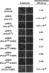
Fig 2. Ability of mutations in Pol IVCD to impede E. coli growth when overexpressed.
E. coli strain MS100 was transformed with the indicated plasmids, and aliquots of each transformation reaction were spread onto M9 minimal media supplemented with either glucose or arabinose, as noted. Representative portions of each plate following overnight incubation at 30°C are shown, as well as the ratio of the transformation frequency observed on plates supplemented with arabinose divided by the frequency observed on plates lacking arabinose. Results shown are representative of 2 separate experiments. The steady state level of Pol IVCD and Pol IVCD-T120P were measured in soluble cell free protein extracts by densitometry of Coomassie Brilliant Blue stained SDS-PAGE following arabinose induction. Based on the density of the region encompassing Pol IVCD, minus the background density observed in the pBAD control, wild type Pol IVCD (7.06±1.55 density units) and Pol IVCD-T120P (7.76±1.09 density units) were present at comparable levels.
Fig 3
Fig 3. Ability of mutant Pol IV proteins to catalyze replication in vitro.
Replication activity of the indicated Pol IV proteins (red) was measured (A) alone or (B) in the presence of the accessory proteins SSB (blue), β clamp (green) and DnaX using a primer extension assay as described in Materials and Methods.
Fig 4
Fig 4. Ability of Pol IV mutants to tolerate MMS-induced DNA damage in vivo.
(A) The dependence of MMS-induced mutagenesis on Pol IV (dinB) and Pol V (umuDC) function using strains RW118, RW120 (ΔumuDC), VB102 (ΔdinB), and VB103 (ΔdinB ΔumuDC), and (B) the ability of ~4-fold higher than SOS-induced levels of wild type or mutant Pol IV proteins (expressed from a plasmid) to suppress the frequency of Pol V-dependent MMS-induced mutagenesis of strain RW118 by competing with Pol V was measured as described in Materials and Methods and [33]. Results represent the average of at least 4 independent experiments ± one standard deviation. Symbols are as follows: **, p<0.0001; *, p<0.05.
Fig 5
Fig 5. The Pol IV-T120P, Pol IVC and Pol IVR strains are sensitized to killing by MMS.
Cultures of isogenic strains expressing the indicated Pol IV protein from the native dinB locus within the chromosome were serially 10-fold diluted using 0.8% saline, and 10 μl aliquots were spotted onto LB agar plates supplemented with the indicated concentrations of MMS. Results shown are representative of 4 independent experiments. The zaf-3633::cat allele, which is linked to the dinB locus and was used in strain construction, does not affect MMS sensitivity of the strains.
Fig 6
Fig 6. The Pol IV-T120P, Pol IVC and Pol IVR strains display an increased frequency of MMS-induced mutagenesis.
(A) The dependence of MMS-induced mutagenesis on Pol IV (dinB) and Pol V (umuDC) function using strains MG1655, MKS101 (ΔumuDC), MKS100 (ΔdinB), and MKS102 (ΔdinB ΔumuDC). (B) The ability of wild type or mutant Pol IV proteins to suppress the frequency of Pol V-dependent MMS-induced mutagenesis when expressed from the chromosome was measured as described in Materials and Methods [33]. Results represent the average of at least 4 independent experiments ± one standard deviation. Symbols are as follows: **, p<0.0001; *, p<0.05.
Fig 7
Fig 7. Pol IVCD-T120P, but not full length Pol IV-T120P, fails to inhibit Pol III processivity in vitro.
(A) Representative trajectories for primer extension of individual DNA molecules by Pol III alone (5 nM), or in the presence of excess Pol IV (300 nM). Processive events are marked in blue (Pol III) or red (Pol IV), with intervening pauses in grey. (B) A reduction in Pol III processivity in the presence of excess Pol IV (circles) can be abrogated by removing the Pol IV CBM (Pol IVC, triangle), but not by the T120P mutation (square, note overlap). Each point represents the mean of 50–470 processive events ± the standard error of the mean. (C) The catalytic domain of Pol IV (Pol IVCD, 900 nM) disrupts active Pol III (5 nM) synthesis, while an equivalent concentration of Pol IVCD-T120P does not. Bars represent the mean of 470, 301 and 657 separate processive events, respectively, ± the standard error of the mean.
Fig 8
Fig 8. Models for the role of residue T120 in Pol IV function.
Models for the structure of the Pol IIIαεθ-β clamp-DNA complex with Pol IV bound to the rim of the β clamp that is immediately adjacent to the β cleft that is bound by either (A) Pol IIIα (red) or (B) Pol IIIε (blue). These models were built using MacPyMol Molecular Graphics System, Version 1.7.4 Schrödinger, LLC and the previously published model for the Pol IIIαεθ-β clamp-DNA complex [84]. Pol IV (PDB 4IR9) was docked onto the rim of the β clamp in complex with Pol IIIαεθ by aligning it with the Pol IVLF domain in PDB 1UNN [12,53]. Residue T120 of Pol IV is in orange, while α-helix 5 corresponding to residues H116-Q135 of Pol IV is in yellow. (C) Model of the Pol III-Pol IV switching mechanism in which Pol IV (cyan) gains access to the DNA template after making contact with one or more subunits of Pol III (green), as well as the rim of the β clamp (purple). The small black circles represent the clefts in the β clamp. See text for further details.

Similar articles

Cited by

References

    1. Friedberg EC, Walker GC, Siede W, Wood RD, Schultz RA, et al. (2006) DNA repair and mutagenesis Washington, D. C.: ASM Press.
    1. Sale JE, Lehmann AR, Woodgate R (2012) Y-family DNA polymerases and their role in tolerance of cellular DNA damage. Nat Rev Mol Cell Biol 13: 141–152. 10.1038/nrm3289 - DOI - PMC - PubMed
    1. Sutton MD (2010) Coordinating DNA polymerase traffic during high and low fidelity synthesis. Biochim Biophys Acta 1804: 1167–1179. 10.1016/j.bbapap.2009.06.010 - DOI - PMC - PubMed
    1. Sanders LH, Devadoss B, Raja GV, O'Connor J, Su S, et al. (2011) Epistatic roles for Pseudomonas aeruginosa MutS and DinB (DNA Pol IV) in coping with reactive oxygen species-induced DNA damage. PLoS One 6: e18824 10.1371/journal.pone.0018824 - DOI - PMC - PubMed
    1. Petrosino JF, Galhardo RS, Morales LD, Rosenberg SM (2009) Stress-induced beta-lactam antibiotic resistance mutation and sequences of stationary-phase mutations in the Escherichia coli chromosome. J Bacteriol 191: 5881–5889. 10.1128/JB.00732-09 - DOI - PMC - PubMed

Publication types

LinkOut - more resources