Clinical Trial
. 2015 Sep 10;373(11):1040-7.
doi: 10.1056/NEJMoa1504542.

Chimeric Antigen Receptor T Cells against CD19 for Multiple Myeloma

Affiliations
Clinical Trial

Chimeric Antigen Receptor T Cells against CD19 for Multiple Myeloma

Alfred L Garfall et al. N Engl J Med. .

Abstract

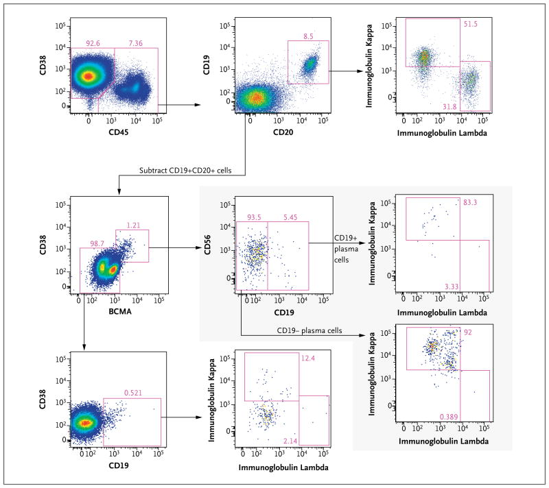
A patient with refractory multiple myeloma received an infusion of CTL019 cells, a cellular therapy consisting of autologous T cells transduced with an anti-CD19 chimeric antigen receptor, after myeloablative chemotherapy (melphalan, 140 mg per square meter of body-surface area) and autologous stem-cell transplantation. Four years earlier, autologous transplantation with a higher melphalan dose (200 mg per square meter) had induced only a partial, transient response. Autologous transplantation followed by treatment with CTL019 cells led to a complete response with no evidence of progression and no measurable serum or urine monoclonal protein at the most recent evaluation, 12 months after treatment. This response was achieved despite the absence of CD19 expression in 99.95% of the patient's neoplastic plasma cells. (Funded by Novartis and others; ClinicalTrials.gov number, NCT02135406.).


Figures

Figure 1
Figure 1. Measures of Multiple Myeloma Disease Burden after Autologous Stem-Cell Transplantations (ASCTs), and Measures of CTL019 Frequency and Activity after the Second Transplantation
Panel A shows the trend in IgA concentrations after the initial ASCT (left), with melphalan conditioning at a dose of 200 mg per square meter of body-surface area (MEL200), and after the second ASCT, with melphalan conditioning at a dose of 140 mg per square meter (MEL140) and CTL019 infusion (right). Additional therapy in the 6 months after the first ASCT included lenalidomide (LEN), bortezomib (BTZ), dexamethasone (DEX), and clarithromycin (CLR). The second ASCT was preceded by two cycles of continuous-infusion cyclophosphamide (CY). The serum monoclonal protein concentration (M spike) is also shown for the period before and after the second ASCT. Panel B shows CTL019 engraftment after the second ASCT, measured by means of flow cytometry as the number of cells per cubic millimeter (in peripheral blood only) and measured by means of a quantitative polymerase-chain-reaction assay (qPCR) as the number of copies of lentiviral vector sequence per microgram of genomic DNA (in peripheral blood and bone marrow), as well as the corresponding B-cell frequencies, measured as the number of cells per cubic millimeter. Panel C shows the serum ferritin, interferon-γ, and interleukin-6 concentrations.
Figure 2
Figure 2. Bone Marrow Core Biopsy Samples Obtained 2 Days before the Second ASCT and on Day 100 after the Second ASCT
The bone marrow sample obtained before the second ASCT (Panel A) shows more than 95% involvement by multiple myeloma on hematoxylin and eosin staining (left) and CD138 immunostaining (right). The sample obtained 100 days after the ASCT (Panel B) shows 1 to 2% overall cellularity and no plasma cells on hematoxylin and eosin staining (left) and CD138 immunostaining (right).
Figure 3
Figure 3. CD19 Expression on Neoplastic Plasma Cells
Panel A shows surface CD19 expression (right) on multiple myeloma plasma cells (CD38+CD45− gate, left) obtained from the pre-ASCT bone marrow aspirate. Brackets (right) indicate fluorescence thresholds that were used for fluorescence-activated cell sorting (FACS) of multiple myeloma plasma cells according to surface CD19 expression for analysis of CD19 messenger RNA levels. Panel B shows immunoglobulin light-chain expression in the overall population of malignant plasma cells (left) and the CD19+ subset of malignant plasma cells (right) from the pre-ASCT bone marrow aspirate. Panel C shows the results of reverse-transcriptase–polymerase-chain-reaction analysis of CD19 expression in FACS subsets of peripheral-blood mononuclear cells (PBMCs) from healthy donors and malignant plasma cells from the pre-ASCT bone marrow aspirate; results are normalized to expression in the CD19+ healthy-donor PBMC sample and plotted on a semi-logarithmic scale. ND denotes not detectable. I bars indicate 95% confidence intervals.
Figure 4
Figure 4. Characterization of CD19+ Cells in Bone Marrow Aspirate
Flow-cytometric analysis of bone marrow aspirate obtained before transplantation shows CD19+ cells outside the dominant CD38+CD45− multiple myeloma plasma-cell population characterized in Figure 3. Before this analysis, gating was performed to exclude doublets, debris, T cells, monocytes, and dead cells (not shown). CD45+ cells (top row, left plot) were found to contain the following CD19+ populations: polyclonal CD19+CD20+ B cells (top row, middle and right plots); a minor population of plasma cells (BCMA+CD38+), a subset of which is CD19+ and all of which are kappa-restricted (shaded inset), indicating that they are a component of the multiple myeloma clone; and a small population of CD45+CD38(dim)+CD20−CD19+ cells without immunoglobulin light-chain expression, most likely representing pro-B cells or early pre-B cells (third row, left and middle plots). BCMA denotes B-cell maturation antigen.

Comment in

  • Chimeric Antigen Receptor T Cells in Myeloma.
    Garfall AL, Stadtmauer EA, June CH. Garfall AL, et al. N Engl J Med. 2016 Jan 14;374(2):194. doi: 10.1056/NEJMc1512760. N Engl J Med. 2016. PMID: 26760100 No abstract available.
  • Chimeric Antigen Receptor T Cells in Myeloma.
    Shimabukuro-Vornhagen A, Schloesser HA, von Bergwelt-Baildon MS. Shimabukuro-Vornhagen A, et al. N Engl J Med. 2016 Jan 14;374(2):193-4. doi: 10.1056/NEJMc1512760. N Engl J Med. 2016. PMID: 26760101 No abstract available.

References

    1. Maus MV, Grupp SA, Porter DL, June CH. Antibody-modified T cells: CARs take the front seat for hematologic malignancies. Blood. 2014;123:2625–35. - PMC - PubMed
    1. Porter DL, Levine BL, Kalos M, Bagg A, June CH. Chimeric antigen receptor–modified T cells in chronic lymphoid leukemia. N Engl J Med. 2011;365:725–33. - PMC - PubMed
    1. Kalos M, Levine BL, Porter DL, et al. T cells with chimeric antigen receptors have potent antitumor effects and can establish memory in patients with advanced leukemia. Sci Transl Med. 2011;3:95ra73. - PMC - PubMed
    1. Maude SL, Frey N, Shaw PA, et al. Chimeric antigen receptor T cells for sustained remissions in leukemia. N Engl J Med. 2014;371:1507–17. - PMC - PubMed
    1. Grupp SA, Kalos M, Barrett D, et al. Chimeric antigen receptor-modified T cells for acute lymphoid leukemia. N Engl J Med. 2013;368:1509–18. - PMC - PubMed

Publication types

Substances

Associated data