. 2015 Sep 9;6(1):170.
doi: 10.1186/s13287-015-0166-2.

Human cord blood-derived platelet lysate enhances the therapeutic activity of adipose-derived mesenchymal stromal cells isolated from Crohn's disease patients in a mouse model of colitis

Affiliations

Human cord blood-derived platelet lysate enhances the therapeutic activity of adipose-derived mesenchymal stromal cells isolated from Crohn's disease patients in a mouse model of colitis

Dorian Forte et al. Stem Cell Res Ther. .

Abstract

Introduction: Due to their immunomodulatory properties, mesenchymal stromal cells (MSCs) have been used for auto-immune disease treatment. Crohn disease (CD) and ulcerative colitis are two major inflammatory bowel diseases (IBDs), resulting from pathological immune responses to environmental or microbial antigens. Preclinical and clinical studies have suggested that MSC-based cellular therapy hold promising potential for IBD treatment. However, open issues include the selection of the proper cell dose, the source and the optimal route of administration of MSCs for more effective results. Platelet lysate has gained clinical interest due to its efficacy in accelerating wound healing. Thus, we propose to combine the administration of MSCs with a human umbilical cord blood-derived platelet lysate (hCBPL) as a novel strategy to improve MSC-based therapy for IBD resolution.

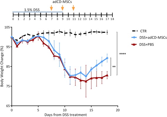
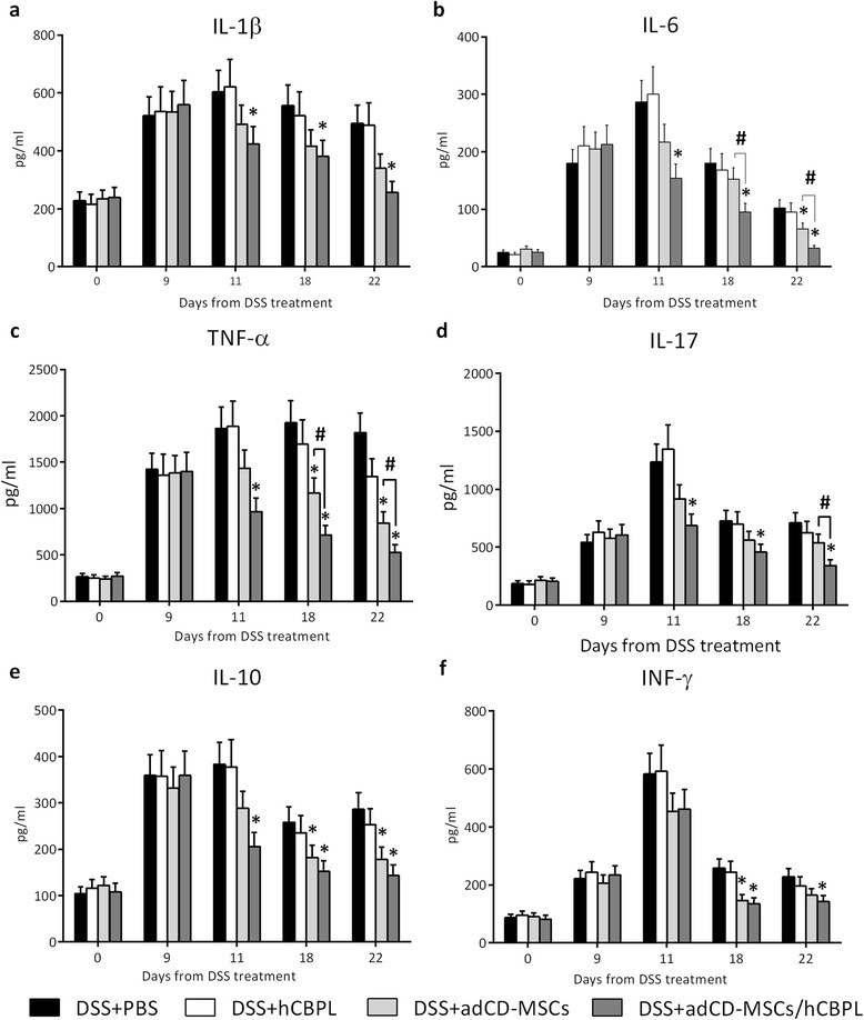
Methods: Colitis was induced in 8-week-old C57BL/6J mice by daily oral administration of dextran sulphate sodium (DSS) (1.5 % w/v in tap water) for 9 days. MSCs were isolated from adipose tissue of CD patients (adCD-MSCs), expanded in proliferation medium, resuspended in hCBPL or PBS and administrated via enema for three times (1 × 10(6) cells/mouse/time) every other day starting on day +7 from DSS induction. The colitis evolution was evaluated by daily monitoring of body weight, stool consistency and bleeding. Histopathological analysis was performed. Inflammatory cytokine plasma levels were determined. adCD-MSCs stained with lipophilic membrane dye Nile Red, were injected in DSS mice as described above. Colon section of mice sacrificed 24 hours after last cell administration, were analyzed by confocal microscopy.

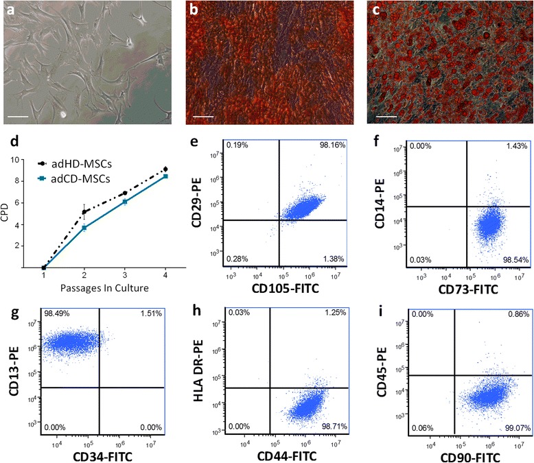
Results: We found that adCD-MSCs could be easily isolated and expanded from CD patients. Upon injection, adCD-MSCs exerted a therapeutic effect on DSS-induced colitis. Moreover, hCBPL increased adCD-MSCs efficacy by significantly reducing colitis scores, extension of the colon inflamed area and plasma levels of inflammatory mediators. Finally, Nile Red staining of MSCs is very efficient, stable and does not impair their vitality and function. Nile Red-labelling was clearly detected in the colitic area of adCD-MSCs injected mice and it was significantly brighter in the colon sections of mice that had received adCD-MSCs/hCBPL.

Conclusions: In summary, with this study we propose a novel and promising adCD-MSC/hCBPL-based therapy for refractory IBDs.


Figures

Fig. 1
Fig. 1
adCD-MSCs show typical MSC biological properties. a Representative field of exponential growing culture of adCD-MSCs. b Alizarin red staining of adCD-MSCs cultured for 3 weeks in osteogenic conditions and c Oil red O staining of adCD-MSCs cultured for 3 weeks in adipogenic conditions. Magnification 10×; scale bar, 100 μm. d Comparison of cumulative population doublings (CPD) calculated in adipose tissue-derived MSC cultures obtained from healthy donors (adHD-MSC) or adipose tissue-derived MSC cultures obtained from CD patients (adCD-MSC) at each passage. Results expressed as the mean ± SEM calculated from data obtained from four independent samples. Differences are not significant (p value not significant). e–i Flow cytometric immunophenotype of adCD-MSCs at passage 3. Representative dot plots. The analysis shows a predominant population of cells positive for CD90, CD29, CD73, CD105, CD13, and CD44, and negative for CD34, CD45, CD14, and HLA-DR. FITC, fluorescein isothiocyanate, PE phycoerythrin
Fig. 2
Fig. 2
Treatment with adCD-MSCs protects against DSS-induced colitis. Mice received 1.5 % of DSS with their drinking water for 9 days. At days +7, +9, and +11 from DSS induction, mice are treated by enema with adCD-MSCs (1 × 10⁶ cells/mice/time) or phosphate saline buffer (PBS) alone. (Top) Schematic of the experiment. The evolution of colitis is monitored by body weight change expressed as a percentage of the initial body weight at day 0 (100 %). CTR, healthy control mice (dotted black line); DSS+adCD-MSCs, DSS-treated mice which received adCD-MSCs (blue line); DSS+PBS, DSS-treated mice which received PBS alone (red line). Data expressed as mean ± standard error of the mean (SEM). n = 4 mice per group. CTR vs. DSS+PBS, ****p <0.0001; DSS+adCD-MSCs vs. DSS+PBS, **p <0.01. adCD-MSC mesenchymal stromal cell isolated from adipose tissue of Crohn’s disease patients, DSS dextran sulfate sodium
Fig. 3
Fig. 3
hCBPL improves the therapeutic efficacy of adCD-MSCs. Mice received 1.5 % of DSS with their drinking water for 9 days. At days +7, +9, and +11 from DSS induction, mice were treated by enema with adCD-MSCs (1 × 10⁶ cells/mice/time) with or without human umbilical cord blood-derived platelet lysate (hCBPL) or phosphate saline buffer (PBS) alone. Clinical evolution of colitis is monitored by body weight change and DAI evaluation. a Body weight changes during the course of the experiment are expressed as the percentage of the initial body weight at day +0. (Top) Schematic of the experiment. CTR, healthy control mice (dotted black line); DSS+adCD-MSCs, DSS-treated mice which received adCD-MSCs (blue line); DSS+adCD-MSCs/hCBPL, DSS-treated mice which received adCD-MSCs resuspended in hCBPL (green line); DSS+hCBPL, DSS-treated mice which received hCBPL alone (pink line); DSS+PBS, DSS-treated mice which received PBS (red line). Data expressed as mean ± SEM. n = 4–12 mice per group. CTR vs. DSS+PBS, ****p <0.0001; DSS+adCD-MSCs/hCBPL vs. DSS+PBS, **p <0.01; hCBPL vs. DSS+PBS, p value not significant. b DAI calculated by the combined score of weight loss, stool consistency, and bleeding, as detailed in Additional file 1. DAI is evaluated daily from day +7 (first administration of adCD-MSCs or adCD-MSCs/hCBPL or PBS or hCBPL) to day +18. Representative time points are shown. Data expressed as mean ± SEM. n = 4–12 mice per group. DSS+adCD-MSCs and DSS+adCD-MSCs/ hCBPL vs. DSS+PBS, *p <0.05. adCD-MSC mesenchymal stromal cell isolated from adipose tissue of Crohn’s disease patients, DSS dextran sulfate sodium
Fig. 4
Fig. 4
adCD-MSCs/hCBPL prevent DSS-induced pathology. Photomicrographs of H&E-stained paraffin sections of mouse colons treated as described before and harvested at day +22. Two representative examples are shown for each condition. Sections from a, b mice that received DSS, c, d mice that received DSS+hCBPL, e, f mice that received DSS+adCD-MSCs, and g, h mice that received adCD-MSCs/hCBPL. Magnification 50×; scale bar, 200 μm. adCD-MSC mesenchymal stromal cell isolated from adipose tissue of Crohn’s disease patients, DSS dextran sulfate sodium, hCBPL human umbilical cord blood-derived platelet lysate, PBS phosphate saline buffer
Fig. 5
Fig. 5
adCD-MSCs/hCBPL decrease systemic inflammatory response in DSS-induced colitis mouse model. Plasma was obtained from mice blood, collected from the caudal vein. a–f Cytokine levels (IL-1β, IL-17, TNFα, IL-6, IL-10, IFNγ) were measured using Bioplex assay at the start of the experiment (day +0), at the end of DSS treatment (day+9), after the administration of PBS (black column), hCBPL alone (white column), adCD-MSCs (light gray column), or adCD-MSCs/hCBPL (dark gray column) (day +11), at day +18, and at the end of the experiment (day +22). Data expressed as mean ± SEM. n = 4–8 mice per group. *p <0.05 with respect to DSS+PBS group; #p <0.05 in the comparison between the MSC groups. adCD-MSC mesenchymal stromal cell isolated from adipose tissue of Crohn’s disease patients, DSS dextran sulfate sodium, hCBPL human umbilical cord blood-derived platelet lysate, IFN-γ interferon gamma, IL interleukin, PBS phosphate saline buffer, TNFα tumor necrosis factor alpha
Fig. 6
Fig. 6
adCD-MSCs can be efficiently labeled by Nile Red staining. Fluorescence intensity and distribution of Nile Red staining. a–d Representative micrograph of stained cell cultures and e–h three-dimensional histograms of confocal analysis of fluorescence intensity (expressed as fluorescence intensity/pixel) for each time point. Fluorescence intensity of Nile Red-labeled adCD-MSCs analyzed by flow cytometry is high at i 15 hours, l 24 hours, and m 72 hours and n decreased significantly at 120 hours post labeling. Magnification 40×; scale bar, 20 μm
Fig. 7
Fig. 7
In vivo administration of Nile Red-labeled adCD-MSCs in DSS-treated colitic mice. DSS-treated mice were subjected to enema administration of Nile Red-stained adCD-MSCs. Colon tissue sections were collected 24 hours after the last MSC administration and were analyzed by confocal or optical microscopy. a DSS-treated mice colon sections show a low fluorescence background, in the absence of adCD-MSCs. Fluorescence signals in the presence of b Nile Red-stained adCD-MSCs or c adCD-MSCs/hCBPL. d Sections from consecutive segment were fixed and stained with H&E. e Merged images show that Nile Red fluorescence is present mainly in the muscular layers surrounding the colon injury. f Fluorescence quantification is presented as mean fluorescence intensity (± SEM). Data are representative of three mice for each experimental group and 15 acquired sections for each experimental condition. *p <0.05. Magnification 20×; scale bar, 150 μm. adCD-MSC mesenchymal stromal cell isolated from adipose tissue of Crohn’s disease patients, DSS dextran sulfate sodium, hCBPL human umbilical cord blood-derived platelet lysate, PBS phosphate saline buffer

References

    1. Caplan AI. Mesenchymal stem cells. J Orthop Res. 1991;9:641–650. doi: 10.1002/jor.1100090504. - DOI - PubMed
    1. Pittenger MF, Mackay AM, Beck SC, Jaiswal RK, Douglas R, Mosca JD, et al. Multilineage potential of adult human mesenchymal stem cells. Science. 1999;284:143–147. doi: 10.1126/science.284.5411.143. - DOI - PubMed
    1. Jiang Y, Jahagirdar BN, Reinhardt RL, Schwartz RE, Keene CD, Ortiz-Gonzalez XR, et al. Pluripotency of mesenchymal stem cells derived from adult marrow. Nature. 2002;418:41–49. doi: 10.1038/nature00870. - DOI - PubMed
    1. Le Blanc K. Immunomodulatory effects of fetal and adult mesenchymal stem cells. Cytotherapy. 2003;5:485–489. doi: 10.1080/14653240310003611. - DOI - PubMed
    1. Fibbe WE, Nauta AJ, Roelofs H. Modulation of immune responses by mesenchymal stem cells. Ann N Y Acad Sci. 2007;1106:272–278. doi: 10.1196/annals.1392.025. - DOI - PubMed

Publication types