Review
. 2015 Oct:116:15-26.
doi: 10.1016/j.eplepsyres.2015.06.010. Epub 2015 Jun 21.

Optogenetic tools for modulating and probing the epileptic network

Affiliations
Review

Optogenetic tools for modulating and probing the epileptic network

Mingrui Zhao et al. Epilepsy Res. 2015 Oct.

Abstract

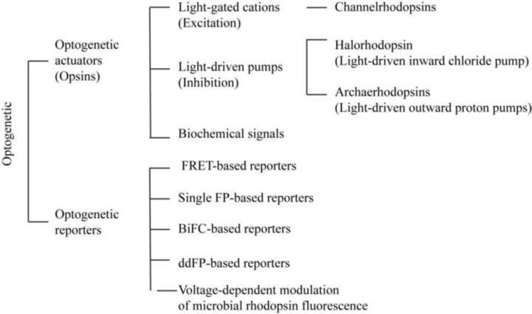
Epilepsy affects roughly 1% of the population worldwide. Although effective treatments with antiepileptic drugs are available, more than 20% of patients have seizures that are refractory to medical therapy and many patients experience adverse effects. Hence, there is a continued need for novel therapies for those patients. A new technique called "optogenetics" may offer a new hope for these refractory patients. Optogenetics is a technology based on the combination of optics and genetics, which can control or record neural activity with light. Following delivery of light-sensitive opsin genes such as channelrhodopsin-2 (ChR2), halorhodopsin (NpHR), and others into brain, excitation or inhibition of specific neurons in precise brain areas can be controlled by illumination at different wavelengths with very high temporal and spatial resolution. Neuromodulation with the optogenetics toolbox have already been shown to be effective at treating seizures in animal models of epilepsy. This review will outline the most recent advances in epilepsy research with optogenetic techniques and discuss how this technology can contribute to our understanding and treatment of epilepsy in the future.

Keywords: Epilepsy model; Optical imaging; Optogenetic; Photostimulation.


Figures

Figure 1
Figure 1
Optogenetic tools. FRET: Förster (Fluorescence) resonance energy transfer. FPs: fluorescence proteins. BiFC: Bimolecular fluorescence complementation.
Figure 2
Figure 2
Targeting optogenetic tools using viral injection. (A) AAVs or lentivirus can be directly injected into cortical and subcortical regions. A typical vector is constructed with a promoter, a opsin, and a reporter. Additional cell-type specificity is attained either by cell-type-specific promoters or via a recombinase-dependent virus, injected in a transgenic animal expressing a recombinase such as Cre in specific cells, leading to specific expression of the transgene only in defined cell types. The opsin gene is combined or fused to a reporter fluorescent protein such as GFP, mCherry, or EYFP. (B) After viral expression, photostimulation drives local excitation or inhibition using different color lights such as laser, LED, or Arc lamp. The detail strategies for spatial optogenetic targeting including local somata or/ and projection (axon) are discussed in previous review paper (Yizhar et al., 2011a).
Figure 3
Figure 3
Optogenetic strategies for controlling epilepsy. A. First optogenetic epilepsy experiment. “Stimulation Train Induced Bursting (STIB) in CA3 is strongly attenuated by orange-light activation of transgene NpHR in organotypic hippocampal cultures. Recordings of 3 consecutive STIB stimulations, with orange-light illumination on second stimulation, in NpHR-transduced slices. Insets: Magnification of traces showing epileptiform bursts after STIB stimulation. Scale bars apply for all traces” Adapted with permission from PNAS (Tonnesen et al., 2009). B. Ipsilateral and contralateral control of seizures in PV-ChR2 mice. (a) Crossing PV-Cre and Cre-dependent ChR2 mouse lines generated mice expressing the excitatory opsin ChR2 in PV-expressing GABAergic cells (PV-ChR2 mice). (b) Example electrographic seizures in a PV-ChR2 mouse (top, no-light) truncated by blue (473 nm) light delivery (bottom, blue line) to the hippocampus. Adapted with permission from Nat Comm (Krook-Magnuson et al., 2013). C. Selective optical inhibition of thalamocortical neurons interrupts ongoing epileptic seizures in awake, freely behaving animals. (a) Diagram of chronic multisite optrode (CMO) implanted into somatosensory thalamus for behaving recordings and optical stimulations. Arrowheads indicate thalamic recording sites (T1–4). (b) Confocal image of coronal brain section taken through the cortical lesion (red dashed line) showing eNPHR-expressing thalamocortical fibers terminating mainly in layer 4 (yellow arrow) from a rat killed after recordings. (c) Representative example of simultaneously recorded cortical EEGs and thalamic LFPs before and during 594-nm light delivery in the thalamus ipsilateral to stroke. Arrows indicate seizure onset and its interruption by light delivery in thalamus. Adapted with permission from Nature Neuroscience (Paz et al., 2013).
Figure 4
Figure 4
Closed-loop system design. EEG input (blue) from the mouse hippocampus is amplified (Amp), digitized (A/D) and relayed to a PC running a custom-designed real-time seizure detection software. The signal is fed into a number of possible detection algorithms, which utilize features of signal power, spikes or frequency. Thresholds for power and spike properties (green) are determined using tunable leaky integrators acting as low-pass filters. Top: Amplitude Correlation (purple, during an example seizure, shown in grey); Middle: spike characteristics (for example, amplitude, rate, regularity and spike width, shown in red); Bottom: power of the signal in specific frequency bands during the same seizure, with warmer colors representing higher energy. Once a seizure has been detected using the selected criteria, for 50% of the events in a random fashion (RND), the software activates the optical output (orange) delivered to the hippocampus of the mouse, via a TTL signal from the digitizer to the laser. All trigger events, however, are flagged for later off-line analysis. COMP, digital comparator. USB, universal serial bus. Adapted with permission from ref Nature Commun (Krook-Magnuson et al., 2013).

References

    1. Adamantidis AR, Zhang F, Aravanis AM, Deisseroth K, de Lecea L. Neural substrates of awakening probed with optogenetic control of hypocretin neurons. Nature. 2007;450:420–424. - PMC - PubMed
    1. Alford Spencer C., Abdelfattah Ahmed S., Ding Y, Campbell Robert E. A Fluorogenic Red Fluorescent Protein Heterodimer. Chemistry & Biology. 2012;19:353–360. - PMC - PubMed
    1. Alford SC, Wu J, Zhao Y, Campbell RE, Knöpfel T. Optogenetic reporters. Biology of the Cell. 2013;105:14–29. - PubMed
    1. Anselmi F, Ventalon C, Bègue A, Ogden D, Emiliani V. Three-dimensional imaging and photostimulation by remote-focusing and holographic light patterning. Proceedings of the National Academy of Sciences of the United States of America. 2011a;108:19504–19509. - PMC - PubMed
    1. Anselmi F, Ventalon C, Bèguea A, Ogdenb D, Emiliani V. Three-dimensional imaging and photostimulation by remote-focusing and holographic light patterning. Proceedings of the National Academy of Sciences of the United States of America. 2011b;108:19504–19509. - PMC - PubMed

Publication types