. 2016 Feb;24(1):26-33.
doi: 10.1038/mt.2015.168. Epub 2015 Sep 10.

Clinical Improvement of Alpha-mannosidosis Cat Following a Single Cisterna Magna Infusion of AAV1

Affiliations

Clinical Improvement of Alpha-mannosidosis Cat Following a Single Cisterna Magna Infusion of AAV1

Sea Young Yoon et al. Mol Ther. 2016 Feb.

Abstract

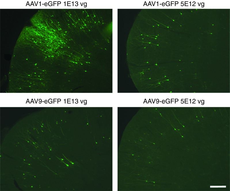
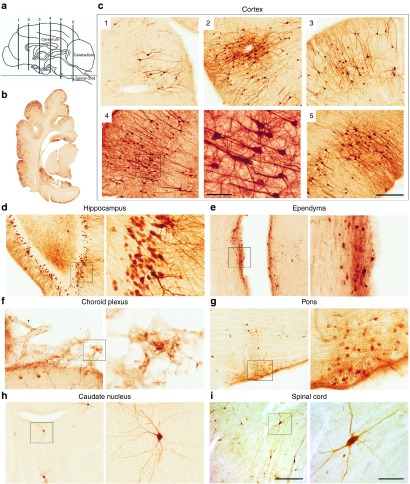
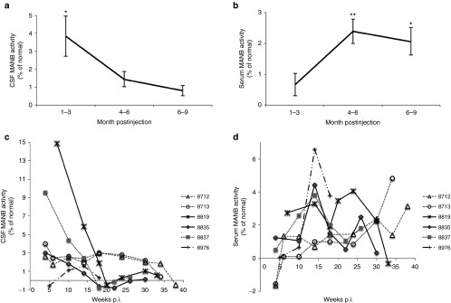
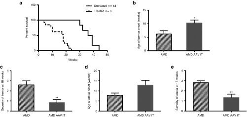
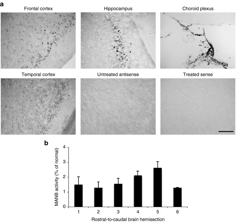
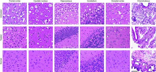
Lysosomal storage diseases (LSDs) are debilitating neurometabolic disorders for most of which long-term effective therapies have not been developed. Gene therapy is a potential treatment but a critical barrier to treating the brain is the need for global correction. We tested the efficacy of cisterna magna infusion of adeno-associated virus type 1 (AAV1) expressing feline alpha-mannosidase gene in the postsymptomatic alpha-mannosidosis (AMD) cat, a homologue of the human disease. Lysosomal alpha-mannosidase (MANB) activity in the cerebrospinal fluid (CSF) and serum were increased above the control values in untreated AMD cats. Clinical neurological signs were delayed in onset and reduced in severity. The lifespan of the treated cats was significantly extended. Postmortem histopathology showed resolution of lysosomal storage lesions throughout the brain. MANB activity in brain tissue was significantly above the levels of untreated tissues. The results demonstrate that a single cisterna magna injection of AAV1 into the CSF can mediate widespread neuronal transduction of the brain and meaningful clinical improvement. Thus, cisterna magna gene delivery by AAV1 appears to be a viable strategy for treatment of the whole brain in AMD and should be applicable to many of the neurotropic LSDs as well as other neurogenetic disorders.


Figures

Figure 1
Figure 1
GFP immunofluorescence in the cerebral cortex of normal cats following cisterna magna injection of AAV1 or AAV9 vector. Normal cats received cisterna magna injections of AAV1 (n = 3, 1013 GC; n = 1, 5 × 1012 GC) or AAV9 vector (n = 2, 1013 GC; n = 1, 5 × 1012 GC) encoding eGFP. Immunofluorescent staining with GFP antibody showed vector transduction in the cerebral cortex of cats 4 weeks postinjection. Bar = 300 µm. AAV, adeno-associated virus; eGFP, enhanced green fluorescent protein.
Figure 2
Figure 2
Widespread gene transduction throughout the central nervous system (CNS) of normal cat following adeno-associated virus type 1 (AAV1) injection into cerebrospinal fluid. Normal cats (n = 3) received cisterna magna injections of AAV1 vector (1013 GC) encoding enhanced green fluorescent protein (eGFP), and GFP expression in the CNS was examined using immunohistochemistry 4 weeks postinjection. (a) The distribution of gene expression was analyzed in coronal sections taken at intervals along the rostral–caudal axis of the brain. (b–d) Extensive vector transduction was achieved throughout the brain, predominantly in cells with the morphology of pyramidal neurons of the cerebral cortex (b,c) and granule cells of the hippocampus (d). GFP expression was also observed in (e) ependymal cells lining the ventricles, (f) choroid plexus, (g) trapezoid body cells in the pons, (h) medium spiny neurons of the caudate nucleus, and (i) spinal cord. Bar = 300 µm (left columns) and 60 µm (right columns).
Figure 3
Figure 3
MANB activity in CSF and serum of alpha-mannosidosis (AMD) cats following cisterna magna injection of adeno-associated virus type 1 (AAV1). AMD cats (n = 6) were treated with a single cisterna magna injection of AAV1 (1013 GC) expressing fMANB into the cisterna magna at 6 weeks of age. CSF and serum samples were collected before and after injection and assayed for MANB enzyme activity. (a) MANB activity in the CSF was significantly elevated above residual background activity at 1–3 months postinjection (p.i.). (b) MANB activity in the serum was significantly elevated after 4 months postinjection. (c,d) MANB activity in the CSF and serum of individual animals are shown. Bars represent means ± SEM. *P < 0.05; **P < 0.01. CSF, cerebrospinal fluid; fMANB, feline alpha-mannosidase; MANB, lysosomal alpha-mannosidase.
Figure 4
Figure 4
Extended lifespan and improvement in neurological signs in treated AMD cats. (a) The treated AMD cats (n = 6) lived significantly longer than untreated cats (n = 13) and were euthanized at 30–44 weeks of age due to disease progression (log-rank test, P < 0.001). Neurological examinations were performed monthly from treatment to assess the therapeutic effects. (b) The treated cats showed a significantly delayed onset of whole body tremor. (d) Onset of ataxia did not reach statistical significance. (c,e) At 18 weeks of age, both the whole body tremor and truncal ataxia were significantly less severe. Bars represent means ± SEM. *P < 0.05; **P < 0.01. AAV, adeno-associated virus; AMD, alpha-mannosidosis.
Figure 5
Figure 5
Widespread distribution of feline alpha-mannosidase (fMANB) mRNA transcripts and MANB enzymatic activity in treated alpha-mannosidosis (AMD) cat brain. (a) In situ hybridization (ISH) against the vector-encoded fMANB mRNA demonstrated a strong ISH signal in the cerebral cortex, hippocampus, and choroid plexus epithelial cells of the treated AMD cat brain. Brains from uninjected animals were negative for fMANB mRNA and the sense probe, used as a control in a treated animal, also detected no signal. The amount of MANB enzymatic activity was measured in coronal sections taken at intervals along the rostral–caudal axis as shown in Figure 2a. (b) The mean activity values for treated AMD cats (n = 4) in transverse slices along the rostral–caudal axis show the wide spatial distribution within the cat brain. Bars represent means ± SEM. Bar = 200 µm. MANB, lysosomal alpha-mannosidase.
Figure 6
Figure 6
Correction of lysosomal storage lesions on hematoxylin and eosin (H&E)-stained brain tissue of treated alpha-mannosidosis cats. H&E-stained brain sections of treated cats showed resolution of lysosomal storage vacuoles throughout the brain, including in the cerebral cortex, caudate nucleus, hippocampus, cerebellum, and choroid plexus. Bar = 60 µm.

References

    1. Auclair, D, Finnie, J, White, J, Nielsen, T, Fuller, M, Kakkis, E et al. (2010). Repeated intrathecal injections of recombinant human 4-sulphatase remove dural storage in mature mucopolysaccharidosis VI cats primed with a short-course tolerisation regimen. Mol Genet Metab 99: 132–141. - PubMed
    1. Chang, M, Cooper, JD, Sleat, DE, Cheng, SH, Dodge, JC, Passini, MA et al. (2008). Intraventricular enzyme replacement improves disease phenotypes in a mouse model of late infantile neuronal ceroid lipofuscinosis. Mol Ther 16: 649–656. - PubMed
    1. Crawley, AC, Marshall, N, Beard, H, Hassiotis, S, Walsh, V, King, B et al. (2011). Enzyme replacement reduces neuropathology in MPS IIIA dogs. Neurobiol Dis 43: 422–434. - PubMed
    1. Dierenfeld, AD, McEntee, MF, Vogler, CA, Vite, CH, Chen, AH, Passage, M et al. (2010). Replacing the enzyme alpha-L-iduronidase at birth ameliorates symptoms in the brain and periphery of dogs with mucopolysaccharidosis type I. Sci Transl Med 2: 60ra89. - PMC - PubMed
    1. Dodge, JC, Clarke, J, Treleaven, CM, Taksir, TV, Griffiths, DA, Yang, W et al. (2009). Intracerebroventricular infusion of acid sphingomyelinase corrects CNS manifestations in a mouse model of Niemann-Pick A disease. Exp Neurol 215: 349–357. - PubMed

Publication types

MeSH terms

Substances

LinkOut - more resources