Review
. 2015 Oct 30;290(44):26422-9.
doi: 10.1074/jbc.R115.665869. Epub 2015 Sep 9.

The linker for activation of T cells (LAT) signaling hub: from signaling complexes to microclusters

Affiliations
Review

The linker for activation of T cells (LAT) signaling hub: from signaling complexes to microclusters

Lakshmi Balagopalan et al. J Biol Chem. .

Abstract

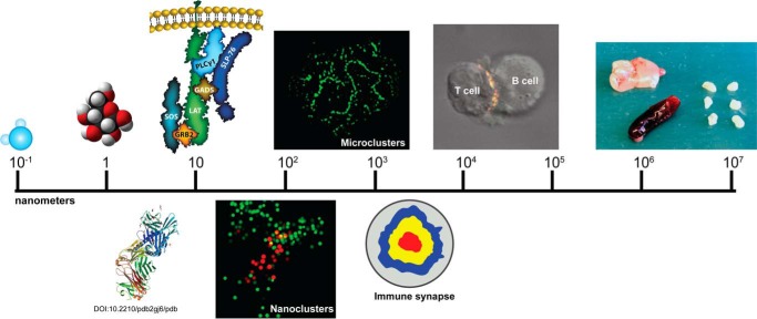
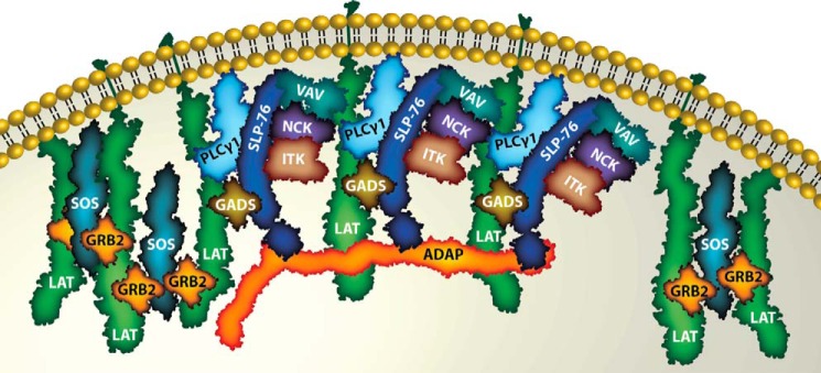
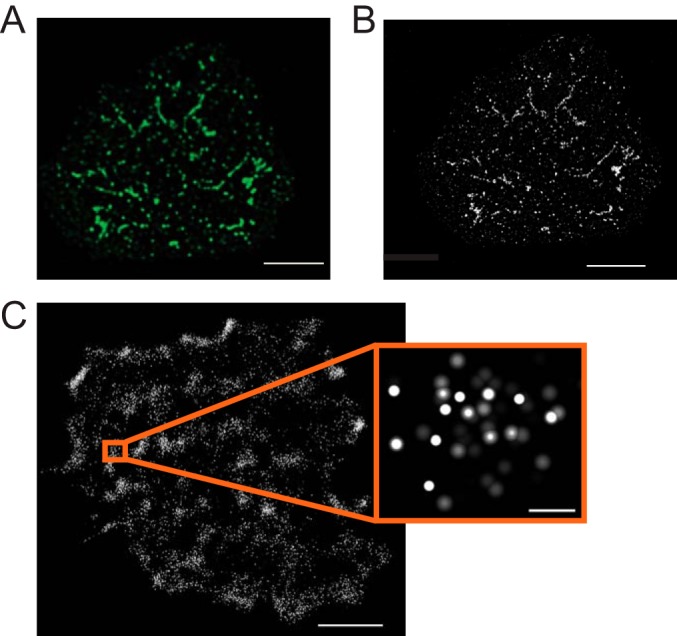
Since the cloning of the critical adapter, LAT (linker for activation of T cells), more than 15 years ago, a combination of multiple scientific approaches and techniques continues to provide valuable insights into the formation, composition, regulation, dynamics, and function of LAT-based signaling complexes. In this review, we will summarize current views on the assembly of signaling complexes nucleated by LAT. LAT forms numerous interactions with other signaling molecules, leading to cooperativity in the system. Furthermore, oligomerization of LAT by adapter complexes enhances intracellular signaling and is physiologically relevant. These results will be related to data from super-resolution microscopy studies that have revealed the smallest LAT-based signaling units and nanostructure.

Keywords: T-cell; adaptor protein; cooperativity; immunology; signal transduction.


Figures

FIGURE 1.
FIGURE 1.
Hierarchy of scale for studying the immune system. Important biological entities vary widely in size. Important small molecules such as water and glucose are shown on the left side of the scale. Next are two representations of protein complexes: a ribbon diagram of the T cell receptor complexed to antigen and MHC (Protein Data Bank entry 2GJ6 (66)) as well as a graphic of a LAT-based signaling complex. These complexes can be arranged into nanoclusters; a localization microscopy image shows LAT molecules in red surrounded by SLP-76 molecules shown in green. Next, a confocal image shows diffraction-limited microclusters in an activated Jurkat cell stained with an antibody to phosphorylated LAT. Signaling molecules can be arranged in large-scale patterns such as the immune synapse shown in graphic form and as part of a two-cell conjugate between a Jurkat cell and a superantigen-pulsed B cell. Finally, these molecules lead to activation of the immune system, represented by various lymphoid organs: thymus (top left), spleen (bottom left), and lymph nodes (right, axillary, brachial, and inguinal).
FIGURE 2.
FIGURE 2.
Formation of LAT oligomers following T cell activation. Binding of two Grb2 molecules to a single Sos1 protein allows the formation of LAT oligomers. Both the GEF activity of Sos1 and multipoint binding are needed for full T cell activation. The binding of ADAP to multiple SLP-76 molecules promotes SLP-76 clustering and could potentially induce LAT oligomers as well.
FIGURE 3.
FIGURE 3.
Imaging LAT-based complexes at different scales. A, diffraction-limited, deconvolved image of a Jurkat T cell activated on a coverslip coated with anti-TCR (anti-CD3ϵ) antibodies and stained for phosphorylated, active LAT. Clusters of activated LAT appear as large puncta, several hundreds of nanometers in size. Scale bar = 3 μm. B, deconvolved STED image of the same cell reveals that microclusters are actually much smaller and that the largest are often several small clusters close together. The smallest of these clusters are near 50 nm in size. Scale bar = 3 μm. C, localization image of an activated Jurkat T cell showing the locations of LAT molecules conjugated to the photoactivatable protein, Dronpa. Scale bar = 2 μm. The inset shows a group of individual LAT molecules from one of the denser areas. Scale bar = 100 nm.

References

    1. Chakraborty A. K., and Weiss A. (2014) Insights into the initiation of TCR signaling. Nat. Immunol. 15, 798–807 - PMC - PubMed
    1. Finco T. S., Kadlecek T., Zhang W., Samelson L. E., and Weiss A. (1998) LAT is required for TCR-mediated activation of PLCγ1 and the Ras pathway. Immunity 9, 617–626 - PubMed
    1. Zhang W., Irvin B. J., Trible R. P., Abraham R. T., and Samelson L. E. (1999) Functional analysis of LAT in TCR-mediated signaling pathways using a LAT-deficient Jurkat cell line. Int. Immunol. 11, 943–950 - PubMed
    1. Shen S., Zhu M., Lau J., Chuck M., and Zhang W. (2009) The essential role of LAT in thymocyte development during transition from the double-positive to single-positive stage. J. Immunol. 182, 5596–5604 - PMC - PubMed
    1. Zhang W., Sommers C. L., Burshtyn D. N., Stebbins C. C., DeJarnette J. B., Trible R. P., Grinberg A., Tsay H. C., Jacobs H. M., Kessler C. M., Long E. O., Love P. E., and Samelson L. E. (1999) Essential role of LAT in T cell development. Immunity 10, 323–332 - PubMed

Publication types

MeSH terms

Associated data