. 2015 Sep 9:13:217.
doi: 10.1186/s12916-015-0454-9.

Chemotherapy reduces PARP1 in cancers of the ovary: implications for future clinical trials involving PARP inhibitors

Affiliations

Chemotherapy reduces PARP1 in cancers of the ovary: implications for future clinical trials involving PARP inhibitors

Maud Marques et al. BMC Med. .

Abstract

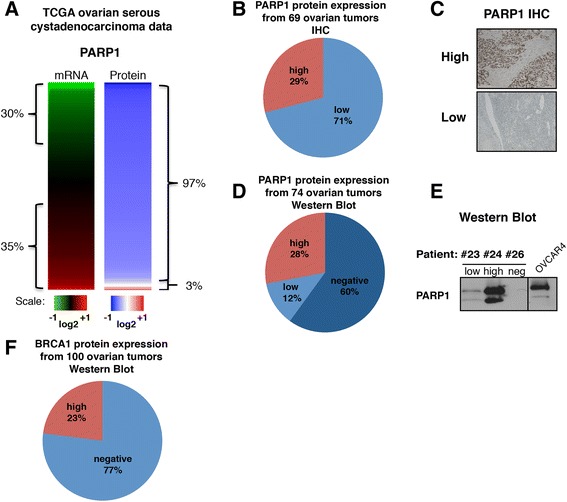
Background: PARP inhibitors have shown promising clinical results in cancer patients carrying BRCA1/2 mutations. Their clinical efficacy could logically be influenced by PARP1 protein levels in patient tumors.

Methods: We screened three cohorts of patients with ovarian cancer, totaling 313 samples, and evaluated PARP1 protein expression by immunohistochemistry with further validation by western blotting.

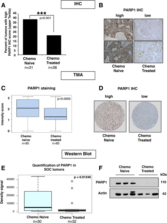
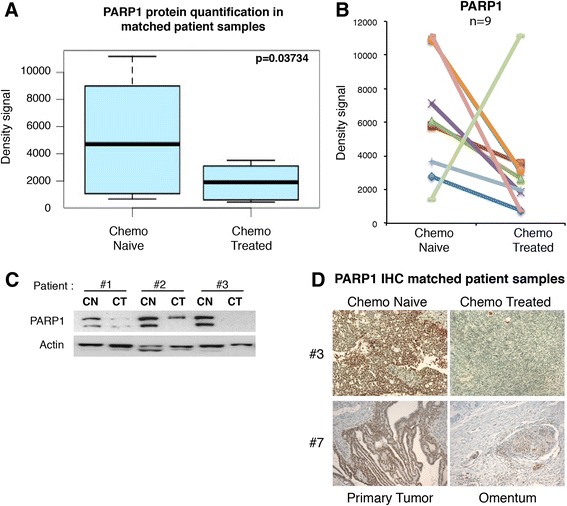
Results: We observed that up to 60 % of tumors showed little PARP1 protein expression. In serous ovarian tumors, comparing intratumoral PARP1 expression between chemo-naïve and post-chemotherapy patients revealed a decrease in intratumoral PARP1 following chemotherapy in all three cohorts (immunohistochemistry: p < 0.001, n = 239; western blot: p = 0.012, n = 74). The findings were further confirmed in a selection of matched samples from the same patients before and after chemotherapy.

Conclusion: Our data suggest that patients should be screened for PARP1 expression prior to therapy with PARP inhibitors. Further, the observed reduction of intratumoral PARP1 post-chemotherapy suggests that treating chemo-naïve patients with PARP inhibitors prior to the administration of chemotherapy, or concurrently, might increase the responsiveness to PARP1 inhibition. Thus, a change in the timing of PARP inhibitor administration may be warranted for future clinical trials.


Figures

Fig. 1
Fig. 1
Low PARP1 protein expression in ovarian tumors. a Analysis of The Cancer Genome Atlas (TCGA) ovarian cystadenocarcinoma database for PARP1 mRNA (n = 570) and protein (n = 412) expression levels. Data were used to generate a heat map with red representing high expression level and green low expression level. b Pie chart representing PARP1 protein level as determined by immunohistochemistry (IHC) in 69 HGSC samples (training cohort). c PARP1 representative IHC for each expression category (low and high). d Pie chart distribution of PARP1 in 100 unselected ovarian tumors from 74 patients (validation cohort 2, several patients had multiple tumors examined) evaluated by western blotting. e PARP1 representative western blot for each expression category (low, high, and negative); OVCAR4 protein extract was used as positive control. f Pie chart representing BRCA1 protein level distribution from 74 patients (100 tumors, validation cohort 2) evaluated by western blotting
Fig. 2
Fig. 2
Chemo-treated tumors have lower levels of PARP1 protein than chemo-naïve tumors. a We used 69 HGSC tumors for immunohistochemistry (IHC) staining of PARP1 (training cohort). Each slide score is the product of the percentage of stained tumors cells and the intensity of the staining as described in the “Methods” section. The bar graph represents the percentage of slides with scores in the upper tertile from chemo-naïve and chemo-treated samples. Chi-square test p < 0.001. b Representative photos of PARP1 IHC from two chemo-naïve tumors and two chemo-treated tumors (20×). c Boxplot showing compilation of PARP1 intensity of staining score for the tissue microarray (TMA). The p-value tested whether there was a significant difference in PARP1 staining intensity between the chemo-naïve and chemo-treated tumors and was calculated using a two-tailed Mann–Whitney test. d Representative TMA cores stain with PARP1 antibody. e PARP1 protein levels in 62 serous ovarian cancer (SOC) tumors (30 chemo-naïve and 32 chemo-treated) were quantified with ImageJ and the density signals obtained were used to generate a boxplot. Wilcox Mann–Whitney test gave p = 0.01246. f Representative western blots of PARP1 in three chemo-naïve tumors and three chemo-treated tumors from cohort shown in e
Fig. 3
Fig. 3
Chemotherapy decreases PARP1 protein levels in matched patient samples. a PARP1 protein levels in tumor samples from the same 15 patients before and after chemotherapy were quantified with ImageJ and the density signals obtained were used to generate a boxplot. Paired Mann–Whitney test p = 0.03734. b PARP1 density signals from nine patients with the presence of PARP1 in the tumors before receiving chemotherapy. c Representative PARP1 and actin western blot of three matched tumor samples before (CN) and after (CT) receiving chemotherapy treatment. d PARP1 immunohistochemistry (IHC) staining in tumor before and after chemotherapy treatment in the same patients (20×)

References

    1. Jemal A, Siegel R, Ward E, Hao Y, Xu J, Murray T, et al. Cancer statistics, 2008. CA Cancer J Clin. 2008;58:71–96. doi: 10.3322/CA.2007.0010. - DOI - PubMed
    1. Permuth-Wey J, Sellers TA. Epidemiology of ovarian cancer. Methods Mol Biol. 2009;472:413–37. doi: 10.1007/978-1-60327-492-0_20. - DOI - PubMed
    1. Miki Y, Swensen J, Shattuck-Eidens D, Futreal PA, Harshman K, Tavtigian S, et al. A strong candidate for the breast and ovarian cancer susceptibility gene BRCA1. Science. 1994;266:66–71. doi: 10.1126/science.7545954. - DOI - PubMed
    1. Wooster R, Bignell G, Lancaster J, Swift S, Seal S, Mangion J, et al. Identification of the breast cancer susceptibility gene BRCA2. Nature. 1995;378:789–92. doi: 10.1038/378789a0. - DOI - PubMed
    1. Fong PC, Boss DS, Yap TA, Tutt A, Wu P, Mergui-Roelvink M, et al. Inhibition of poly(ADP-ribose) polymerase in tumors from BRCA mutation carriers. N Engl J Med. 2009;361:123–34. doi: 10.1056/NEJMoa0900212. - DOI - PubMed

Publication types

MeSH terms

Substances