. 2015 Sep 9;35(36):12425-31.
doi: 10.1523/JNEUROSCI.1330-15.2015.

Neurotrophin-3 Enhances the Synaptic Organizing Function of TrkC-Protein Tyrosine Phosphatase σ in Rat Hippocampal Neurons

Affiliations

Neurotrophin-3 Enhances the Synaptic Organizing Function of TrkC-Protein Tyrosine Phosphatase σ in Rat Hippocampal Neurons

Ina Ammendrup-Johnsen et al. J Neurosci. .

Abstract

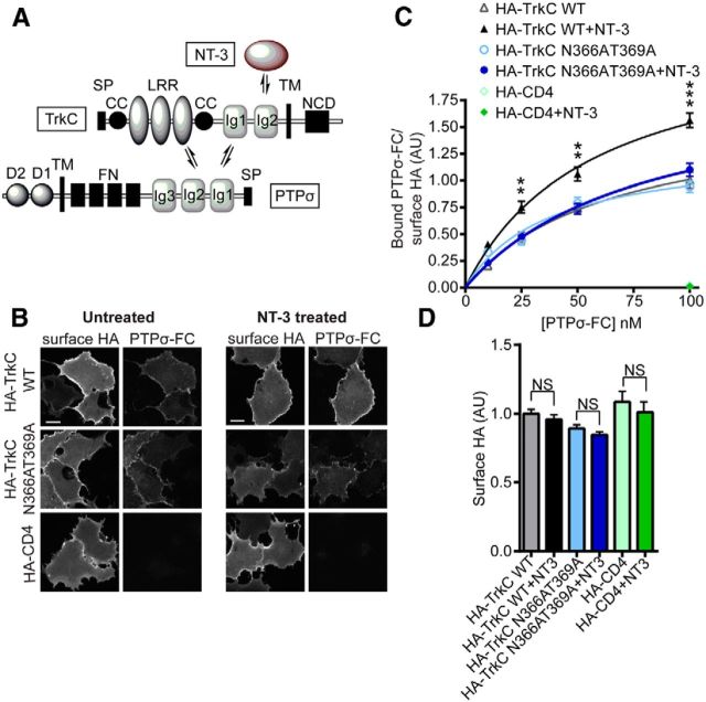
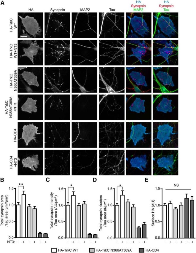
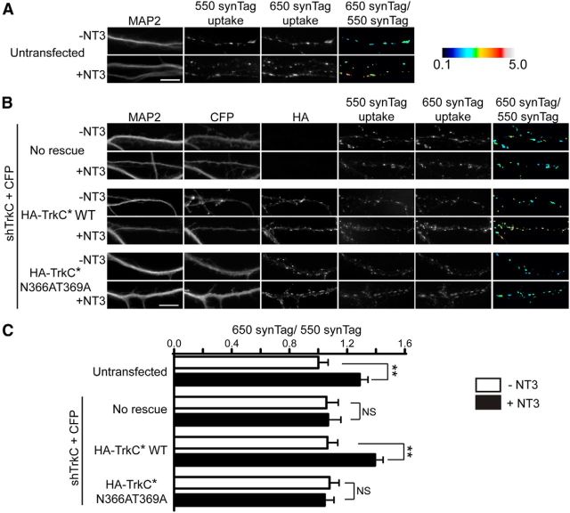
Neurotrophin-3 (NT-3) and its high-affinity receptor TrkC play crucial trophic roles in neuronal differentiation, axon outgrowth, and synapse development and plasticity in the nervous system. We demonstrated previously that postsynaptic TrkC functions as a glutamatergic synapse-inducing (synaptogenic) cell adhesion molecule trans-interacting with presynaptic protein tyrosine phosphatase σ (PTPσ). Given that NT-3 and PTPσ bind distinct domains of the TrkC extracellular region, here we tested the hypothesis that NT-3 modulates TrkC/PTPσ binding and synaptogenic activity. NT-3 enhanced PTPσ binding to cell surface-expressed TrkC and facilitated the presynapse-inducing activity of TrkC in rat hippocampal neurons. Imaging of recycling presynaptic vesicles combined with TrkC knockdown and rescue approaches demonstrated that NT-3 rapidly potentiates presynaptic function via binding endogenous postsynaptic TrkC in a tyrosine kinase-independent manner. Thus, NT-3 positively modulates the TrkC-PTPσ complex for glutamatergic presynaptic assembly and function independently from TrkC kinase activation. Our findings provide new insight into synaptic roles of neurotrophin signaling and mechanisms controlling synaptic organizing complexes. Significance statement: Although many synaptogenic adhesion complexes have been identified in recent years, little is known about modulatory mechanisms. Here, we demonstrate a novel role of neurotrophin-3 in synaptic assembly and function as a positive modulator of the TrkC-protein tyrosine phosphatase σ complex. This study provides new insight into the involvement of neurotrophin signaling in synapse development and plasticity, presenting a molecular mechanism that may underlie previous observations of short- and long-term enhancement of presynaptic function by neurotrophin. Given the links of synaptogenic adhesion molecules to autism and schizophrenia, this study might also contribute to a better understanding of the pathogenesis of these disorders and provide a new direction for ameliorating imbalances in synaptic signaling networks.

Keywords: PTPσ; TrkC; neurotrophin-3; synapse organization; synaptic plasticity.


Figures

Figure 1.
Figure 1.
NT-3 enhances the binding of PTPσ to cell-surface TrkC. A, Schematic domain structures and protein interactions of TrkC noncatalytic isoform, PTPσ, and NT-3. TrkC LRR and Ig1 are required for PTPσ binding, whereas TrkC Ig2 is responsible for NT-3 binding. SP, Signal peptide; CC, cysteine cluster; TM, transmembrane; NCD, noncatalytic domain; FN, fibronectin type-III; D1 and D2, phosphatase domains. B, Representative images of PTPσ–Fc binding to COS-7 cells expressing HA–TrkC WT, HA–TrkC N366AT369A (NT-3-binding dead mutant), or a negative control HA–CD4, in the absence (left) and presence (right) of 100 ng/ml NT-3. PTPσ–Fc protein at 100 nm was applied. Scale bars, 10 μm. C, Quantification of bound PTPσ–Fc per surface HA expression. NT-3 treatment increased the intensity of PTPσ–Fc bound to HA–TrkC WT but not HA–TrkC N366AT369A. **p < 0.01, ***p < 0.001 in two-way ANOVA with Bonferroni's multiple comparisons, n = 20–40 cells from two independent experiments. D, Quantification of surface HA expression in cells analyzed in C. NS, Not significant. One-way ANOVA with Bonferroni's multiple comparisons.
Figure 2.
Figure 2.
NT-3 enhances presynaptic induction by TrkC. A, Representative coculture images showing induced synapsin clustering in hippocampal neurons by COS-7 cells expressing HA–TrkC WT, HA–TrkC N366AT369A, or HA–CD4 in the presence and absence of 100 ng/ml NT-3. Scale bar, 20 μm. B–D, Quantification of induced synapsin clustering. The area (B), total integrated intensity (C), and number (D) of synapsin clusters per axon–COS-7 cell contact area were measured. *p < 0.05, **p < 0.01 in one-way ANOVA with Bonferroni's multiple comparisons, n = 25–30 cells from three independent experiments. E, Quantification of surface HA expression on COS-7 cells analyzed in B–D. NS, Not significant. One-way ANOVA (p = 0.115).
Figure 3.
Figure 3.
NT-3 increases recycling presynaptic vesicles via endogenous postsynaptic TrkC. Hippocampal neurons were assayed for two rounds of vesicle recycling using anti-SynTag, first with Oyster-550–SynTag to establish baseline and then with Oyster-650–SynTag to assess the effects of NT-3. A, Representative images of differential uptake of SynTag in untransfected hippocampal neurons treated with or without NT-3 (+NT-3 or −NT-3). The right column shows the intensity ratio of Oyster-650/Oyster-550 as a heat map with the defined range of 0.1–5 to visualize changes in presynaptic vesicle recycling. B, Representative images of differential uptake of SynTag in neurons transfected with shRNA-expressing vector for TrkC knockdown coexpressing CFP (sh-TrkC + CFP) either alone or together with shRNA-resistant HA–TrkC* WT or HA–TrkC* N366AT369A and treated with or without NT-3. C, Quantification of changes in presynaptic vesicle recycling expressed as the intensity ratio of Oyster-650/Oyster-550. **p < 0.01 in one-way ANOVA with Bonferroni's multiple comparisons, n = 15–30 neurons from three independent experiments. Scale bars, 10 μm.

Similar articles

Cited by

References

    1. Barbacid M. The Trk family of neurotrophin receptors. J Neurobiol. 1994;25:1386–1403. doi: 10.1002/neu.480251107. - DOI - PubMed
    1. Chao MV. Neurotrophins and their receptors: a convergence point for many signalling pathways. Nat Rev Neurosci. 2003;4:299–309. doi: 10.1038/nrn1078. - DOI - PubMed
    1. Coles CH, Mitakidis N, Zhang P, Elegheert J, Lu W, Stoker AW, Nakagawa T, Craig AM, Jones EY, Aricescu AR. Structural basis for extracellular cis and trans RPTPsigma signal competition in synaptogenesis. Nat Commun. 2014;5:5209. doi: 10.1038/ncomms6209. - DOI - PMC - PubMed
    1. Collin C, Vicario-Abejon C, Rubio ME, Wenthold RJ, McKay RD, Segal M. Neurotrophins act at presynaptic terminals to activate synapses among cultured hippocampal neurons. Eur J Neurosci. 2001;13:1273–1282. doi: 10.1046/j.0953-816x.2001.01500.x. - DOI - PubMed
    1. Dean C, Scholl FG, Choih J, DeMaria S, Berger J, Isacoff E, Scheiffele P. Neurexin mediates the assembly of presynaptic terminals. Nat Neurosci. 2003;6:708–716. doi: 10.1038/nn1074. - DOI - PMC - PubMed

Publication types

Substances