. 2015 Nov;89(22):11654-67.
doi: 10.1128/JVI.01815-15. Epub 2015 Sep 9.

A Highly Immunogenic and Protective Middle East Respiratory Syndrome Coronavirus Vaccine Based on a Recombinant Measles Virus Vaccine Platform

Affiliations

A Highly Immunogenic and Protective Middle East Respiratory Syndrome Coronavirus Vaccine Based on a Recombinant Measles Virus Vaccine Platform

Anna H Malczyk et al. J Virol. 2015 Nov.

Abstract

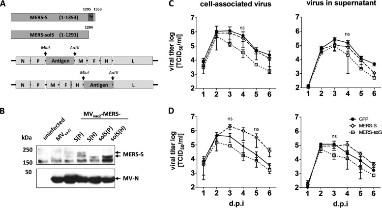
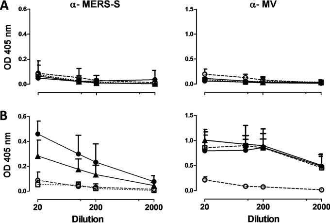
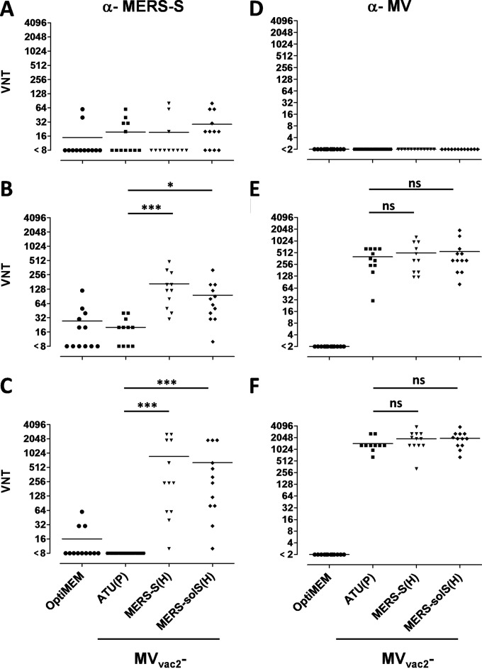
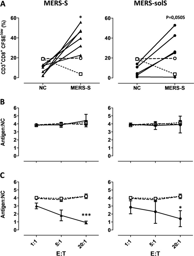
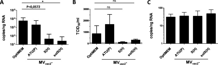
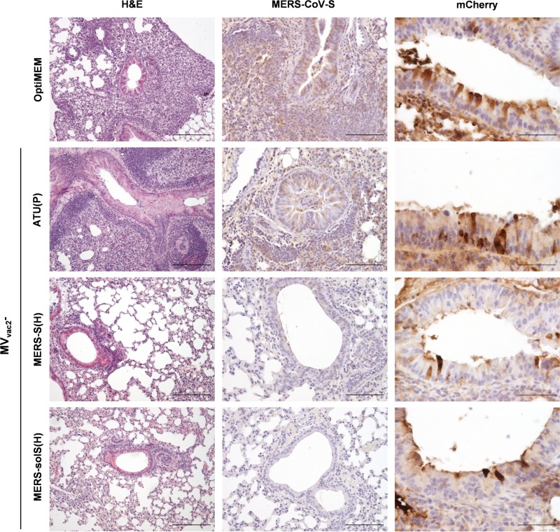
In 2012, the first cases of infection with the Middle East respiratory syndrome coronavirus (MERS-CoV) were identified. Since then, more than 1,000 cases of MERS-CoV infection have been confirmed; infection is typically associated with considerable morbidity and, in approximately 30% of cases, mortality. Currently, there is no protective vaccine available. Replication-competent recombinant measles virus (MV) expressing foreign antigens constitutes a promising tool to induce protective immunity against corresponding pathogens. Therefore, we generated MVs expressing the spike glycoprotein of MERS-CoV in its full-length (MERS-S) or a truncated, soluble variant of MERS-S (MERS-solS). The genes encoding MERS-S and MERS-solS were cloned into the vaccine strain MVvac2 genome, and the respective viruses were rescued (MVvac2-CoV-S and MVvac2-CoV-solS). These recombinant MVs were amplified and characterized at passages 3 and 10. The replication of MVvac2-CoV-S in Vero cells turned out to be comparable to that of the control virus MVvac2-GFP (encoding green fluorescent protein), while titers of MVvac2-CoV-solS were impaired approximately 3-fold. The genomic stability and expression of the inserted antigens were confirmed via sequencing of viral cDNA and immunoblot analysis. In vivo, immunization of type I interferon receptor-deficient (IFNAR(-/-))-CD46Ge mice with 2 × 10(5) 50% tissue culture infective doses of MVvac2-CoV-S(H) or MVvac2-CoV-solS(H) in a prime-boost regimen induced robust levels of both MV- and MERS-CoV-neutralizing antibodies. Additionally, induction of specific T cells was demonstrated by T cell proliferation, antigen-specific T cell cytotoxicity, and gamma interferon secretion after stimulation of splenocytes with MERS-CoV-S presented by murine dendritic cells. MERS-CoV challenge experiments indicated the protective capacity of these immune responses in vaccinated mice.

Importance: Although MERS-CoV has not yet acquired extensive distribution, being mainly confined to the Arabic and Korean peninsulas, it could adapt to spread more readily among humans and thereby become pandemic. Therefore, the development of a vaccine is mandatory. The integration of antigen-coding genes into recombinant MV resulting in coexpression of MV and foreign antigens can efficiently be achieved. Thus, in combination with the excellent safety profile of the MV vaccine, recombinant MV seems to constitute an ideal vaccine platform. The present study shows that a recombinant MV expressing MERS-S is genetically stable and induces strong humoral and cellular immunity against MERS-CoV in vaccinated mice. Subsequent challenge experiments indicated protection of vaccinated animals, illustrating the potential of MV as a vaccine platform with the potential to target emerging infections, such as MERS-CoV.


Figures

FIG 1
FIG 1
Generation and characterization of MVvac2-MERS-S and MVvac2-MERS-solS. (A) Schematic depiction of full-length MERS-S and a soluble variant lacking the transmembrane and cytoplasmatic region (MERS-solS) (upper schemes) and recombinant MVvac2 genomes used for their expression (lower schemes). Antigen or antigen-encoding genes are depicted in dark gray; MV viral gene cassettes (light gray) are annotated. MluI and AatII restriction sites used for cloning of antigen-encoding genes into post-P or post-H ATU are highlighted (B) Immunoblot analysis of Vero cells infected at an MOI of 0.03 with MVvac2-MERS-S, MVvac2-MERS-solS, or MVvac2-GFP(H) (MVvac2), as depicted above the lanes. Uninfected cells served as mock controls. Blots were probed using rabbit serum reactive against MERS-CoV (upper blot) or mAb reactive against MV-N (lower blot). Arrows indicate specific bands. (C and D) Growth kinetics of recombinant MV on Vero cells infected at an MOI of 0.02 with MVvac2-MERS-S (MERS-S), MVvac2-MERS-solS (MERS-solS), or MVvac2-GFP encoding extra genes in post-H (C) or post-P (D) ATU. Titers of samples prepared at the indicated time points postinfection were determined on Vero cells. Means and standard deviations of three independent experiments are presented. ns, not significant.
FIG 2
FIG 2
Induction of antibodies that specifically bind MERS-S (α-MERS-S) or MV (α-MV) antigens. Sera of mice vaccinated on days 0 and 28 with indicated viruses were sampled on days −7 (prebleed, A) and 49 (B) and analyzed for antibodies that bound MERS-S or MV bulk antigens by ELISA. Medium-inoculated mice served as mock controls. Antibodies binding to recombinant MERS-S or MV bulk antigens were detected at an optical density of 405 nm in the ELISA. Means and standard deviations of each group are depicted (n = 6). Filled triangles, MVvac2-MERS-S(H); filled circles, MVvac2-MERS-solS(H); open circles, mock controls; open squares, MVvac2-ATU(P).
FIG 3
FIG 3
Analysis of neutralizing antibodies. VNTs for animals vaccinated on days 0 and 28 with the indicated viruses and sampled on day −7 (A and D), 28 (B and E), or 49 (C and F) for complete neutralization of 200 TCID50 of MERS-CoV or 50 PFU of MV. Medium-inoculated mice served as mock controls. VNTs were calculated as reciprocals of the highest dilution abolishing infectivity. Dots represent single animals (n = 10); horizontal lines represents mean per group. The y axis starts at the detection limit; all mice with VNTs at the detection limit had no detectable VNT. ns, not significant; *, P < 0.05; ***, P < 0.0001.
FIG 4
FIG 4
Secretion of IFN-γ after antigen-specific restimulation of splenocytes. (A to C) IFN-γ ELISpot analysis results with splenocytes of mice vaccinated on days 0 and 28 with indicated viruses, isolated 4 days after boost immunization and after coculture with JAWSII (A), DC2.4 (B), or DC3.2 (C) dendritic cell lines transgenic for MERS-S or untransduced controls (NC). (D) To analyze cellular responses directed against MV, splenocytes were stimulated with 10 μg/ml MV bulk antigens (MV-N) or left unstimulated (unst.). The reactivity of splenocytes was confirmed by ConA treatment (10 μg/ml). Presented are means and standard deviation per group (n = 6). ns, not significant; *, P < 0.05; **, P < 0.01.
FIG 5
FIG 5
Induction of MERS-S-specific CTLs. (A) Proliferation assay using splenocytes of mice vaccinated on days 0 and 28 with MVvac2-MERS-S(H) or MVvac2-MERS-solS(H) and isolated 21 days after boost immunization, after coculture with JAWSII dendritic cell lines transgenic for MERS-S (right, filled triangles), or untransduced controls (left, filled circles). Depicted are the percentages of CD8+ T cells with low CFSE staiing, indicating proliferation in the samples. Results for splenocytes of vaccinated mice are displayed individually and the trend between paired unstimulated and restimulated samples is outlined. Splenocytes of control vaccinated mice [open circles, mock; open squares, MVvac2-ATU(P)] were pooled. (B and C) Killing assay using splenocytes of mice vaccinated on days 0 and 28 and isolated 4 days after boost immunization. Splenocytes were cocultured with untransduced JAWSII (B) or with antigen-presenting JAWSII-MERS-S (C) or for 6 days. Activated CTLs were then cocultured with EL-4-MERS-S target cells (antigen) and EL-4red control cells (NC) at the indicated effector:target (E:T) ratios for 4 h. Ratios of living target versus nontarget cells (antigen:NC) were determined by flow cytometry. Filled triangles, MVvac2-MERS-S(H); filled circles, MVvac2-MERS-solS(H); open circles, mock; open squares, MVvac2-ATU(P). Results shown are means and standard deviation of each group (n = 6). ns, not significant; *, P < 0.05; ***, P < 0.0001.
FIG 6
FIG 6
Viral loads after MERS-CoV challenge in vivo. (A and B) Viral loads, determined as genome copies per nanogram of RNA (A) or infectious virus titers (B) in the lungs of prevaccinated mice after transduction with DPP4-encoding AdV 21 days after boost and challenge with MERS-CoV 25 days after boost. Two independent experiments (n = 4 to 5 per group). Error bars, standard errors of the means (SEM). Dotted line, limit of detection (LOD of qPCR, <1.7 copies/ng RNA). ns, not significant; *, P < 0.05. (C) AdV transduction control mCherry mRNA results (in copies per nanogram of RNA). Error bars, SEM.
FIG 7
FIG 7
Histopathological changes and immunohistochemical analysis of lungs after challenge. Analysis of lung tissue of representative prevaccinated mice (as indicated) after transduction with hDPP4-encoding AdV and challenge with MERS-CoV. Pictures arranged in a row were from samples of the same individual mouse. Paraffin-fixed tissue was stained with H&E (first column; scale bar, 200 μm), with Ab against MERS-CoV-S antigen (middle column; scale bar, 100 μm), and as a control of AdV transduction against mCherry (right column; scale bar, 50 μm).

References

    1. Zaki AM, van Boheemen S, Bestebroer TM, Osterhaus AD, Fouchier RM. 2012. Isolation of a novel coronavirus from a man with pneumonia in Saudi Arabia. N Engl J Med 367:1814–1820. doi:10.1056/NEJMoa1211721. - DOI - PubMed
    1. World Health Organization. 2014. Middle East respiratory syndrome coronavirus (MERS-CoV)—Saudi Arabia. World Health Organization, Geneva, Switzerland: http://www.who.int/csr/don/17-december-2014-mers/en/.
    1. Drosten C, Meyer B, Müller MA, Corman VM, Al-Masri M, Hossain R, Madani H, Sieberg A, Bosch BJ, Lattwein E, Alhakeem RF, Assiri AM, Hajomar W, Albarrak AM, Al-Tawfiq JA, Zumla AI, Memish ZA. 2014. Transmission of MERS-coronavirus in household contacts. N Engl J Med 371:828–835. doi:10.1056/NEJMoa1405858. - DOI - PubMed
    1. Petersen E, Hui DS, Perlman S, Zumla A. 2015. Middle East respiratory syndrome: advancing the public health and research agenda on MERS. Lessons from the South Korea outbreak. Int J Infect Dis 36:54–55. doi:10.1016/j.ijid.2015.06.004. - DOI - PMC - PubMed
    1. Cowling BJ, Park M, Fang VJ, Wu P, Leung GM, Wu JT. 2015. Preliminary epidemiological assessment of MERS-CoV outbreak in South Korea, May to June. Euro Surveill 20:pii=2113 http://dx.doi.org/10.28907/1560-7917.ES2015.25.21163. - DOI - PMC - PubMed

Publication types

MeSH terms

LinkOut - more resources