. 2015 Oct 15;195(8):3828-37.
doi: 10.4049/jimmunol.1501139. Epub 2015 Sep 9.

IFN-γ Prevents Adenosine Receptor (A2bR) Upregulation To Sustain the Macrophage Activation Response

Affiliations

IFN-γ Prevents Adenosine Receptor (A2bR) Upregulation To Sustain the Macrophage Activation Response

Heather B Cohen et al. J Immunol. .

Abstract

The priming of macrophages with IFN-γ prior to TLR stimulation results in enhanced and prolonged inflammatory cytokine production. In this study, we demonstrate that, following TLR stimulation, macrophages upregulate the adenosine 2b receptor (A2bR) to enhance their sensitivity to immunosuppressive extracellular adenosine. This upregulation of A2bR leads to the induction of macrophages with an immunoregulatory phenotype and the downregulation of inflammation. IFN-γ priming of macrophages selectively prevents the induction of the A2bR in macrophages to mitigate sensitivity to adenosine and to prevent this regulatory transition. IFN-γ-mediated A2bR blockade leads to a prolonged production of TNF-α and IL-12 in response to TLR ligation. The pharmacologic inhibition or the genetic deletion of the A2bR results in a hyperinflammatory response to TLR ligation, similar to IFN-γ treatment of macrophages. Conversely, the overexpression of A2bR on macrophages blunts the IFN-γ effects and promotes the development of immunoregulatory macrophages. Thus, we propose a novel mechanism whereby IFN-γ contributes to host defense by desensitizing macrophages to the immunoregulatory effects of adenosine. This mechanism overcomes the transient nature of TLR activation, and prolongs the antimicrobial state of the classically activated macrophage. This study may offer promising new targets to improve the clinical outcome of inflammatory diseases in which macrophage activation is dysregulated.


Figures

Figure 1
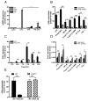
Figure 1. IFN-γ prolongs the macrophage activation state
(A,C) Cytokine production by LPS stimulated unprimed (black squares, solid line) or IFN-γ primed (gray circles, solid line) BMDMs over time. TNFα (A) and IL-12/23p40 (C) was detected by ELISA. The means +/− SEM of 3 independent experiments are shown. (B,D) Unprimed (black squares, solid line) or IFN-γ primed (gray circles, solid line) BMDMs were stimulated with LPS then washed with PBS (break in y-axis) and replaced with LPS-free media and monitored over the next 1–6 hours for TNFα (B), IL-12/23p40 (D). (E) BMDMs were stimulated with LPS alone or in the presence of increasing doses of POM-1. 8 hours post stimulation, cytokine production was assessed by ELISA for TNFα (blue line), IL-12/23p40 (red line), and IL-10 (black line). The means +/− SEM of 3 experiments are shown. (F–I) Unprimed BMDMs (black bars) and IFN-γ primed BMDMs (gray bars) were stimulated with LPS for 4 hrs. Il-10 (F), Arg1 (G), Il-33 (H), and Hmox-1 (I) mRNA levels were determined by qPCR. The means +/−SEM of at least 3 independent experiments are shown. (J) WT (black bars) or STAT1−/− BMDMs (striped bars) were left unprimed or primed with IFN-γ then stimulated with LPS. TNFα (left) and IL-12/23p40 (right) were measured in the supernatants 8 hours after stimulation. Protein was measured by ELISA. The means +/− SD of triplicates are shown.
Figure 2
Figure 2. IFN-γ blocks ATP-induced immunosuppression downstream of eATP hydrolysis
(A,B) Unprimed (black bars) and IFN-γ primed (gray bars) BMDMs were stimulated with LPS in the absence or presence of 10μM ATP for 8 hours. TNFα (A), and IL-12/23p40 (B) production was measured by ELISA. The means +/− SEM of 3 independent experiments are shown. (C,D) Unprimed (black bars) and IFN-γ primed (gray bars) BMDMs were stimulated with LPS in the absence or presence of 10μM adenosine for 8 hours. TNFα (C), IL-12/23p40 (D) production was measured by ELISA. The means +/− SEM of 3 independent experiments are shown. (E–G) Unprimed BMDMs (black bars) and IFN-γ primed BMDMs (gray bars) were stimulated with LPS and adenosine (Ado) for 4 hours mRNA levels were determined by qPCR. The means +/−SEM of at least 3 independent experiments are shown.
Figure 3
Figure 3. LPS-induced ATP release and hydrolysis is intact in the presence of IFN-γ
(A) Levels of ATP released from LPS-stimulated unprimed (black line) and IFN-γ primed (gray line) RAW264.7 cells was measured over time. The means +/− SEM of 2 independent experiments are shown. (B) Flow cytometry analysis of CD39 surface expression (black histograms) on unprimed (left) and IFN-γ primed (right) BMDMs compared to isotype controls (gray histograms). Data are representative of 3 independent experiments. (C) The hydrolysis of 500μM ATP by unprimed BMDMs (black squares, solid line) over time compared to IFN-γ primed BMDMs (gray circles, solid line). Percent of hydrolysis was compared to levels of ATP detected in the absence of cells and determined by the ATPlite assay. The means +/−SEM from 3 independent experiments is shown.
Figure 4
Figure 4. IFN-γ preferentially inhibits LPS-induced A2bR expression in macrophages
(A) mRNA expression of A1r (dotted bars), A2ar (horizontally striped bars), A2br (diagonally striped bars), and A3r (solid black bars) in unprimed and IFN-γ primed BMDMs stimulated for 4 hours. The means +/− SEM of 3 independent experiments are shown. (B,D) Unprimed BMDMs (black bars) and IFN-γ primed BMDMs (gray bars) were stimulated with the indicated TLR agonists for 4 hours. A2bR (B) and A2aR (D) mRNA levels were determined by qPCR. The means +/−SEM of at least 3 independent determinations are shown. (C) Unprimed BMDMs (black bars) and IFN-γ primed BMDMs (gray bars) were stimulated with LPS over time. A2bR mRNA levels were determined by qPCR. The means +/−SEM of 2 independent experiments are shown. (D) Unprimed BMDMs (black bars) and IFN-γ primed BMDMs (gray bars) were stimulated with the indicated TLR agonists for 4hrs. A2aR mRNA levels were determined by qPCR. The means +/−SEM of at least 3 independent determinations are shown. (E) WT BMDMs (black bars) or STAT1−/− BMDMs (striped bars) were left unprimed or IFN-γ primed, then stimulated with LPS for 4 hours. A2bR mRNA levels were determined by qPCR. The mean +/−SEM of at least 3 independent determinations are shown.
Figure 5
Figure 5. Specific antagonism of the A2bR recapitulates IFN-γ-mediated adenosine insensitivity
(A, B) Unprimed (black bars) and MRS1754 primed (gray striped bars) BMDMs were stimulated with LPS in the absence or presence of 10μM adenosine for 8 hours. TNFα (A) and IL-12/23p40 (B) production was measured by ELISA. The means +/− SEM from at least 3 independent experiments are shown. (C) Unprimed (black squares, solid line), IFN-γ primed (gray circles, solid line), and MRS1754 (10μM) primed (gray open squares, dotted line) BMDMs were stimulated with LPS in the presence or absence of adenosine at various concentrations. 8 hours post stimulation TNFα levels were detected by ELISA. Percent of TNFα produced is based on TNFα levels produced by LPS alone for each condition. The means +/−SEM from at least 3 independent experiments are shown. (D,E) Unprimed (black bars) and IFN-γ primed (gray bars) BMDMs were stimulated with LPS in the absence or presence of 1nM of the A2bR-specific agonist NECA (D) or BAY 60-6583 (E). TNFα production was measured 8 hours after stimulation by ELISA. Percent of TNFα produced is based on TNFα levels produced by LPS alone for each condition. The means +/− SEM from 3 independent experiments are shown.
Figure 6
Figure 6. IFN-γ inhibits A2bR-dependent immunosuppression in macrophages
(A,B) Unprimed (black bars) and IFN-γ primed (gray bars) wild-type (A) or A2br−/− (B) BMDMs were stimulated with LPS in the absence or presence of 10μM adenosine for 8 hours. TNFα production was measured by ELISA. The means +/− SEM from at least 3 independent experiments are shown. (C, D) Unprimed (black bars) and IFN-γ primed (gray bars) wild-type (C) or A2ar−/− (D) BMDMs were stimulated with LPS in the absence or presence of 10μM adenosine (Ado) for 8 hours. TNFα production was measured by ELISA. The means +/− SEM from 3 independent experiments are shown.
Figure 7
Figure 7. A2bR overexpression permits the development of immunosuppressive macrophages in the presence of IFN-γ
(A) BMDMs were transfected with plasmids containing GFP (top row) or A2bR-EYFP (bottom row) cDNA. 24 hours later, cells were either left unprimed or primed with IFN-γ followed by stimulation with LPS alone or in the presence of 10μM adenosine (Ado) and BFA. After 4 hours of stimulation, TNFα production was assessed by intracellular FACS. Data are representative from 3 independent experiments. (B, C) Histograms representing TNFα production within transfected (GFP/EYFP+) cells (B). Quantification of TNFα levels within stimulated transfected macrophages as determined by intracellular FACS (C). Data are mean +/−SEM of 3 independent experiments.

Similar articles

Cited by

References

    1. Maisonneuve C, Bertholet S, Philpott DJ, De Gregorio E. Unleashing the potential of NOD- and Toll-like agonists as vaccine adjuvants. Proc Natl Acad Sci U S A. 2014;111:12294–12299. - PMC - PubMed
    1. Mosser DM, Edwards JP. Exploring the full spectrum of macrophage activation. Nat Rev Immunol. 2008;8:958–969. - PMC - PubMed
    1. Cohen HB, Briggs KT, Marino JP, Ravid K, Robson SC, Mosser DM. TLR stimulation initiates a CD39-based autoregulatory mechanism that limits macrophage inflammatory responses. Blood. 2013;122:1935–1945. - PMC - PubMed
    1. Hasko G, Kuhel DG, Chen JF, Schwarzschild MA, Deitch EA, Mabley JG, Marton A, Szabo C. Adenosine inhibits IL-12 and TNF-[alpha] production via adenosine A2a receptor-dependent and independent mechanisms. FASEB J. 2000;14:2065–2074. - PubMed
    1. Hasko G, Pacher P. Regulation of macrophage function by adenosine. Arterioscler Thromb Vasc Biol. 2012;32:865–869. - PMC - PubMed

Publication types

MeSH terms

LinkOut - more resources