Whole-Body MRI in Children: Current Imaging Techniques and Clinical Applications
- PMID: 26355493
- PMCID: PMC4559794
- DOI: 10.3348/kjr.2015.16.5.973
Whole-Body MRI in Children: Current Imaging Techniques and Clinical Applications
Abstract
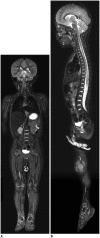
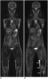
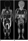
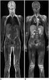
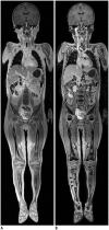
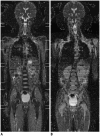
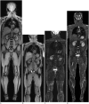
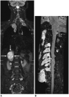
Whole-body magnetic resonance imaging (MRI) is increasingly used in children to evaluate the extent and distribution of various neoplastic and non-neoplastic diseases. Not using ionizing radiation is a major advantage of pediatric whole-body MRI. Coronal and sagittal short tau inversion recovery imaging is most commonly used as the fundamental whole-body MRI protocol. Diffusion-weighted imaging and Dixon-based imaging, which has been recently incorporated into whole-body MRI, are promising pulse sequences, particularly for pediatric oncology. Other pulse sequences may be added to increase diagnostic capability of whole-body MRI. Of importance, the overall whole-body MRI examination time should be less than 30-60 minutes in children, regardless of the imaging protocol. Established and potentially useful clinical applications of pediatric whole-body MRI are described.
Keywords: Infants and children; Systemic disease; Tumor; Whole-body MRI.
Figures




Similar articles
-
Current utilization and procedural practices in pediatric whole-body MRI.Pediatr Radiol. 2018 Aug;48(8):1101-1107. doi: 10.1007/s00247-018-4145-5. Epub 2018 May 2. Pediatr Radiol. 2018. PMID: 29721598
-
Current and Emerging Roles of Whole-Body MRI in Evaluation of Pediatric Cancer Patients.Radiographics. 2019 Mar-Apr;39(2):516-534. doi: 10.1148/rg.2019180130. Epub 2019 Jan 25. Radiographics. 2019. PMID: 30681900 Review.
-
Whole-body magnetic resonance imaging in children: technique and clinical applications.Pediatr Radiol. 2016 May;46(6):858-72. doi: 10.1007/s00247-016-3586-y. Epub 2016 May 26. Pediatr Radiol. 2016. PMID: 27229503 Review.
-
Current role of body MRI in pediatric oncology.Pediatr Radiol. 2016 May;46(6):873-80. doi: 10.1007/s00247-016-3560-8. Epub 2016 May 26. Pediatr Radiol. 2016. PMID: 27229504 Review.
-
Whole-body magnetic resonance imaging and positron emission tomography-computed tomography in oncology.Top Magn Reson Imaging. 2007 Jun;18(3):193-202. doi: 10.1097/RMR.0b013e318093e6bo. Top Magn Reson Imaging. 2007. PMID: 17762383 Review.
Cited by
-
Evaluating prostate cancer bone metastasis using accelerated whole-body isotropic 3D T1-weighted Dixon MRI with compressed SENSE: a feasibility study.Eur Radiol. 2023 Mar;33(3):1719-1728. doi: 10.1007/s00330-022-09181-9. Epub 2022 Oct 21. Eur Radiol. 2023. PMID: 36269371
-
Magnetic resonance imaging of the musculoskeletal system in the diagnosis of rheumatic diseases in the pediatric population.Reumatologia. 2024;62(3):196-206. doi: 10.5114/reum/190262. Epub 2024 Jul 12. Reumatologia. 2024. PMID: 39055724 Free PMC article. Review.
-
Whole-Body Magnetic Resonance Tomography and Whole-Body Computed Tomography in Pediatric Polytrauma Diagnostics-A Retrospective Long-Term Two-Center Study.Diagnostics (Basel). 2023 Mar 23;13(7):1218. doi: 10.3390/diagnostics13071218. Diagnostics (Basel). 2023. PMID: 37046436 Free PMC article.
-
Medullary Thyroid Carcinoma: An Update on Imaging.J Thyroid Res. 2019 Jul 7;2019:1893047. doi: 10.1155/2019/1893047. eCollection 2019. J Thyroid Res. 2019. PMID: 31360432 Free PMC article. Review.
-
Diffusion-weighted MRI and intravoxel incoherent motion model for diagnosis of pediatric solid abdominal tumors.J Magn Reson Imaging. 2018 Jun;47(6):1475-1486. doi: 10.1002/jmri.25901. Epub 2017 Nov 21. J Magn Reson Imaging. 2018. PMID: 29159937 Free PMC article.
References
-
- Daldrup-Link HE, Franzius C, Link TM, Laukamp D, Sciuk J, Jürgens H, et al. Whole-body MR imaging for detection of bone metastases in children and young adults: comparison with skeletal scintigraphy and FDG PET. AJR Am J Roentgenol. 2001;177:229–236. - PubMed
-
- Goo HW, Choi SH, Ghim T, Moon HN, Seo JJ. Whole-body MRI of paediatric malignant tumours: comparison with conventional oncological imaging methods. Pediatr Radiol. 2005;35:766–773. - PubMed
-
- Goo HW, Yang DH, Ra YS, Song JS, Im HJ, Seo JJ, et al. Whole-body MRI of Langerhans cell histiocytosis: comparison with radiography and bone scintigraphy. Pediatr Radiol. 2006;36:1019–1031. - PubMed
-
- Punwani S, Taylor SA, Bainbridge A, Prakash V, Bandula S, De Vita E, et al. Pediatric and adolescent lymphoma: comparison of whole-body STIR half-Fourier RARE MR imaging with an enhanced PET/CT reference for initial staging. Radiology. 2010;255:182–190. - PubMed
-
- Goo HW. Whole-body MRI of neuroblastoma. Eur J Radiol. 2010;75:306–314. - PubMed
Publication types
MeSH terms
LinkOut - more resources
Full Text Sources
Other Literature Sources
Medical

