

Sex and age differences in brain-derived neurotrophic factor and vimentin in the zebra finch song system: Relationships to newly generated cells
- PMID: 26355496
- PMCID: PMC4731248
- DOI: 10.1002/cne.23893
Sex and age differences in brain-derived neurotrophic factor and vimentin in the zebra finch song system: Relationships to newly generated cells
Abstract
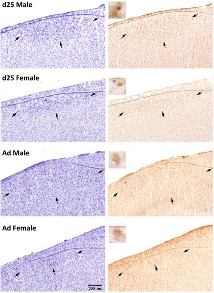
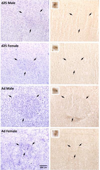
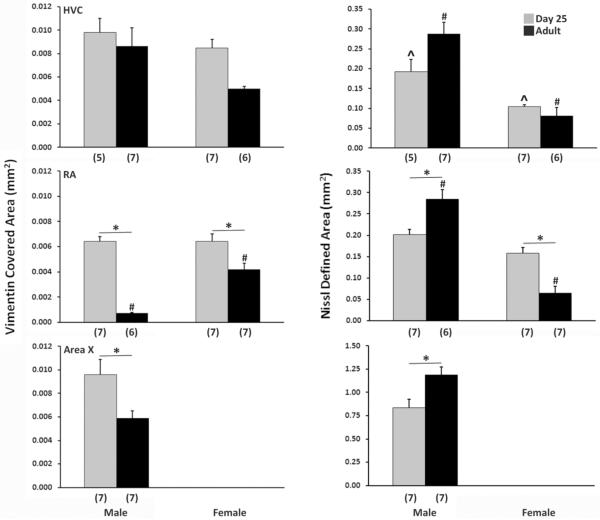
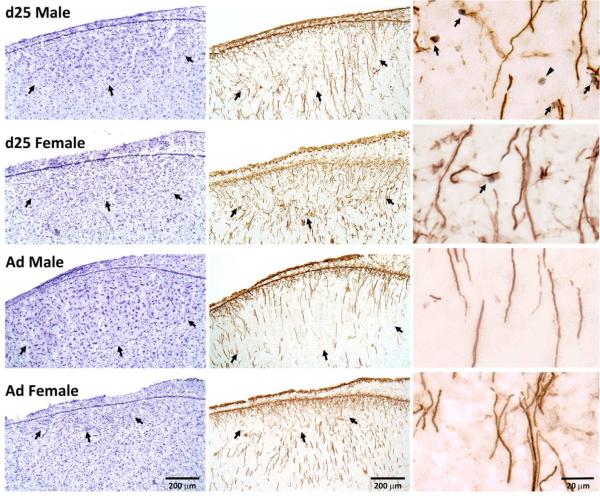
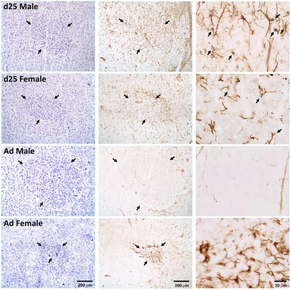
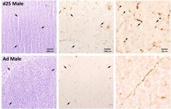
The neural song circuit is enhanced in male compared with female zebra finches due to differential rates of incorporation and survival of cells between the sexes. Two double-label immunohistochemical experiments were conducted to increase the understanding of relationships between newly generated cells (marked with bromodeoxyuridine [BrdU]) and those expressing brain-derived neurotrophic factor (BDNF) and vimentin, a marker for radial glia. The song systems of males and females were investigated at posthatching day 25 during a heightened period of sexual differentiation (following BrdU injections on days 6-10) and in adulthood (following a parallel injection paradigm). In both HVC (proper name) and the robust nucleus of the arcopallium (RA), about half of the BrdU-positive cells expressed BDNF across sexes and ages. Less than 10% of the BDNF-positive cells expressed BrdU, but this percentage was greater in juveniles than adults. Across both brain regions, more BDNF-positive cells were detected in males compared with females. In RA, the number of these cells was also greater in juveniles than adults. In HVC, the average cross-sectional area covered by the vimentin labeling was greater in males than females and in juveniles compared with adults. In RA, more vimentin was detected in juveniles than adults, and within adults it was greater in females. In juveniles only, BrdU-positive cells appeared in contact with vimentin-labeled fibers in HVC, RA, and Area X. Collectively, the results are consistent with roles of BDNF- and vimentin-labeled cells influencing sexually differentiated plasticity of the song circuit.
Keywords: AB_514483; AB_528504; AB_630940; neurogenesis; radial glia; sexual differentiation; songbird.
© 2015 Wiley Periodicals, Inc.
Figures

Similar articles
-
Developmental changes in BDNF protein in the song control nuclei of zebra finches.Neuroscience. 2013 Oct 10;250:578-87. doi: 10.1016/j.neuroscience.2013.07.062. Epub 2013 Aug 3. Neuroscience. 2013. PMID: 23920158 Free PMC article.
-
Sex differences in the telencephalic song control circuitry in Bengalese finches (Lonchura striata var. domestica).Zoolog Sci. 2005 Oct;22(10):1089-94. doi: 10.2108/zsj.22.1089. Zoolog Sci. 2005. PMID: 16286720
-
Sexual Differences in Cell Loss during the Post-Hatch Development of Song Control Nuclei in the Bengalese Finch.PLoS One. 2015 May 4;10(5):e0125802. doi: 10.1371/journal.pone.0125802. eCollection 2015. PLoS One. 2015. PMID: 25938674 Free PMC article.
-
Testosterone and brain-derived neurotrophic factor interactions in the avian song control system.Neuroscience. 2013 Jun 3;239:115-23. doi: 10.1016/j.neuroscience.2012.09.023. Epub 2012 Oct 30. Neuroscience. 2013. PMID: 23123886 Free PMC article. Review.
-
Sexual differentiation of the zebra finch song system.Ann N Y Acad Sci. 2004 Jun;1016:540-59. doi: 10.1196/annals.1298.015. Ann N Y Acad Sci. 2004. PMID: 15313794 Review.
Cited by
-
BDNF Expression in Larval and Adult Zebrafish Brain: Distribution and Cell Identification.PLoS One. 2016 Jun 23;11(6):e0158057. doi: 10.1371/journal.pone.0158057. eCollection 2016. PLoS One. 2016. PMID: 27336917 Free PMC article.
-
Emergence of sex-specific transcriptomes in a sexually dimorphic brain nucleus.Cell Rep. 2022 Aug 2;40(5):111152. doi: 10.1016/j.celrep.2022.111152. Cell Rep. 2022. PMID: 35926465 Free PMC article.
-
BDNF, Brain, and Regeneration: Insights from Zebrafish.Int J Mol Sci. 2018 Oct 13;19(10):3155. doi: 10.3390/ijms19103155. Int J Mol Sci. 2018. PMID: 30322169 Free PMC article. Review.
-
Brain-Derived Neurotrophic Factor Has a Transsynaptic Trophic Effect on Neural Activity in an Adult Forebrain Circuit.J Neurosci. 2020 Feb 5;40(6):1226-1231. doi: 10.1523/JNEUROSCI.2375-19.2019. Epub 2019 Dec 19. J Neurosci. 2020. PMID: 31857358 Free PMC article.
-
Daily high doses of atorvastatin alter neuronal morphology in a juvenile songbird model.PLoS One. 2025 Apr 28;20(4):e0314690. doi: 10.1371/journal.pone.0314690. eCollection 2025. PLoS One. 2025. PMID: 40294005 Free PMC article.
References
-
- Alonso G. NG2 proteoglycan-expressing cells of the adult rat brain: possible involvement in the formation of glial scar astrocytes following stab wound. Glia. 2005;49(3):318–338. - PubMed
-
- Alvarez-Buylla A. Mechanism of neurogenesis in adult avian brain. Experientia. 1990;46(9):948–955. - PubMed
-
- Alvarez-Buylla A. Neurogenesis and plasticity in the CNS of adult birds. Exp Neurol. 1992;115(1):110–114. - PubMed
-
- Alvarez-Buylla A, Buskirk DR, Nottebohm F. Monoclonal antibody reveals radial glia in adult avian brain. J Comp Neurol. 1987;264(2):159–170. - PubMed
Publication types
MeSH terms
Substances
Grants and funding
LinkOut - more resources
Full Text Sources
Other Literature Sources
Miscellaneous
