. 2015 Aug 28:9:4997-5018.
doi: 10.2147/DDDT.S89876. eCollection 2015.

Pharmacokinetic and nephroprotective benefits of using Schisandra chinensis extracts in a cyclosporine A-based immune-suppressive regime

Affiliations

Pharmacokinetic and nephroprotective benefits of using Schisandra chinensis extracts in a cyclosporine A-based immune-suppressive regime

Qiao Lai et al. Drug Des Devel Ther. .

Abstract

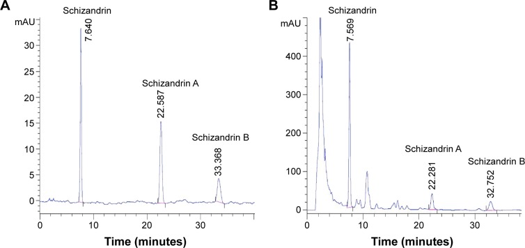
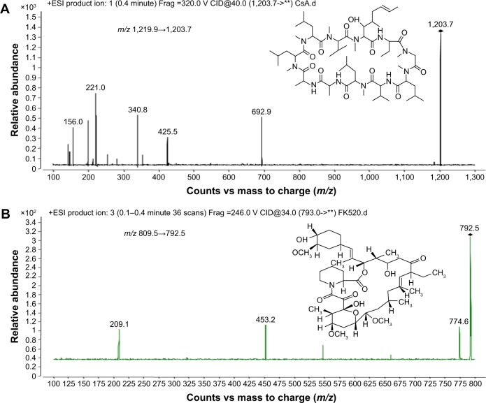
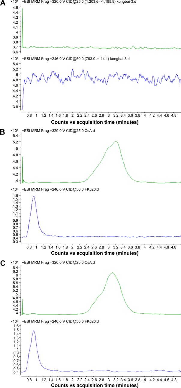
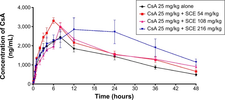
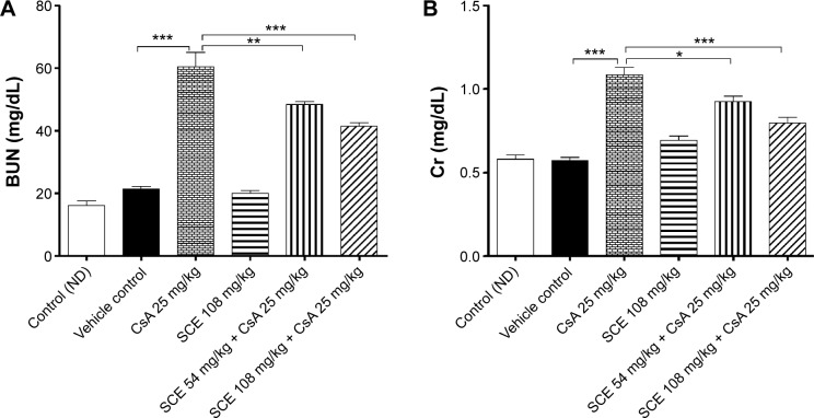
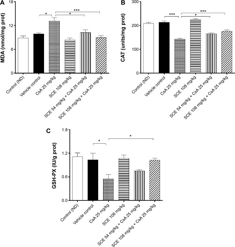
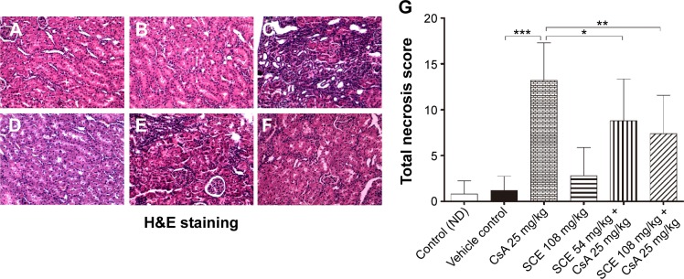
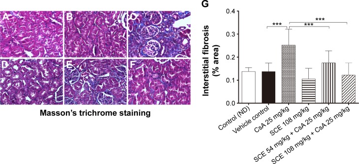
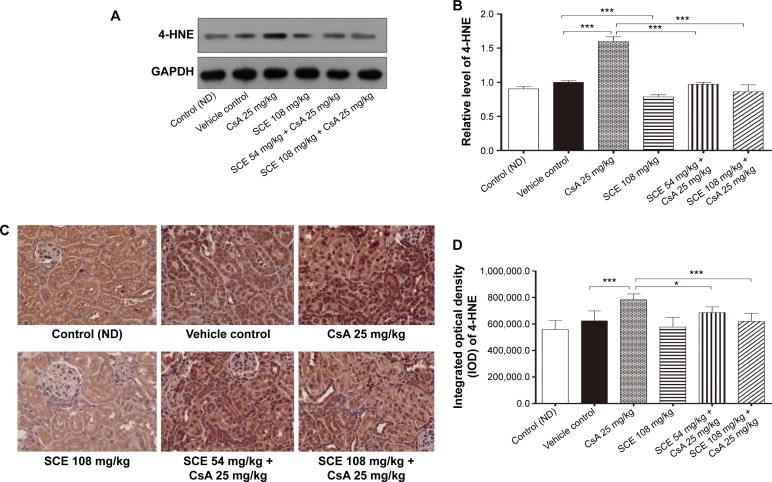
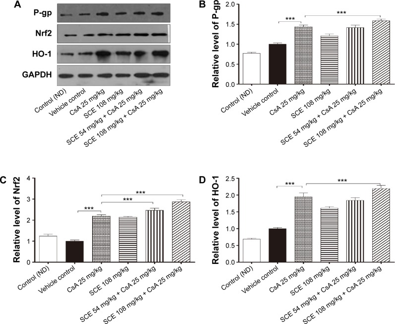
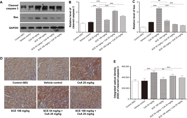
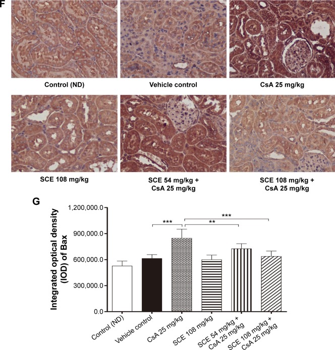
Cyclosporine A (CsA) is a powerful immunosuppressive drug. However, nephrotoxicity resulting from its long-term usage has hampered its prolonged therapeutic usage. Schisandra chinensis extracts (SCE) have previously been used in traditional Chinese medicine and more recently coadministered with Western medicine for the treatment of CsA-induced side effects in the People's Republic of China. This study aimed to investigate the possible effects of SCE on the pharmacokinetics of CsA in rats and elucidate the potential mechanisms by which it hinders the development of CsA-induced nephrotoxicity. A liquid chromatography/tandem mass spectrometry method was developed and validated for determining the effect of SCE on the pharmacokinetics of CsA. Male Sprague Dawley rats, which were administered with CsA (25 mg/kg/d) alone or in combination with SCE (54 mg/kg/d and 108 mg/kg/d) for 28 days, were used to evaluate the nephroprotective effects of SCE. Our study showed that SCE increased the mean blood concentration of CsA. Furthermore, we found that the concomitant administration of SCE alongside CsA prevented the disruption of catalase activity and reduction in creatinine, urea, renal malondialdehyde, and glutathione peroxidase levels that would have otherwise occurred in the absence of SCE administration. SCE treatment markedly suppressed the expression of 4-hydroxynonenal, Bcl-2-associated X protein, cleaved caspase 3, and autophagy-related protein LC3 A/B. On the other hand, the expression of heme oxygenase-1, nuclear factor erythroid 2-related factor 2 (Nrf2), and P-glycoprotein was enhanced by the very same addition of SCE. SCE was also able to increase the systemic exposure of CsA in rats. The renoprotective effects of SCE were thought to be mediated by its antiapoptotic and antioxidant abilities, which caused the attenuation of CsA-induced autophagic cell death. All in all, these findings suggest the prospective use of SCE as an effective adjunct in a CsA-based immunosuppressive regimen.

Keywords: Schisandra chinensis extracts; apoptosis; autophagy; cyclosporine A; nephroprotective; oxidative stress; pharmacokinetics.


Figures

Figure 1
Figure 1
Evaluation of the components in Schisandra chinensis extracts (SCE) using high-performance liquid chromatography assay. Notes: (A) The mixture of the three reference compounds. (B) The sample of SCE. Chromatographic separation was performed on a Kromasil C18 column (250 mm ×4.6 mm id, 5 μm) at 35°C. The mobile phase consisted of eluents A (water) and B (methanol). The elution program was optimized and conducted as follows: a linear gradient of 68–70% B (0–5 minutes), a linear gradient of 70–75% B (5–15 minutes), an isocratic elution of 75% B (15–38 minutes), a linear gradient of 75–68% B (38–39 minutes), an isocratic elution of 68% B (39–45 minutes). The peaks were recorded using PDA absorbance at 254 nm and the solvent flow rate was kept at 1.0 mL/min.
Figure 2
Figure 2
Full-scan product ion of precursor of (A) CsA and (B) FK520. Notes: The chromatographic peak was confirmed by LC-MS experiment using the Agilent 6460 triple-quadrupole mass spectrometer under a positive electrospray ionization (ESI) mode, with the spray voltage set at 3500 V. Desolvation gas (nitrogen) was heated to 300°C and delivered at a flow rate of 5 L/min. The nitrogen nebulizing gas was set at 45 psi. Abbreviation: CsA, cyclosporine A.
Figure 3
Figure 3
Representative multiple reaction monitoring (MRM) chromatograms of CsA and FK520 in rat whole blood. Notes: (A) A blank whole blood sample. (B) A blank whole blood sample spiked with CsA and FK520 at 50 ng/mL. (C) A whole blood sample from a rat after a single oral administration of CsA (25 mg/kg). Abbreviation: CsA, cyclosporine A.
Figure 4
Figure 4
Whole blood concentration–time curves of CsA after a single oral dose of CsA (25 mg/kg) to rats with and without an oral dose of 54 mg/kg, 108 mg/kg, and 216 mg/kg SCE, respectively. Note: Each point represents the mean ± SD (n=6). Abbreviations: CsA, cyclosporine A; SCE, Schisandra chinensis extract; SD, standard deviation.
Figure 5
Figure 5
Effect of SCE on cyclosporine A (CsA)-induced nephrotoxicity in BUN (A) and Cr (B) levels in serum of different experimental groups. Notes: Data were analyzed by one-way ANOVA and presented as mean ± SD (n=6). *P<0.05, **P<0.01, and ***P<0.001. Abbreviations: SCE, Schisandra chinensis extract; BUN, blood urea nitrogen; Cr, creatinine; ANOVA, analysis of variance; SD, standard deviation; ND, normal diet.
Figure 6
Figure 6
Effect of SCE on cyclosporine A (CsA)-induced nephrotoxicity in MDA (A), CAT (B), and GSH-Px (C) in kidney of different experimental groups. Notes: Data were analyzed by one-way ANOVA and presented as mean ± SD (n=6). *P<0.05, ***P<0.001. Abbreviations: SCE, Schisandra chinensis extract; MDA, malondialdehyde; CAT, catalase; GSH-Px, glutathione peroxidase; ANOVA, analysis of variance; SD, standard deviation; ND, normal diet.
Figure 7
Figure 7
Photomicrographs (×200, H&E) of renal tubules showing progressive stages of tubular necrosis and respective necrosis score. Notes: Twelve random sections (×200) from each kidney were examined, and a score from 0 to 3 was given to the tubular profile according to the following arbitrary scale: the damage affecting no >5% of the field was scored 0; mild damage affecting 5%–25% of the field was scored 1; moderate damage affecting 25%–75% of the field was scored 2; and severe damage exceeding 75% of the field was scored 3. All 12 scores were added to give the total necrosis score for each kidney. (A) Control group. (B) Vehicle control. (C) 25 mg/kg CsA group. (D) 108 mg/kg SCE group. (E) 54 mg/kg SCE + 25 mg/kg CsA group. (F) 108 mg/kg SCE + 25 mg/kg CsA group. (G) Semiquantitative scores revealed that the scores of total tubular necrosis were significant in the CsA exposed but reduced by SCE. Data were analyzed by nonparameters and expressed as mean number in the high power field of each group (mean ± SD, n=6). *P<0.05, **P<0.01, and ***P<0.001. Abbreviations: H&E, hematoxylin–eosin; CsA, cyclosporine A; SCE, Schisandra chinensis extract; SD, standard deviation.
Figure 8
Figure 8
Tubulointerstitial fibrosis was detected by Masson’s trichrome staining. Notes: Photomicrographs (×200) of renal cortex obtain from Sprague Dawley rats for 28 days with (A) control group. (B) Vehicle control. (C) 25 mg/kg CsA group. (D) 108 mg/kg SCE group. (E) 54 mg/kg SCE + 25 mg/kg CsA group. (F) 108 mg/kg SCE + 25 mg/kg CsA group. (G) Semiquantitative scores revealed that the percentage of stained area per high power filed of each group (mean ± SD, n=6). ***P<0.001. Abbreviations: CsA, cyclosporine A; SCE, Schisandra chinensis extract; SD, standard deviation.
Figure 9
Figure 9
Effect of SCE on expression of 4-HNE in CsA-induced nephrotoxicity. Notes: (A) Immunoblotting for the target molecules of 4-HNE showing the effects of SCE in CsA-induced nephrotoxicity. (B) Bar graphs show the expression level of 4-HNE. GAPDH was used as the internal control. (C) The immunohistochemical stain of 4-HNE in renal tissue between different study groups (×200). (D) Quantification of 4-HNE integral optical density (IOD). Data were analyzed by one-way ANOVA and presented as mean ± SD (n≥3). *P<0.05 and ***P<0.001. Abbreviations: SCE, Schisandra chinensis extract; 4-HNE, 4-hydroxynonenal; CsA, cyclosporine A; GAPDH, glyceraldehyde-3-phosphate dehydrogenase; ANOVA, analysis of variance; SD, standard deviation; ND, normal diet.
Figure 10
Figure 10
Effect of SCE on expression of P-gp, Nrf2, and HO-1 in CsA-induced nephrotoxicity. Notes: (A) Immunoblotting for the target molecule of P-gp, Nrf2, and HO-1 showing the effects of SCE in CsA-induced nephrotoxicity. (BD) Bar graphs show the expression levels of P-gp, Nrf2, and HO-1. GAPDH was used as the internal control. Data were analyzed by one-way ANOVA and presented as mean ± SD (n≥3). ***P<0.001. Abbreviations: SCE, Schisandra chinensis extract; P-gp, P-glycoprotein; Nrf2, nuclear factor erythroid 2-related factor 2; HO-1, heme oxygenase 1; CsA, cyclosporine A; GAPDH, glyceraldehyde-3-phosphate dehydrogenase; ANOVA, analysis of variance; SD, standard deviation; ND, normal diet.
Figure 11
Figure 11
Effect of SCE on the expressions of cleaved caspase 3 and Bax in CsA-induced nephrotoxicity. Notes: (A) Immunoblotting for the target molecules of cleaved caspase 3 and Bax showing the effects of SCEs in CsA-induced nephrotoxicity. (B and C) Bar graphs show the expression levels of cleaved caspase 3 and Bax. GAPDH was used as the internal control. (D) The immunohistochemical stain of cleaved caspase 3 in renal tissue between different study groups (×200). (E) Quantification of cleaved caspase 3 integral optical density (IOD). (F) The immunohistochemical stain of Bax in renal tissue between different study groups (×200). (G) Quantification of Bax integral optical density (IOD). Data were analyzed by one-way ANOVA and presented as mean ± SD (n≥3). **P<0.01 and ***P<0.001. Abbreviations: SCE, Schisandra chinensis extract; CsA, cyclosporine A; GAPDH, glyceraldehyde-3-phosphate dehydrogenase; Bax, Bcl-2-associated X protein; ANOVA, analysis of variance; SD, standard deviation; ND, normal diet.
Figure 11
Figure 11
Effect of SCE on the expressions of cleaved caspase 3 and Bax in CsA-induced nephrotoxicity. Notes: (A) Immunoblotting for the target molecules of cleaved caspase 3 and Bax showing the effects of SCEs in CsA-induced nephrotoxicity. (B and C) Bar graphs show the expression levels of cleaved caspase 3 and Bax. GAPDH was used as the internal control. (D) The immunohistochemical stain of cleaved caspase 3 in renal tissue between different study groups (×200). (E) Quantification of cleaved caspase 3 integral optical density (IOD). (F) The immunohistochemical stain of Bax in renal tissue between different study groups (×200). (G) Quantification of Bax integral optical density (IOD). Data were analyzed by one-way ANOVA and presented as mean ± SD (n≥3). **P<0.01 and ***P<0.001. Abbreviations: SCE, Schisandra chinensis extract; CsA, cyclosporine A; GAPDH, glyceraldehyde-3-phosphate dehydrogenase; Bax, Bcl-2-associated X protein; ANOVA, analysis of variance; SD, standard deviation; ND, normal diet.
Figure 12
Figure 12
Effect of SCE on the expression of LC3A/B in CsA-induced nephrotoxicity. Notes: (A) Immunoblotting for the target molecules of LC3A/B showing the effects of SCE in CsA-induced nephrotoxicity. (B) Bar graphs show the expression levels of LC3A/B. GAPDH was used as the internal control. (C) The immunohistochemical stain of LC3A/B in renal tissue between different study groups (×200). (D) Quantification of LC3A/B integral optical density (IOD). Data were analyzed by one-way ANOVA and presented as mean ± SD (n≥3). ***P<0.001. Abbreviations: SCE, Schisandra chinensis extract; LC3A/B, autophagy-related protein LC3A/B; CsA, cyclosporine A; GAPDH, glyceraldehyde-3-phosphate dehydrogenase; ANOVA, analysis of variance; SD, standard deviation; ND, normal diet.

Similar articles

Cited by

References

    1. Grinyo JM, Cruzado JM. Cyclosporine nephrotoxicity. Transplant Proc. 2004;36(2):240S–242S. - PubMed
    1. Mariee AD, Abd-Ellah MF. Protective effect of docosahexaenoic acid against cyclosporine A-induced nephrotoxicity in rats: a possible mechanism of action. Ren Fail. 2011;33(1):66–71. - PubMed
    1. Tavares P, Fontes Ribeiro CA, Teixeira F. Cyclosporin effect on noradrenaline release from the sympathetic nervous endings of rat aorta. Pharmacol Res. 2003;47(1):27–33. - PubMed
    1. Tedesco D, Haragsim L. Cyclosporine: a review. J Transplant. 2012;2012:230386. - PMC - PubMed
    1. Capasso G, Di Gennaro CI, Della Ragione F, et al. In vivo effect of the natural antioxidant hydroxytyrosol on cyclosporine nephrotoxicity in rats. Nephrol Dial Transplant. 2008;23(4):1186–1195. - PubMed

Publication types

MeSH terms