

Neutrophil Responses to Sterile Implant Materials
- PMID: 26355958
- PMCID: PMC4565661
- DOI: 10.1371/journal.pone.0137550
Neutrophil Responses to Sterile Implant Materials
Abstract
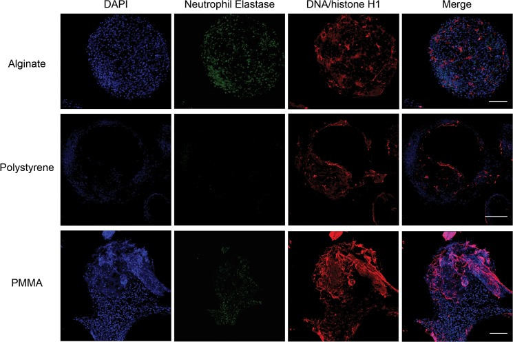
In vivo implantation of sterile materials and devices results in a foreign body immune response leading to fibrosis of implanted material. Neutrophils, one of the first immune cells to be recruited to implantation sites, have been suggested to contribute to the establishment of the inflammatory microenvironment that initiates the fibrotic response. However, the precise numbers and roles of neutrophils in response to implanted devices remains unclear. Using a mouse model of peritoneal microcapsule implantation, we show 30-500 fold increased neutrophil presence in the peritoneal exudates in response to implants. We demonstrate that these neutrophils secrete increased amounts of a variety of inflammatory cytokines and chemokines. Further, we observe that they participate in the foreign body response through the formation of neutrophil extracellular traps (NETs) on implant surfaces. Our results provide new insight into neutrophil function during a foreign body response to peritoneal implants which has implications for the development of biologically compatible medical devices.
Conflict of interest statement
Figures
Similar articles
-
Frontline Science: Splenic progenitors aid in maintaining high neutrophil numbers at sites of sterile chronic inflammation.J Leukoc Biol. 2016 Aug;100(2):253-60. doi: 10.1189/jlb.1HI0615-248RR. Epub 2016 Mar 10. J Leukoc Biol. 2016. PMID: 26965635 Free PMC article.
-
Neutrophils, NETs, NETosis - old or new factors in sepsis and septic shock?Anaesthesiol Intensive Ther. 2017;49(3):235-240. doi: 10.5603/AIT.2017.0041. Anaesthesiol Intensive Ther. 2017. PMID: 28803442 Review.
-
Aggregated neutrophil extracellular traps resolve inflammation by proteolysis of cytokines and chemokines and protection from antiproteases.FASEB J. 2019 Jan;33(1):1401-1414. doi: 10.1096/fj.201800752R. Epub 2018 Aug 21. FASEB J. 2019. PMID: 30130433 Free PMC article.
-
Damage-associated molecular pattern-activated neutrophil extracellular trap exacerbates sterile inflammatory liver injury.Hepatology. 2015 Aug;62(2):600-14. doi: 10.1002/hep.27841. Epub 2015 May 29. Hepatology. 2015. PMID: 25855125 Free PMC article.
-
The Regulatory Effects of Interleukin-4 Receptor Signaling on Neutrophils in Type 2 Immune Responses.Front Immunol. 2019 Oct 24;10:2507. doi: 10.3389/fimmu.2019.02507. eCollection 2019. Front Immunol. 2019. PMID: 31708926 Free PMC article. Review.
Cited by
-
Shifting landscapes: dynamic changes from pro- to anti-inflammatory leukocyte phenotype in myocardial ischemia/reperfusion injury.Front Cardiovasc Med. 2025 Jun 30;12:1596538. doi: 10.3389/fcvm.2025.1596538. eCollection 2025. Front Cardiovasc Med. 2025. PMID: 40662132 Free PMC article.
-
The Emerging Role of Neutrophils in the Pathogenesis of Thrombosis in COVID-19.Int J Mol Sci. 2021 May 20;22(10):5368. doi: 10.3390/ijms22105368. Int J Mol Sci. 2021. PMID: 34065210 Free PMC article. Review.
-
Micrococcal Nuclease stimulates Staphylococcus aureus Biofilm Formation in a Murine Implant Infection Model.Front Cell Infect Microbiol. 2022 Jan 17;11:799845. doi: 10.3389/fcimb.2021.799845. eCollection 2021. Front Cell Infect Microbiol. 2022. PMID: 35111695 Free PMC article.
-
Inflammation via myeloid differentiation primary response gene 88 signaling mediates the fibrotic response to implantable synthetic poly(ethylene glycol) hydrogels.Acta Biomater. 2019 Dec;100:105-117. doi: 10.1016/j.actbio.2019.09.043. Epub 2019 Sep 27. Acta Biomater. 2019. PMID: 31568879 Free PMC article.
-
Evaluation of encapsulating and microporous nondegradable hydrogel scaffold designs on islet engraftment in rodent models of diabetes.Biotechnol Bioeng. 2018 Sep;115(9):2356-2364. doi: 10.1002/bit.26741. Epub 2018 Jun 25. Biotechnol Bioeng. 2018. PMID: 29873059 Free PMC article.
References
-
- Langer R, Tirrell DA. Designing materials for biology and medicine. Nature. 2004; 428: 487–492. - PubMed
-
- Pashuck ET, Stevens MM. Designing regenerative biomaterial therapies for the clinic. Sci Transl Med. 2012; 4: 160sr164. - PubMed
-
- Peppas NA, Langer R. New challenges in biomaterials. Science. 1994; 263: 1715–1720. - PubMed
-
- Ratner BD, Hoffman AS, Schoen FJ, Lemons JE. Biomaterials Science. Amsterdam: Elsevier; 2012.
Publication types
MeSH terms
Substances
Grants and funding
- DE013023/DE/NIDCR NIH HHS/United States
- R01 DE016516/DE/NIDCR NIH HHS/United States
- T32 HL066987/HL/NHLBI NIH HHS/United States
- R01 AI111595/AI/NIAID NIH HHS/United States
- P01AI111595/AI/NIAID NIH HHS/United States
- R01 DE013023/DE/NIDCR NIH HHS/United States
- EB000351/EB/NIBIB NIH HHS/United States
- R01 EB000244/EB/NIBIB NIH HHS/United States
- U54 CA151884/CA/NCI NIH HHS/United States
- CA151884/CA/NCI NIH HHS/United States
- U19 AI095261/AI/NIAID NIH HHS/United States
- R37 EB000244/EB/NIBIB NIH HHS/United States
- 5T32 HL066987/HL/NHLBI NIH HHS/United States
- EB000244/EB/NIBIB NIH HHS/United States
- R01 EB000351/EB/NIBIB NIH HHS/United States
- U19AI095261/AI/NIAID NIH HHS/United States
LinkOut - more resources
Full Text Sources
Other Literature Sources
