. 2016 Apr 15;524(6):1208-21.
doi: 10.1002/cne.23899. Epub 2015 Sep 28.

Disruption in dopaminergic innervation during photoreceptor degeneration

Affiliations

Disruption in dopaminergic innervation during photoreceptor degeneration

Elena Ivanova et al. J Comp Neurol. .

Abstract

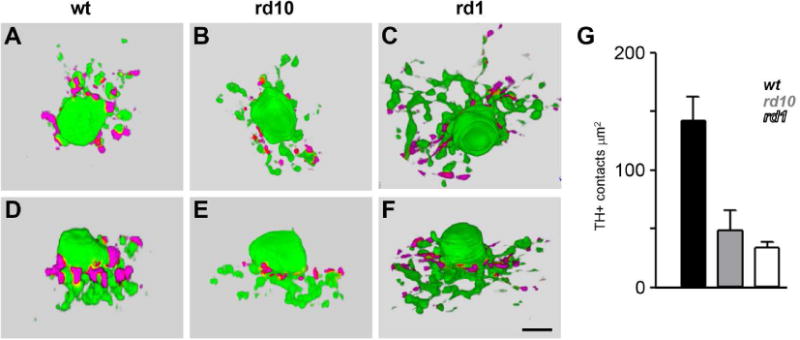
Dopaminergic amacrine cells (DACs) release dopamine in response to light-driven synaptic inputs, and are critical to retinal light adaptation. Retinal degeneration (RD) compromises the light responsiveness of the retina and, subsequently, dopamine metabolism is impaired. As RD progresses, retinal neurons exhibit aberrant activity, driven by AII amacrine cells, a primary target of the retinal dopaminergic network. Surprisingly, DACs are an exception to this physiological change; DACs exhibit rhythmic activity in healthy retina, but do not burst in RD. The underlying mechanism of this divergent behavior is not known. It is also unclear whether RD leads to structural changes in DACs, impairing functional regulation of AII amacrine cells. Here we examine the anatomical details of DACs in three mouse models of human RD to determine how changes to the dopaminergic network may underlie physiological changes in RD. By using rd10, rd1, and rd1/C57 mice we were able to dissect the impacts of genetic background and the degenerative process on DAC structure in RD retina. We found that DACs density, soma size, and primary dendrite length are all significantly reduced. Using a novel adeno-associated virus-mediated technique to label AII amacrine cells in mouse retina, we observed diminished dopaminergic contacts to AII amacrine cells in RD mice. This was accompanied by changes to the components responsible for dopamine synthesis and release. Together, these data suggest that structural alterations of the retinal dopaminergic network underlie physiological changes during RD.

Keywords: RRID:AB_2315595; RRID:AB_572268; RRID:Jax:00002; RRID:Jax:000659; RRID:Jax:000664; RRID:Jax:004297; RRID:nif-0000-30467; RRID:nlx_157306; RRID:rid_000042; amacrine cell; dopamine; gap junctions; retinal degeneration.


Conflict of interest statement

Conflict of interest

All authors declare they have no conflict of interest.

Figures

Figure 1
Figure 1
Analysis of the retina for specific features. Cell counts of DACs were performed in each of four quadrants (Ventral, Nasal, Dorsal, and Temporal; outlined in gray), denoted by the directional symbol (lower right). The temporal quadrant is marked by an incision (arrow, left). Soma sizes and primary dendrite densities were measured in areas marked by white boxes. Measurements of the axonal system were performed within the areas marked by black boxes. Scale bar, 1 mm.
Figure 2
Figure 2
Distribution of DACs in wildtype and RD retinas. (A–D) DACs were immunolabeled for tyrosine hydroxylase. Insets show locations of the cell somas in red. Ventral, Nasal, Dorsal, and Temporal quadrants denoted by directional symbol; for rd1C57 D pole is marked. Scale bar, 1 mm. (E) Total number of DACs in rd1 retinas was significantly less than wt (p < 0.0001).
Figure 3
Figure 3
Morphological changes of somas and primary dendrites of DACs vary across retinal quadrants in RD. Magnified areas from ventral (A–H) and dorsal (I–P) regions from the same retinas shown in Figure 2. Representative somas are shown in upper insets (×2.52 further magnification). Tracings of primary dendrites are shown with highlighted soma in lower insets. (Q) Soma size was reduced in RD (p < 0.0001). (R) Primary dendrite density was reduced in RD (p < 0.0001). The density of rd1 dendrites was also significantly less than rd10 (p = 0.002). Scale bar, 0.1 mm.
Figure 4
Figure 4
Chromatin distribution, TH expression, and cytoplasm volume of DACs were different in RD. Single confocal images of DAC somas in the retinal wholemounts from wt, rd10, rd1 and rd1/C57 mice, aged P180. Retinas were double labeled for TO-PRO-3 (A–D, blue) and tyrosine hydroxylase (TH, E–H, red). All samples were taken from central part of the temporal quadrant. Scale bar, 5 μm.
Figure 5
Figure 5
Morphological changes within the axonal system of DACs. (A–L) Single confocal images of TH+ processes (cyan) in retinal wholemounts double-labeled for VMAT2 (green). Arrowheads point to the primary dendrites. In wt, the intensity of TH labeling was comparable between varicosities and slender connecting processes. In RD the TH-staining was concentrated in varicosities. (M) Reduction in TH+ particle size in rd10 and rd1 (p < 0.0001). (N) Increase in rd1 VMAT2 particle size (p < 0.0001). Scale bar 10 μm.
Figure 6
Figure 6
Dopaminergic rings around AII amacrine cells are reduced in RD. (A–C) Viral construct for targeting expression of GFP in AII amacrine cells (top). Somas of GFP-labeled AII amacrine cells in retinal wholemounts. Inset shows a single reconstructed AII amacrine cell. (D–F) Dopaminergic rings are visible in TH+ processes (cyan) around AII somas labeled by GFP (green). Scale bars, 50 μm in A, 10 μm in B.
Figure 7
Figure 7
Dopaminergic contacts to AII amacrine cells are reduced in RD. (A–F) Somas and lobular appendages of representative AII amacrine cells (green) with the adjacent TH+ processes (cyan). Note that arboreal dendrites of AII amacrine cells are not shown. (G) Surface area of TH+ contacts to AII soma measured are reduced in rd10 (p = 0.002) and rd1 (p < 0.001). Scale bar, 5 μm.
None
Combining immunolabeling for tyrosine hydroxylase to quantify dopaminergic cells (magenta) and AAV-mediated GFP-labeling of target AII amacrine cells (green) in the retina, the authors demonstrate deconstruction of the dopaminergic amacrine cell network during photoreceptor degeneration.

References

    1. Atkinson CL, Feng J, Zhang DQ. Functional integrity and modification of retinal dopaminergic neurons in the rd1 mutant mouse: roles of melanopsin and GABA. J Neurophysiol. 2013;109(6):1589–1599. - PubMed
    1. Bloomfield SA, Volgyi B. The diverse functional roles and regulation of neuronal gap junctions in the retina. Nat Rev Neurosci. 2009;10(7):495–506. - PMC - PubMed
    1. Borowska J, Trenholm S, Awatramani GB. An intrinsic neural oscillator in the degenerating mouse retina. J Neurosci. 2011;31(13):5000–5012. - PMC - PubMed
    1. Cembrowski MS, Logan SM, Tian M, Jia L, Li W, Kath WL, Riecke H, Singer JH. The mechanisms of repetitive spike generation in an axonless retinal interneuron. Cell Rep. 2012;1(2):155–166. - PMC - PubMed
    1. Chang GQ, Gaitan A, Hao Y, Wong F. Correlation of DNA fragmentation and chromatin condensation in apoptotic nuclei of the Ser 6 mouse retina. Microsc Res Tech. 1997;36(2):123–129. - PubMed

Publication types