. 2015 Oct 13;6(31):30859-75.
doi: 10.18632/oncotarget.5087.

Novel Axl-driven signaling pathway and molecular signature characterize high-grade ovarian cancer patients with poor clinical outcome

Affiliations

Novel Axl-driven signaling pathway and molecular signature characterize high-grade ovarian cancer patients with poor clinical outcome

Katia Rea et al. Oncotarget. .

Abstract

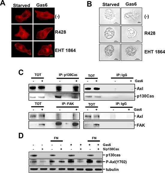
High-grade epithelial ovarian cancer (HGEOC) is a clinically diverse and molecularly heterogeneous disease comprising subtypes with distinct biological features and outcomes. The receptor tyrosine kinases, expressed by EOC cells, and their ligands, present in the microenvironment, activate signaling pathways, which promote EOC cells dissemination. Herein, we established a molecular link between the presence of Gas6 ligand in the ascites of HGEOCs, the expression and activation of its receptor Axl in ovarian cancer cell lines and biopsies, and the progression of these tumors. We demonstrated that Gas6/Axl signalling converges on the integrin β3 pathway in the presence of the adaptor protein p130Cas, thus inducing tumor cell adhesion to the extracellular matrix and invasion. Accordingly, Axl and p130Cas were significantly co-expressed in HGEOC samples. Clinically, we identified an Axl-associated signature of 62 genes able to portray the HGEOCs with the shortest overall survival. These data biologically characterize a group of HGEOCs and could help guide a more effective therapeutic approach to be taken for these patients.

Keywords: Axl; Gas6; extracellular matrix; ovarian cancer; p130Cas.


Conflict of interest statement

CONFLICTS OF INTEREST

The authors declare no conflict of interest.

Figures

Figure 1
Figure 1. Gas6 is expressed in EOC cells and activates the TAM RTK Axl
A. Real-time PCR showing the levels of mRNA for Gas6, AXL, MERTK, and TYRO-3 in six EOC cell lines. Results are presented as relative expression normalized to GAPDH mRNA levels. B. Western blotting on the total cell lysates from the same six EOC cell lines. C. IP with Anti-P-Tyrosine (P-Tyr) performed on lysates from starved or Gas6-stimulated SKOV3 and NL3507 cells. Immunoprecipitated samples were analyzed by western blotting with Abs against the proteins reported on the right. D. Western blotting on the total cell lysates from starved SKOV3 and NL3507 cells pre-treated with Axl-Fc (2.5 μg/ml) and stimulated or not with Gas6 (500 ng/ml). Abs are reported on the right. β-actin was used as the gel loading control.
Figure 2
Figure 2. Gas6-stimulated promotion of invasion through the interaction between ovarian cancer cells and ECM
A. Invasion assay of starved or Gas6-stimulated SKOV3 and NL3507 cells grown in Matrigel. Representative images of three independent experiments are shown. B. Left panel, phase contrast microscopy of starved or Gas6-stimulated SKOV3 cells grown and stimulated with Gas6. C. Upper panel, western blotting on the total cell lysates from starved or Gas6-stimulated SKOV3 and NL3507 cells seeded on FN for 30 min. A representative experiment of the three performed is shown. Abs are reported on the right. β-actin was used as the gel loading control. Lower panel, quantitative evaluation of phosphorylated Axl in Gas6-stimulated cells upon adhesion on FN. The graph reports the mean ± SD from the three independent experiments. D. Upper panel, membrane staining of the integrin β3 receptor determined by flow cytometry on the NL3507 cell line. The expression of the other integrin β receptors on both the SKOV3 and NL3505 cells was also evaluated and the results are reported in Supplementary Fig. 3. The red and light blue peaks, respectively, represent the fluorescence of the cells incubated with the secondary antibody alone as control (α-mouse) and the anti-integrin β3 Ab (Anti-β3). Lower panel, western blotting on the total cell lysates from starved or Gas6-stimulated NL3507 cells, in the absence or in the presence of the anti-integrin β3 Ab. A representative experiment is shown. Abs are reported on the right. β-actin was used as the gel loading control.
Figure 3
Figure 3. Gas6/Axl signaling triggers PI3K/AKT/rac activation and requires the scaffold protein p130Cas
A. IF performed on SKOV3 cells grown on FN, starved and then induced to migrate through a wound in the presence or not of Gas6. Cells were treated with the reported inhibitors, and F-actin was stained with phalloidin (red). B. Invasion assay of starved or Gas6-stimulated SKOV3 and NL3507 cells grown in Matrigel. Cells were treated with the reported inhibitors. Representative images of the three independent experiments are shown. C. IP performed with anti-p130Cas (upper panel) or with anti-FAK (lower panel), respectively, on lysates of starved or Gas6-stimulated SKOV3 cells. Normal mouse or rabbit (IgG) sera were used as the negative control, respectively. The immunoprecipitated samples were analyzed by western blotting. D. Western blotting on the total cell lysates from SKOV3 cells transiently transfected with a control siRNA (−) or with a pool of siRNA against p130Cas (sip130Cas). SiRNA-transfected SKOV3 cells were starved or Gas6-stimulated and seeded on FN for 30 min. Immunoblottings were performed with Abs against the proteins reported on the right. Alpha (tubulin) was used as the gel loading control.
Figure 4
Figure 4. Reduction of Gas6-dependent adhesion and invasion following the impairment of p130Cas/Axl interactions
A. Live cell imaging performed on untreated (−) or Gas6-stimulated control siRNA (siCO)- or p130 siRNA (sip130Cas)-transfected SKOV3 cells during adhesion on FN (see Supplementary Video 1 and 2, and Methods). Representative frames at 0, 10 min, and 20 min are reported. Scale bar, 100 μm. B. Graph reporting the number of FN adherent siCo- or sip130Cas-transfected SKOV3 cells, from the live imaging experiment, taken at different time points. Asteriscs indicate the significant differences between the two curves (paired t test, p = 0.0004) C. IF performed on cells as above after 20 min of adhesion on FN. The F-actin was stained with phalloidin (green). D. Invasion assay performed in Matrigel on starved or Gas6-stimulated SKOV3 cells stably p130Cas-silenced (shp130Cas) or infected with an empty shRNA vector (Mock). Representative images of the three independent experiments are shown.
Figure 5
Figure 5. In vivo validation on HGEOC samples
A. Gas6 levels measured by ELISA on 22 EOC ascites and 22 non-malignant ascites. B. Evaluation in the EOC ascites of the sequestration by the Axl ectodomain of Gas6. The dosage of the two molecules was performed as described in Methods. The graph reports the molar ratio between Gas6 and Axl ectodomain (sAxl). The red line represents the equimolar ratio. C. Invasion assay performed on starved or Gas6-stimulated MCAs from two representative Axl-positive MCAs from EOC ascites grown in Matrigel. EOC samples containing 80% of HGEOC cells, as evaluated by the cytopathologist, were processed. Satellite cells present in the starved sample are likely to be some immune cells present in the sample D. IP performed on the total cell lysates obtained from the EOC cells of patient #1 with anti-p130Cas. Immunoprecipitated samples were analyzed by western blotting with Abs against the proteins reported on the right. A longer exposure of the immunoblot with both Abs on the total lysates is shown on the right. E. Representative staining with anti-Axl and anti-p130Cas Abs on FFPE sections from two (a and b) HGEOC patients. Scale bar, 50 μm. F. Graph reporting the correlation between the p130Cas and Axl expressions in Type I and Type II (HGEOCs) tumors. The results of the contingency analysis are reported (Fisher's exact test, p = 0,0009).
Figure 6
Figure 6. A. Identification of an Axl–driven signature in serous HGEOCs
A. Evaluation of the expression changes of the genes included in the Axl-associated gene set in each of the HGEOC subtypes according to the TCGA data set [5]. A total of 114 out of the 121 genes identified were plotted, due to differences in the gene symbols. Only the genes reported by Verhaak et al. [5] in the Supplementary Table 8A are plotted. The bars corresponding to Axl are reported in red. B. Graphical representation of the network identified by IPA. The top five networks, reported in Supplementary Table 5B, were identified by loading the genes reported in bold in Table 1. The Axl-correlated genes included in the network are highlighted in red.
Figure 7
Figure 7. Axl-driven signature identifies the HGEOC patients with poor overall survival
Kaplan-Meyer curves, log-rank p values and HR are used to compare OS between HGEOC patients with ‘high’ (red lines) and ‘low’ (green lines) expression intensities of the metagene consisting of the Axl-driven signature. The tables below each curve reports the median survival of each group of patients.

References

    1. Coleman RL, Monk BJ, Sood AK, Herzog TJ. Latest research and treatment of advanced-stage epithelial ovarian cancer. Nat Rev Clin Oncol. 2013;10:211–224. - PMC - PubMed
    1. Nick AM, Coleman RL, Ramirez PT, Sood AK. A framework for a personalized surgical approach to ovarian cancer. Nat Rev Clin Oncol. 2015;12:239–245. - PMC - PubMed
    1. Bashashati A, Ha G, Tone A, Ding J, Prentice LM, Roth A, et al. Distinct evolutionary trajectories of primary high-grade serous ovarian cancers revealed through spatial mutational profiling. J Pathol. 2013;231:21–34. - PMC - PubMed
    1. Tothill RW, Tinker AV, George J, Brown R, Fox SB, Lade S, Johnson DS, Trivett MK, Etemadmoghadam D, Locandro B, Traficante N, Fereday S, Hung JA, Chiew YE, Haviv I, Gertig D, DeFazio A, Bowtell DD. Novel molecular subtypes of serous and endometrioid ovarian cancer linked to clinical outcome. Clin Cancer Res. 2008;14:5198–5208. - PubMed
    1. Verhaak RG, Tamayo P, Yang JY, Hubbard D, Zhang H, Creighton CJ, et al. Prognostically relevant gene signatures of high-grade serous ovarian carcinoma. J Clin Invest. 2013;123:517–525. - PMC - PubMed

Publication types

MeSH terms

Substances