. 2015 Sep 10;10(9):e0137512.
doi: 10.1371/journal.pone.0137512. eCollection 2015.

Chronic Compression of the Dorsal Root Ganglion Enhances Mechanically Evoked Pain Behavior and the Activity of Cutaneous Nociceptors in Mice

Affiliations

Chronic Compression of the Dorsal Root Ganglion Enhances Mechanically Evoked Pain Behavior and the Activity of Cutaneous Nociceptors in Mice

Tao Wang et al. PLoS One. .

Abstract

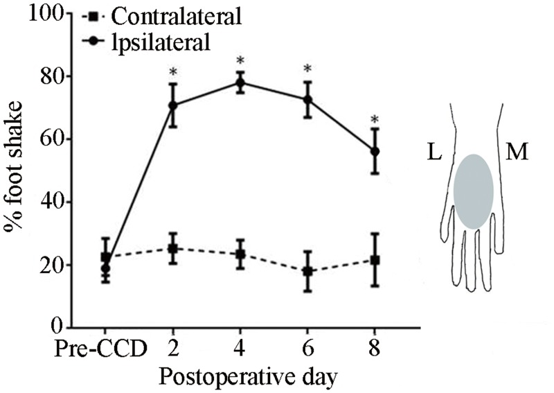
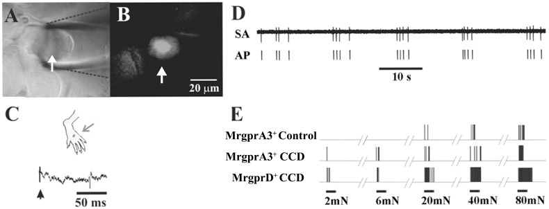
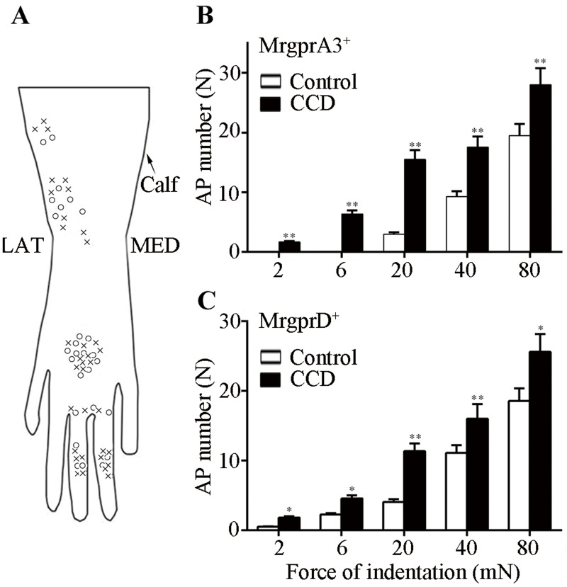
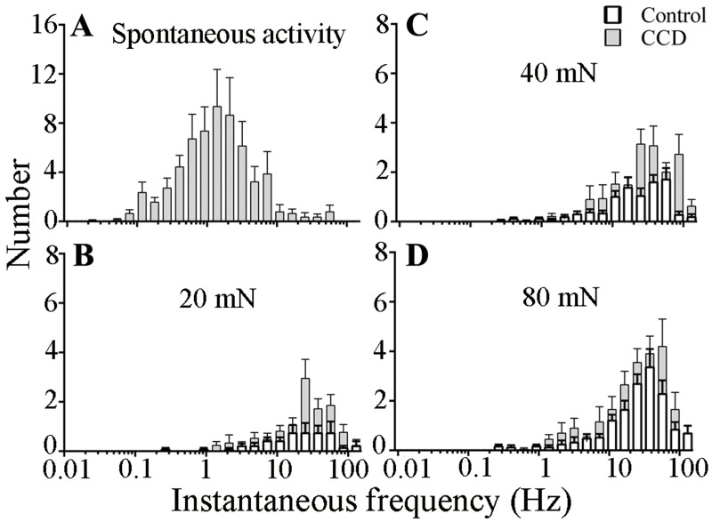
Radicular pain in humans is usually caused by intraforaminal stenosis and other diseases affecting the spinal nerve, root, or dorsal root ganglion (DRG). Previous studies discovered that a chronic compression of the DRG (CCD) induced mechanical allodynia in rats and mice, with enhanced excitability of DRG neurons. We investigated whether CCD altered the pain-like behavior and also the responses of cutaneous nociceptors with unmyelinated axons (C-fibers) to a normally aversive punctate mechanical stimulus delivered to the hairy skin of the hind limb of the mouse. The incidence of a foot shaking evoked by indentation of the dorsum of foot with an aversive von Frey filament (tip diameter 200 μm, bending force 20 mN) was significantly higher in the foot ipsilateral to the CCD surgery as compared to the contralateral side on post-operative days 2 to 8. Mechanically-evoked action potentials were electrophysiologically recorded from the L3 DRG, in vivo, from cell bodies visually identified as expressing a transgenically labeled fluorescent marker (neurons expressing either the receptor MrgprA3 or MrgprD). After CCD, 26.7% of MrgprA3+ and 32.1% MrgprD+ neurons exhibited spontaneous activity (SA), while none of the unoperated control neurons had SA. MrgprA3+ and MrgprD+ neurons in the compressed DRG exhibited, in comparison with neurons from unoperated control mice, an increased response to the punctate mechanical stimuli for each force applied (6, 20, 40, and 80 mN). We conclude that CCD produced both a behavioral hyperalgesia and an enhanced response of cutaneous C-nociceptors to aversive punctate mechanical stimuli.


Conflict of interest statement

Competing Interests: The authors have declared that no competing interests exist.

Figures

Fig 1
Fig 1. Effects of CCD on the incidence of foot-shaking evoked by punctate mechanical stimulation of the hairy skin.
A von Frey filament with a tip diameter 200 μm, and delivering a bending force of 20 mN was applied to the dorsum of each foot before the CCD surgery ("Pre-CCD") and up to 8 days after the surgery. Mice (n = 11), *P < 0.05 indicates the statistical significance in comparisons of the mean incidence obtained for the foot ipsilateral to the CCD surgery (solid line) compared to both the contralateral foot (dashed line) and to the pre-operative ipsilateral baseline. Inset: Schematic diagram of the region where the von-Frey filament was applied (shaded area) on the hairy skin (dorsum) of the right hind paw. L: lateral, M: medial.
Fig 2
Fig 2. Spontaneous and mechanically evoked action potential activity electrophysiologically recorded in vivo from cell bodies of C-nociceptors.
(A) Bright-field image of the cell body of a neuron whose action potentials are extracellularly recorded via a glass pipette electrode (outlined with dashed black lines) in the DRG of a control mouse. (B) Epifluorescent image of GFP indicated that the neuron expressed MrgprA3. The scale bar in B also applies to A. (C) The neuron was identified as a C-mechanoheat nociceptor (CHM) with a receptive field (RF) on the dorsum of the hairy skin (black dot) that was electrically stimulated (arrow) to obtain its conduction velocity (CV) (0.63 m/s). (D) Spontaneous activity of a CMH nociceptor recorded after CCD (RF on ankle, CV = 0.69 m/s). (E) Responses of three C-mechanosensitive nociceptive neurons to von Frey filaments each with the same tip diameter of 200-μm but delivering different bending forces of 2, 6, 20, 40, and 80 mN. Each also responded to noxious heat (responses not shown). These neurons include the neuron referred to in panels A-C ("MrgprA3+ control") and two neurons recorded from CCD mice, one expressing MrgprA3 with RF on the ankle and CV of 0.69 m/s ("MrgprA3 CCD") and the other expressing MrgprD+ with RF on 3rd toe and CV of 0.5 m/s ("MrgprD CCD").
Fig 3
Fig 3. Responses of nociceptors to punctate mechanical stimulation as a function of force of indentation.
Von Frey filaments (200 μm tip diameter) with differing bending forces (2-80mN) were each applied to the receptive field for 1 s. (A) Schematic diagram of the locations of receptive fields for each of the MrgprA3+ (circles) and MrgprD+ (crosses) expressing nociceptors on the dorsum (hairy) skin of the hind paw. L: lateral, M: medial. (B) Mean number of action potentials evoked in MrgprA3+ neurons, control mice (n = 13, open bars) and CCD mice (n = 14, closed bars). (C) Mean number of action potentials evoked in MrgprD+ neurons from control- (n = 15) and CCD mice (n = 20) (*P<0.05, **P<0.01).
Fig 4
Fig 4. Histograms of instantaneous frequencies of spontaneous and mechanically evoked responses of C mechanosensitive nociceptors.
The number of instantaneous frequencies that occurred within each bin (log increments, factor √2) is combined for MrgprA3+ and MrgprD+ nociceptors and plotted for control mice (closed bars, n = 28) and CCD mice (open bars, n = 34). (A) Spontaneous activity, observed only after CCD. (B–D) Responses to 20, 40 and 80 mN forces delivered by von Frey filaments each with the same tip diameter of 200 μm.

References

    1. Zhang JM, Munir M. Radicular low back pain: what have we learned from recent animal research? Anesthesiology. 2008;108(1):5–6. 10.1097/01.anes.0000296148.09016.d5 . - DOI - PubMed
    1. Briggs CA, Chandraraj S. Variations in the lumbosacral ligament and associated changes in the lumbosacral region resulting in compression of the fifth dorsal root ganglion and spinal nerve. Clinical anatomy. 1995;8(5):339–46. 10.1002/ca.980080506 . - DOI - PubMed
    1. Marziniak M, Phan NQ, Raap U, Siepmann D, Schurmeyer-Horst F, Pogatzki-Zahn E, et al. Brachioradial pruritus as a result of cervical spine pathology: the results of a magnetic resonance ctomography study. Journal of the American Academy of Dermatology. 2011;65(4):756–62. 10.1016/j.jaad.2010.07.036 . - DOI - PubMed
    1. Olmarker K, Rydevik B, Nordborg C. Autologous nucleus pulposus induces neurophysiologic and histologic changes in porcine cauda equina nerve roots. Spine. 1993;18(11):1425–32. . - PubMed
    1. Kawakami M, Tamaki T, Weinstein JN, Hashizume H, Nishi H, Meller ST. Pathomechanism of pain-related behavior produced by allografts of intervertebral disc in the rat. Spine. 1996;21(18):2101–7. . - PubMed

Publication types