. 2016 Apr 1;524(5):1033-61.
doi: 10.1002/cne.23890. Epub 2015 Sep 29.

Dynamic expression of transcription factor Brn3b during mouse cranial nerve development

Affiliations

Dynamic expression of transcription factor Brn3b during mouse cranial nerve development

Szilard Sajgo et al. J Comp Neurol. .

Abstract

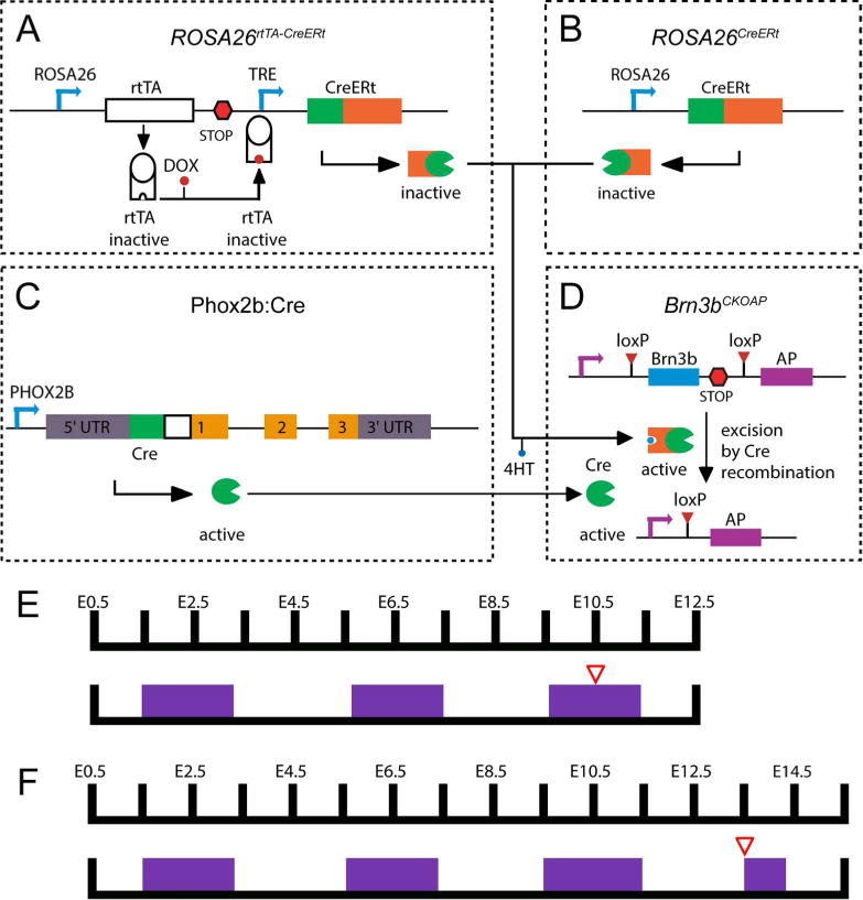
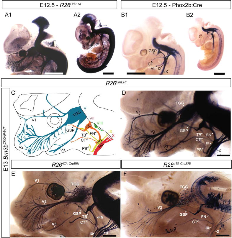
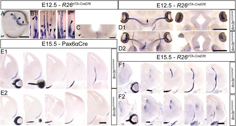
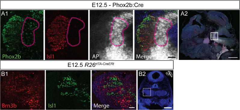
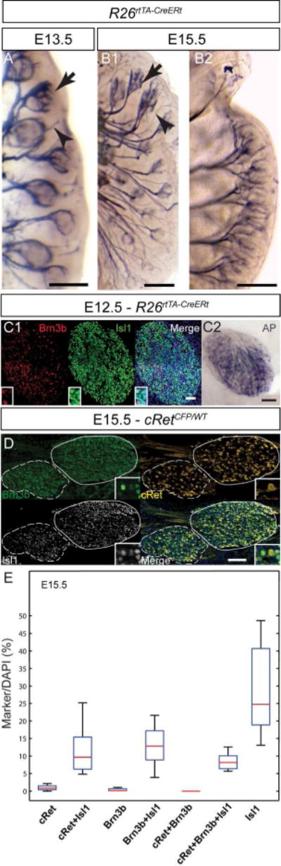
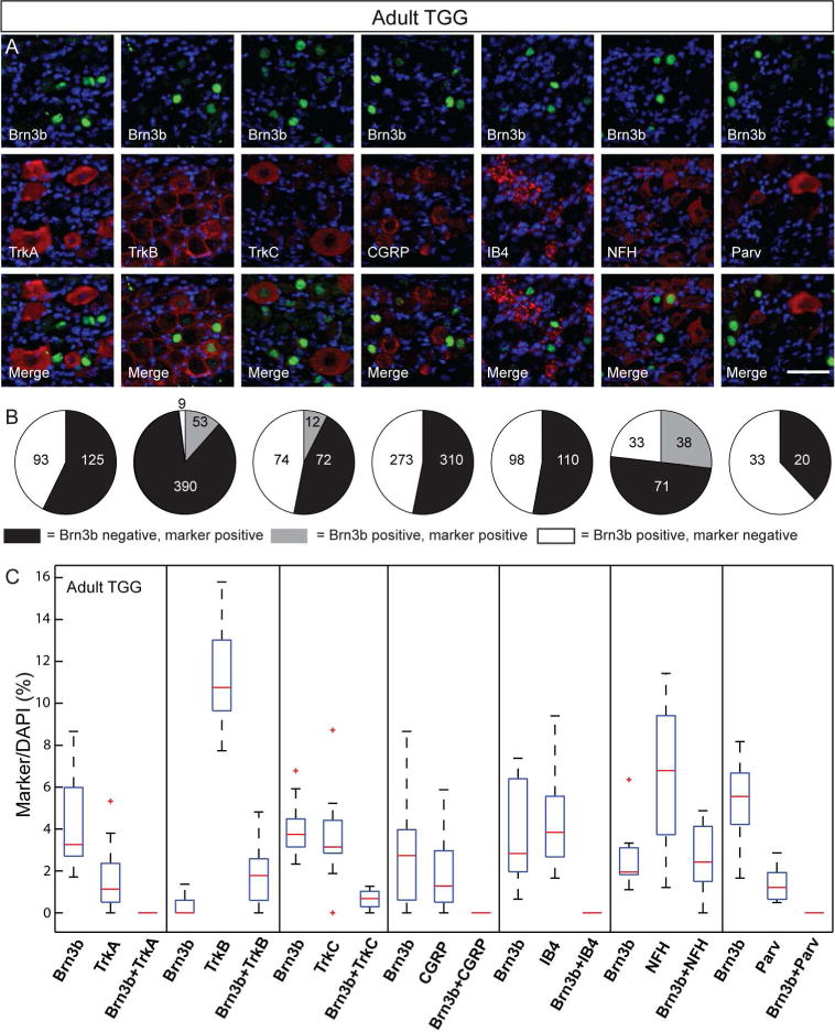
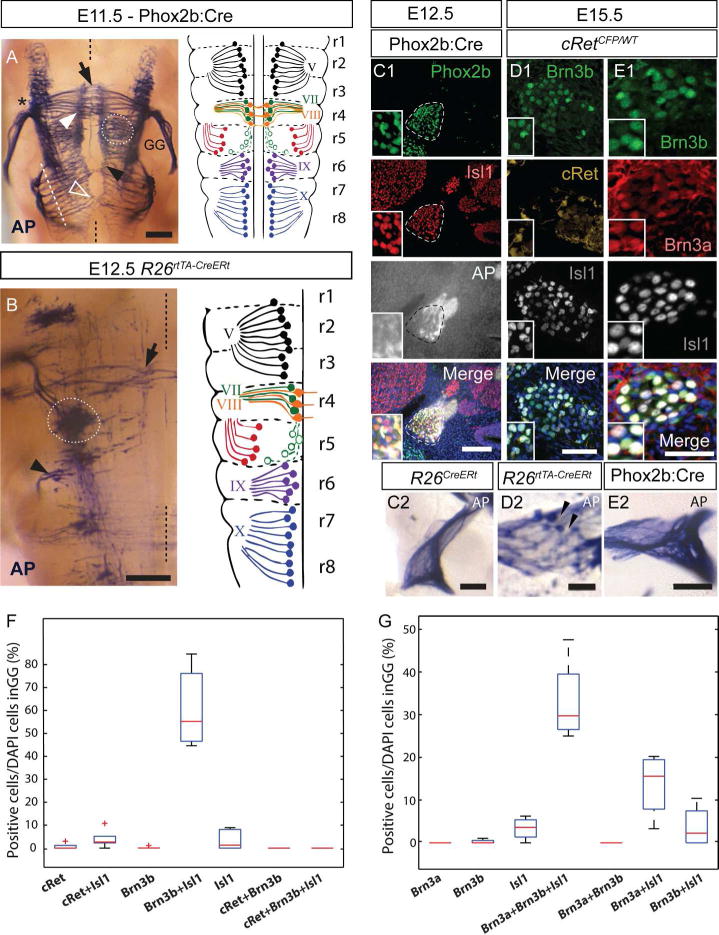
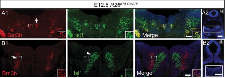
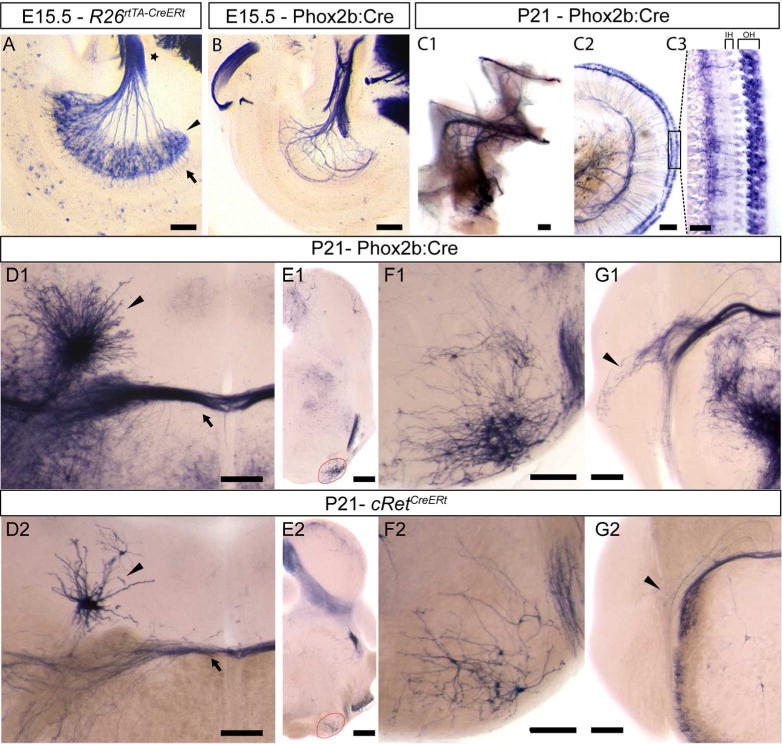
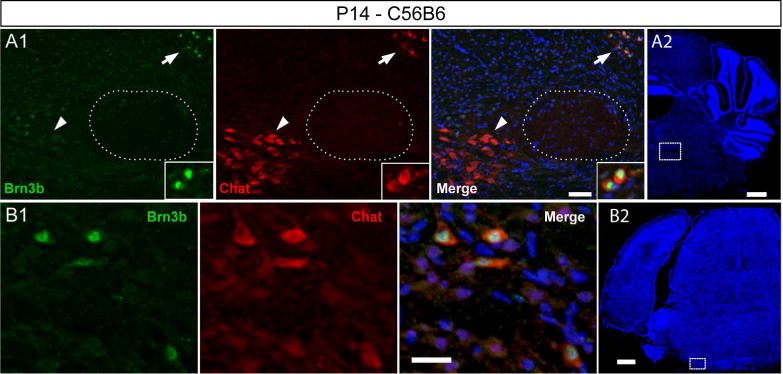
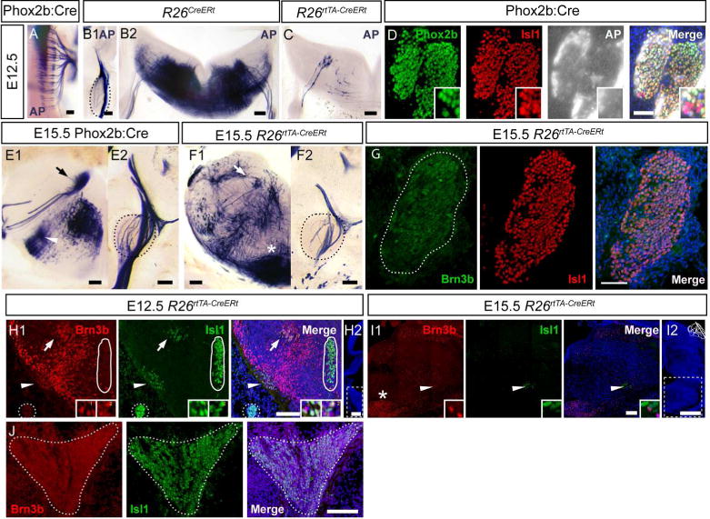
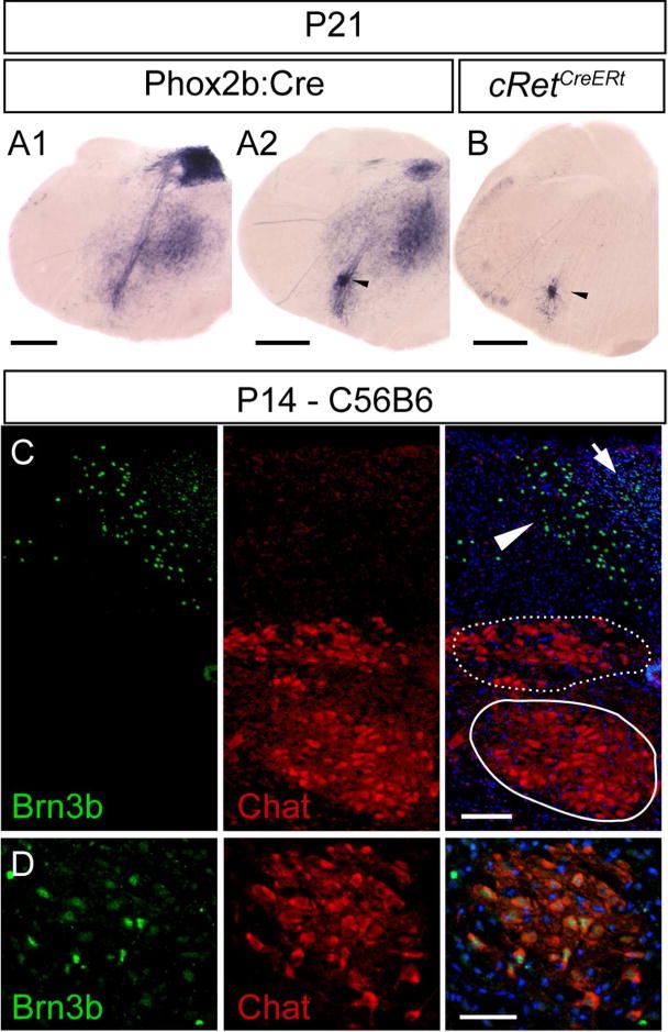
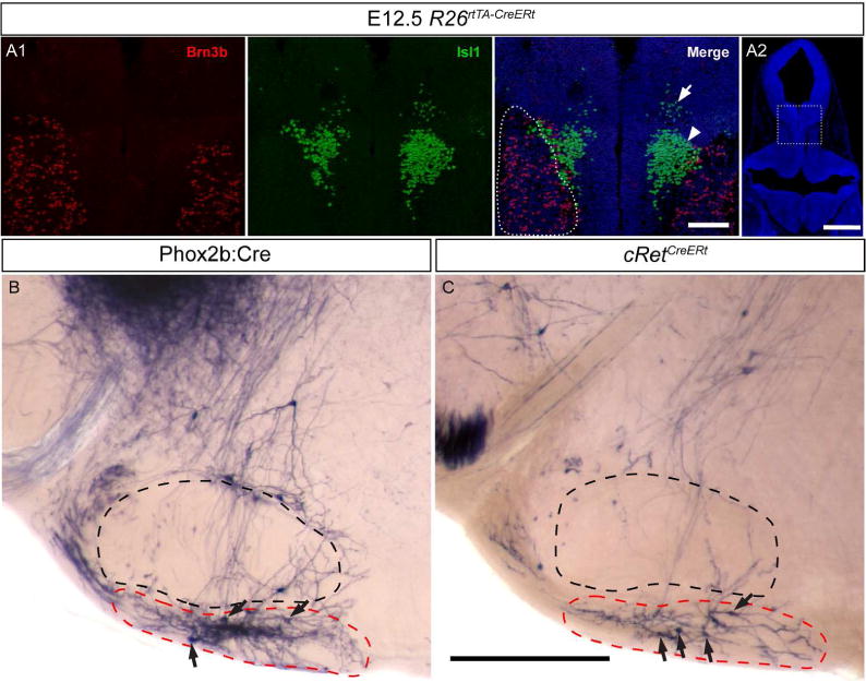
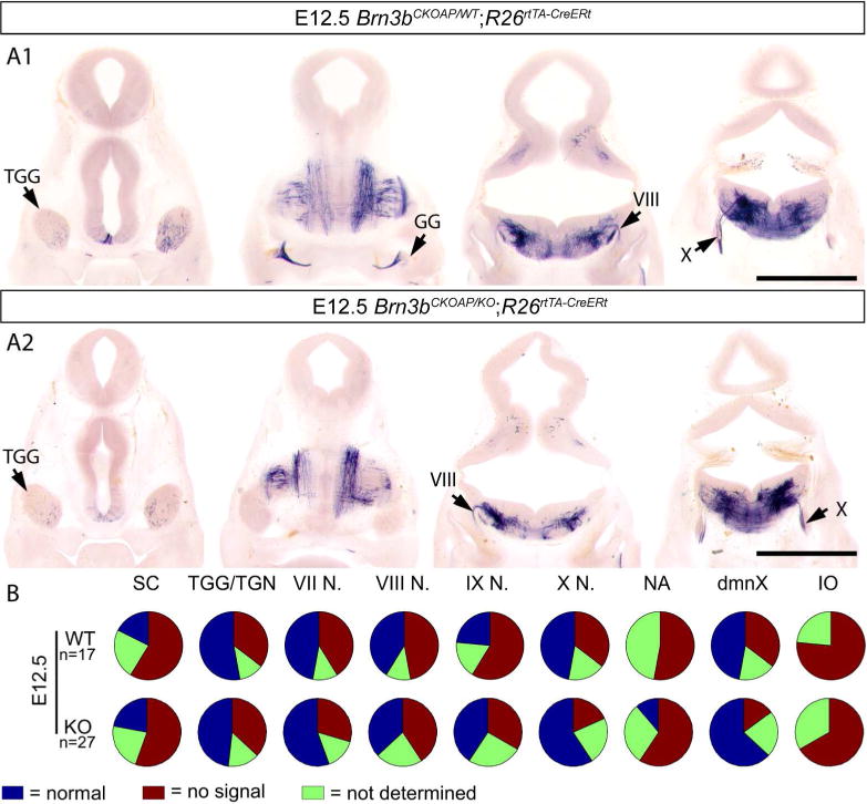
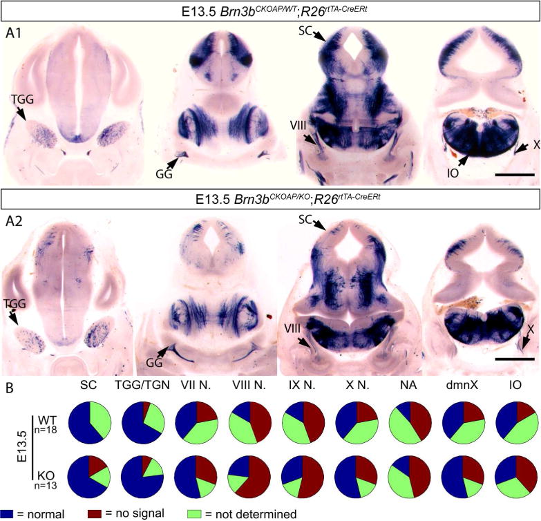
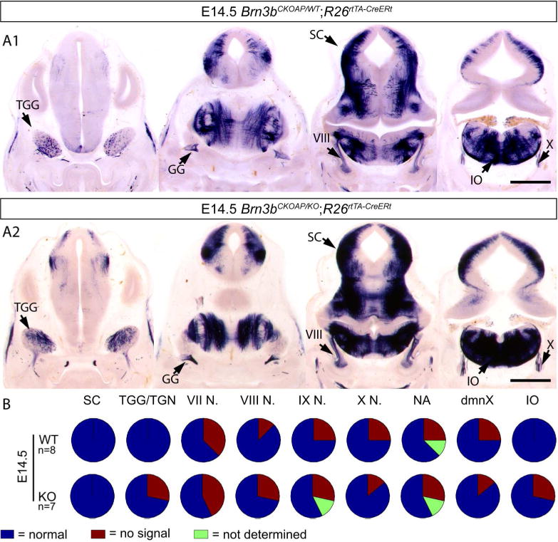
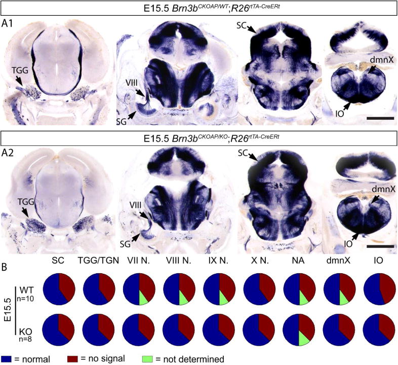
During development, transcription factor combinatorial codes define a large variety of morphologically and physiologically distinct neurons. Such a combinatorial code has been proposed for the differentiation of projection neurons of the somatic and visceral components of cranial nerves. It is possible that individual neuronal cell types are not specified by unique transcription factors but rather emerge through the intersection of their expression domains. Brn3a, Brn3b, and Brn3c, in combination with each other and/or transcription factors of other families, can define subgroups of retinal ganglion cells (RGC), spiral and vestibular ganglia, inner ear and vestibular hair cell neurons in the vestibuloacoustic system, and groups of somatosensory neurons in the dorsal root ganglia. The present study investigates the expression and potential role of the Brn3b transcription factor in cranial nerves and associated nuclei of the brainstem. We report the dynamic expression of Brn3b in the somatosensory component of cranial nerves II, V, VII, and VIII and visceromotor nuclei of nerves VII, IX, and X as well as other brainstem nuclei during different stages of development into adult stage. We find that genetically identified Brn3b(KO) RGC axons show correct but delayed pathfinding during the early stages of embryonic development. However, loss of Brn3b does not affect the anatomy of the other cranial nerves normally expressing this transcription factor.

Keywords: POU domain Brn3b; cranial nerves; facial nerve; glossopharyngeal nerve; optic nerve; transcription; trigeminal nerve; vagus nerve.


Conflict of interest statement

No known conflicts of interest.

Figures

Figure 1
Figure 1. Genetic-pharmacological and intersectional genetic strategies used to label Brn3b-positive cranial nerve neurons
The Cre dependent Brn3b conditional knock-in AP reporter mouse line (A) was crossed to three alternative genetic drivers in order to obtain ubiquitous dense (B), neuronal subpopulation specific (C) or ubiquitous sparse (D) Cre activity. A, Conditional Knock-in reporter construct targeted at the Brn3b locus (Brn3bCKOAP). B, Near complete recombination was induced by 4HT induction of the CreERt activity from the R26CreERt knock-in allele. C, BAC transgenic line expressing constitutively active Cre in Phox2b positive neurons. D, Sparse random recombination was achieved by dual pharmacological control of a R26rtTA-CreERt knock-in construct. E, F Examples of dual pharmacological control regime used for Cre activity control in R26rtTA-CreERt; Brn3bCKOAP crosses (D × A). Plugged females were given regular feed alternating with 0.2 mg/g Dox chow (purple boxes) every two days, beginning with the day the plug was found (E0.5). 12.5 – 50 μg 4HT injected intraperitoneally two days prior to embryo harvesting (red triangle), at either E12.5 (E) or E15.5 (F).
Figure 2
Figure 2. Brn3bAP labeling in cranial nerves II, V, VII, VIII, IX and X during development
Full expression pattern of Brn3b determined by staining R26CreERt; Brn3bCKOAP embryos at E12.5 (A1, A2) and E13 (D). AP can be detected in the retina, spinal cord, dorsal root ganglia (DRG), brainstem, mesencephalon, trigeminal ganglion (TGG), trigeminal nerve (V), facial (VII), glossopharyngeal (IX) and vagal nerves (X) (schematized in C). B1, B2 Phox2b:Cre; Brn3bCKOAP E12.5 embryo shows AP positive signal in the cranial nerves VII, IX, X. C, Schematic of AP expressing nerves: 3 branches of Trigeminal nerve (V): ophthalmic – V1, maxillary V2 and mandibular V3. Facial nerve (VII) with Greater Superficial Petrosal (GSP), Chorda Tympani (CT), and main branch (FN). Vestibulocochlear nerve (VIII) Glossopharyngeal nerve (IX) with tympanic (TB) and pharyngeal (PB) branches. Vagus nerve (X). E,F R26rtTA-CreERt; Brn3bCKOAP embryos from pregnant females treated with 0.2mg/g doxycycline (DOX) and 50μg (E) or 17.5μg (F) of 4 hydroxytamoxifen (4HT). Different doses of 4HT – labeling off all or few nerve fibers in the developing cranial nerves (E – F). Scale bars: A1, A2, B1, B2=1mm, D – F=500μm.
Figure 3
Figure 3. Brn3b expression in developing retinal ganglion cells
A, Optic cup section with sparsely labeled Brn3bAP RGCs at E12.5. RGCs at different developmental time points: columnar stage (hollow arrow), cells in the RGC layer with retracted ventricular end feet (black arrow head), cells projecting into the optic nerve (black arrow). B, Brn3bAP RGCs in intermediate stages of migration towards the GCL and ventricular (arrow head) process retraction (1). Note the presence of pairs of Brn3bAP RGCs (star) whose ventricular processes are cofasciculated (2) or closely apposed (3). End foot retraction (black arrowhead) and axon extension (black arrow) (4). C, Coronal section at the level of the optic chiasm at E12.5, ipsilateral Brn3bAP RGC axon (black arrow head) climbing on exterior wall of the thalamus. D1, D2, Serial horizontal sections at the level of the developing optic chiasm in R26rtTA-CreERt; Brn3bCKOAP/WT (D1) and R26rtTA-CreERt; Brn3bCKOAP/KO (D2) E12.5 littermates. E1, E2, Serial coronal sections at the level of the developing optic tract in Pax6α:Cre; Brn3bCKOAP/WT; (E1) and Pax6α:Cre; Brn3bCKOAP/KO (E2) E15.5 littermates. F1, F2, Serial coronal sections at the level of the developing optic tract in R26rtTA-CreERt; Brn3bCKOAP/WT (E1) and R26rtTA-CreERt; Brn3bCKOAP/KO; (E2) E15.5 littermates. Scale bars: A,C=100μm, B=10μm, D1,D2,E1–F2=500μm
Figure 4
Figure 4. Brn3b expression in peritrigeminal neurons
A1 A2, AP negative Phox2b and Isl1 positive MoV neurons at E12.5. AP indicating Phox2b and Brn3b coexpression is present in peritrigeminal neurons (purple lines). B1 B2 Coronal section at the level of the MoV showing mutual exclusive expression of Brn3b positive neurons and Isl1 positive motoneurons. Scale bars: A1,B2=50μm, A2,B2=500μm
Figure 5
Figure 5. Brn3b expression in embryonic trigeminal neurons
A, Brn3bAP positive deep vibrissal (arrow) and intervibrissal (arrow head) fibers in the mystacial pad are visible as early as E13.5. B1, Brn3bAP positive deep vibrissal (arrow) and intervibrissal (arrow head) as well as general sensory fibers (B2) innervating the lips of E15.5 embryo. C1, Extensive overlap of Brn3b and Isl1 in E12.5 embryos. C2, Brn3bAP positive TGG cells in ROSA26rtTACreERt; Brn3bCKOAP/WT E12.5 mice. D, Sagittal section of a E15.5 cRetCFP/WT maxillary (lines) and mandibular territories (doted lines) of TGG. Insets show combinatorial expression of Brn3b, Isl1, and cRet. E, Box whisker plot for cell populations of E15.5 TGG (D). Scale bars: A,B1,B2,C2,D=100μm, C1=50μm
Figure 6
Figure 6. Cell type distribution of Brn3b in adult trigeminal neurons
A, Double immunostaining for Brn3b (green) and indicated molecular markers (red) in the adult TGG. B Pie charts showing the number of TGG cells single and double positive for the various TGG markers and Brn3b stained in A. C, Box whisker plot for cell populations of the adult TGG neurons. Scale bars: A=50μm
Figure 7
Figure 7. Brn3b expression in the developing facial nerve (VII) system
A, Left panel, Brn3bAP histochemistry of the E11.5 brainstem of Phox2b:Cre; Brn3bCKOAP embryo in whole mount dorsal perspective. AP Positive cell bodies include visceral motor neurons of the superior salivatory nucleus (SSN) (white stippled circle) and branchiomotor neurons (white arrow head). Inner ear efferent fibers (black arrow) are crossing the midline (black stippled line). Exit point of the nerves VII and VIII are indicated by black asterisk. Cell bodies of the glossopharyngeal (black arrow head) and vagal (white hollow arrow) nuclei exiting the neuraxis through multiple rootlets, arranged in a continuous fashion (white stippled line). Right panel, schematic of the rhombomeric distribution of neuronal cell bodies, axonal processes and exit points of cranial nerves V, VII, VIII, IX and X, as they map onto the rhombomeres (adapted from Lumsden and Krumlauf, 1996). B, Whole mount Brn3bAP histochemistry same perspective as A, in an E12.5 R26rtTA-CreER; Brn3bCKOAP embryo. Left side of the animal, with the midline indicated by a black stippled line. Isolated cell bodies or small groups of the nuclei indicated in A and their axonal projections are visible. Black arrow indicates inner ear efferent neuron axons extending contralaterally across the midline. Superior salivary nucleus (white stippled circle). Group of cells with axons projecting in the glosspharyngeal nerve (black arrow head). C1, Combined Brn3bAP histochemistry and IIF of the GG (stippled lines) in the Phox2b:Cre; Brn3bCKOAP E12.5 embryo, showing extensive Phox2b and Isl1 double positive neurons in the GG. C2 Coronal section at the level of the GG of the R26CreERt; Brn3bCKOAP E12.5 mouse, with no visible AP positive cell bodies. D1,E1 Immunostaining of the GG in cRetCFP/WT E15.5 embryos. D2, Coronal section at the level of the GG of the R26rtTA-CreERt; Brn3bCKOAP E15.5 embryo, showing Brn3bAP positive cells (black arrowheads). E2 AP positive fibers crossing the GG of Phox2b:Cre; Brn3bCKOAP E15.5 embryo with no visible AP positive cell bodies. F box whisker plots for cell populations in D1. G box whisker plots for cell populations in E1. Scale bars: A = 250μm, B = 200μm, C1,C2, D1, D2, E2 = 100μm, E1=50μm
Figure 8
Figure 8. Brn3b expression in the developing facial nucleus
A1, A2 Coronal section at the level of genu of the facial nerve (arrow) containing Brn3b (red) and Isl1 (green) positive cells. B1, B2, Coronal section at the level of the migrating stream of facial branchiomotor (Isl1 positive, Brn3b negative, white arrowhead). Scale bars: A1, B1 = 100μm, A2 B2 = 500μm
Figure 9
Figure 9. Brn3b expression in the afferent and efferent projecting cells of the developing cochlear nerve
A, Spiral ganglion neuron cell bodies (black arrowhead), axons forming the cochlear nerve (star) and dendritic processes innervating the cochlea (arrow) in a R26rtTA-CreERt; Brn3bCKOAP E15.5 embryo. B, Efferent axon innervations of the cochlea, in a Phox2b:Cre; Brn3bCKOAP E15.5 embryo and adult (C1,C2). C3 Efferent innervation of the three rows of outer hair cells. D1–G2 coronal sections through the adult pons and brainstem. D1, D2 Dense and sparse labeling of Eve (nucleus of origin of efferents of the vestibular nerve) (arrow head) and OCB (Olivocochlear Bundle) (arrow) in adult Phox2b:Cre; Brn3bCKOAP (D1) and cRetCreERt; Brn3bCKOAP (D2) animals. E1, F1 Brn3b/Phox2b and E2, F2 Brn3b/cRet double positive neurons of the ventral nucleus of the trapezoid body (VNTB), at the level of the superior olivary complex (SOC) (red circle). G1, G2 Phox2b+ Brn3b+ positive (G1) and cRet+ Brn3b+ positive (G2) VNTB projections to the cochlear nucleus (black arrow head). Scale bars: A,B,C1,C2 = 100μm, C3=25μm, D1,D2,F1,F2,G1,G2,=200μm, E1,E2=500μm
Figure 10
Figure 10. Brn3b expression in the Eve and VNTB of P14 animals
A1 A2 Double immunostaining at the level of the genu (stippled circle) in P14 animals. Two Chat positive nuclei visible, Brn3b positive Eve (white arrow), and Brn3b negative nucleus abducens (white arrow head). B1, B2 Brn3b Chat double positive cells in the VNTB. Scale bars: A1,B1=25μm, A2,B2=500μm
Figure 11
Figure 11. Brn3b expression in the developing glossopharyngeal and vagal systems
A, Dorsal view of the brainstem of a E12.5 Phox2b:Cre; Brn3bCKOAP embryo showing Brn3bAP positive cell bodies at the level of the dorsal motor nucleus of the vagus. B1, B2, AP histochemistry of coronal sections through R26CreERt; Brn3bCKOAP E12.5 embryos. B1, Brn3bAP positive fibers wrapping around the N/P ganglia complex. B2, Full Brn3bAP histochemistry of brainstem at the level of the dmnX. C, Hemisection through a sparsely labeled R26rtTA-CreERt; Brn3bCKOAP E12.5 embryo, showing a few dmnX cell bodies and their projections into the Xth nerve. D, Immunostaining for Phox2b and Isl1 combined with histochemistry for Brn3bAP, in sections of Nodose and Petrose Ganglia (N/P) of E12.5 Phox2b:Cre; Brn3bCKOAP embryo. E1, Brn3bAP histochemistry of brainstem sections in E15.5 Phox2b:Cre; Brn3bCKOAP embryos. Note the precise isolation of the dmnX (arrow) and NA (arrowhead), from several other Brn3bAP positive nuclei. E2, N/P ganglia from the same animal as in E1, wrapped in Brn3bAP fibers, but devoid of AP positive nuclei. F1, F2, Brn3bAP histochemistry in E15.5 R26rtTA-CreERt; Brn3bCKOAP embryos labeled at intermediate sparse levels, showing AP positivity in the dmnX (arrow), and lack of AP positive cell bodies in the N/P (black stippled lines). G Immunostaining of N/P ganglia shows Isl1 but no Brn3b positivity at E15.5. H1,H2 Coexpression of Brn3b (red) and Isl1 (green) in the dmnX (white arrow) and nucleus ambiguus (NA, white arrowhead). Insets in bottom right corner show high magnification of Brn3b and Isl1 co-expression in NA (1) and dmnX (2). I1,I2, Expression of Brn3b in the NA at E15.5. H2,I2 Stippled lines show the regions in H1 and I1. Asterisk in F1 and I1 marks E15.5 expression of Brn3b (I1) and Brn3bAP (F1) in the inferior olive. J Sagittal section at the level of superior and jugular ganglia (stippled line) of E12.5 embryo show now Brn3b expression. Scale bars: A,B1,B2,C,E1,E2,F1,F2,G,H1,I1,J=100μm,D=50μm, H2,I2=500μm.
Figure 12
Figure 12. Brn3b expression in the area postrema of the postnatal mouse
A1, Brn3AP positive areas include central stations of the vagus nerve (AP, NTS, and dmnX), in an adult Phox2b:Cre; Brn3bCKOAP animal. A2 B, AP signal in the anatomic position of the NA (black arrow head) in Phox2b:Cre; Brn3bCKOAP (A2) and cRetCreERt; Brn3bCKOAP (B) mouse. C, D Immunostaining analysis did not reveal any Brn3b expression in the Chat positive dmnX (Figure 12C stippled circle) or hypoglossal nuclei (12C circle). Expression was limited to the Area postrema (arrow), subdividions of the nucleus of the solitary tract (C arrow head) and NA (D). Scale bars: A1,A2,B=500μm, C,D=50μm.
Figure 13
Figure 13. Other Brainstem nuclei
A1 A2, Isl1 positive oculomotor (arrow) and trochlear (arrow head) nuclei do not express Brn3b. Brn3b positive cells in proximity of to the trochlear nucleus (stippled line). B, C AP positive cell bodies (black arrows) ventral to the facial nucleus (black stippled lines) in the retrotrapezoid area (red lines) indicating Phox2b/Brn3b (B) and Brn3b/cRet (C) coexpression. Scale bar A1=100μm, A2, B, C = 500μm
Figure 14
Figure 14
Analysis of Brn3bAP positive structures in E12.5 R26rtTA-CreERt; Brn3bCKOAP/WT (A1) R26rtTA-CreERt; Brn3bCKOAP/KO (A2). B, n = the total number of analyzed embryos per condition. Scale bars A1, A2= 1mm
Figure 15
Figure 15
Analysis of Brn3bAP positive structures in E13.5 R26rtTA-CreERt; Brn3bCKOAP/WT (A1) R26rtTA-CreERt; Brn3bCKOAP/KO (A2). B, n = the total number of analyzed embryos per condition. Scale bars A1, A2= 1mm
Figure 16
Figure 16
Analysis of Brn3bAP positive structures in E14.5 R26rtTA-CreERt; Brn3bCKOAP/WT (A1) R26rtTA-CreERt; Brn3bCKOAP/KO (A2). B, n = the total number of analyzed embryos per condition. Scale bars A1, A2= 1mm
Figure 17
Figure 17
Analysis of Brn3bAP positive structures in E15.5 R26rtTA-CreERt; Brn3bCKOAP/WT (A1) R26rtTA-CreERt; Brn3bCKOAP/KO (A2). B, n = the total number of analyzed embryos per condition. Scale bars A1, A2= 1mm

Similar articles

Cited by

References

    1. Altschuler SM, Bao X, Bieger D, Hopkins DA, Miselis RR. Viscerotopic representation of the upper alimentary tract in the rat: sensory ganglia and nuclei of the solitary and spinal trigeminal tracts. Journal of Comparative Neurology. 1989;283(2):248–268. - PubMed
    1. Alvarado-Mallart MR, Batini C, Buisseret-Delmas C, Corvisier J. Trigeminal representations of the masticatory and extraocular proprioceptors as revealed by horseradish peroxidase retrograde transport. Experimental brain research. 1975;23(2):167–179. - PubMed
    1. Ashwell KW. The adult mouse facial nerve nucleus: morphology and musculotopic organization. Journal of anatomy. 1982;135(Pt 3):531–531. - PMC - PubMed
    1. Auclair F, Valdés N, Marchand R. Rhombomere-specific origin of branchial and visceral motoneurons of the facial nerve in the rat embryo. Journal of Comparative Neurology. 1996;369(3):451–461. - PubMed
    1. Ayer-Le Lievre, CS Le, Douarin NM. The early development of cranial sensory ganglia and the potentialities of their component cells studied in quail-chick chimeras. Developmental biology. 1982;94(2):291–310. - PubMed

Publication types

Substances