. 2016 Feb;142(2):423-35.
doi: 10.1007/s00432-015-2044-9. Epub 2015 Sep 10.

Norepinephrine induced epithelial-mesenchymal transition in HT-29 and A549 cells in vitro

Affiliations

Norepinephrine induced epithelial-mesenchymal transition in HT-29 and A549 cells in vitro

Jie Zhang et al. J Cancer Res Clin Oncol. 2016 Feb.

Abstract

Purpose: Norepinephrine (NE) has been implicated in epithelial-mesenchymal transition (EMT) of cancer cells. However, the underlying mechanism is poorly understood. The goal of this study was to explore the effect of NE on cancer cell EMT and to investigate the potential mechanism.

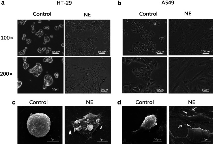
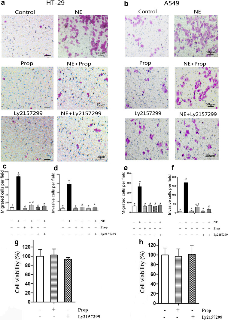
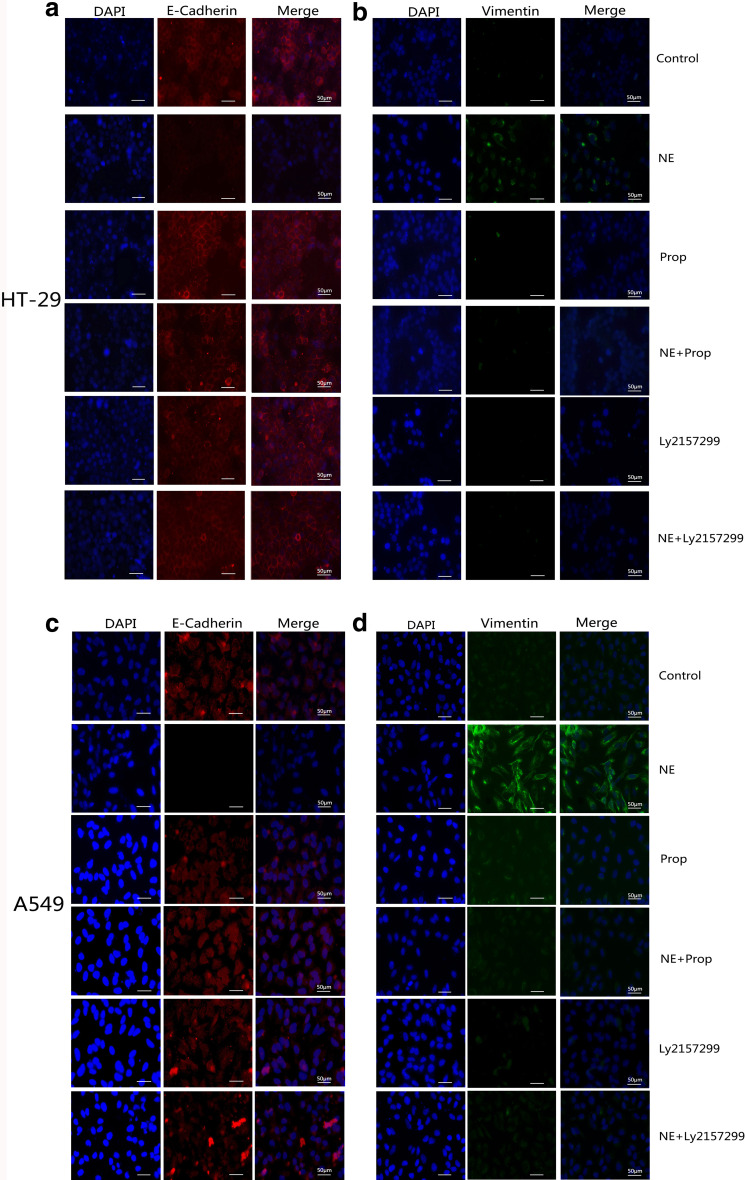
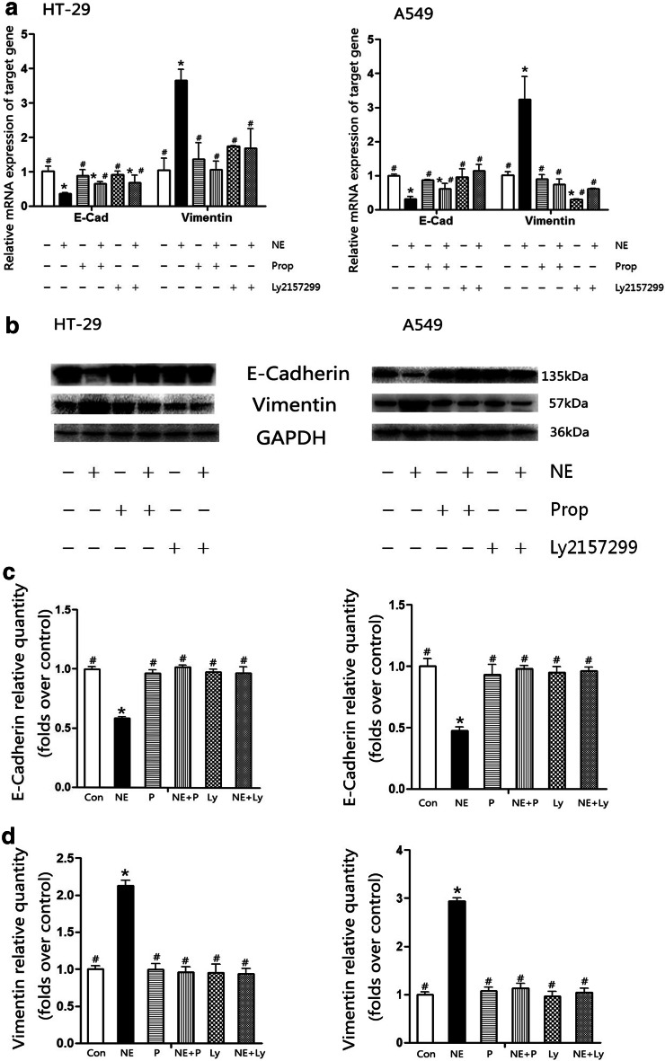
Methods: HT-29 and A549 cells were treated with NE, β-adrenergic receptor (β-AR) antagonist (propranolol) or inhibitor of transforming growth factor-β (TGF-β) receptor type I kinase (Ly2157299). Morphology of cells was observed with optical and electron microscope and immunofluorescence staining. Cellular migration and invasion were tested with transwell migration assay and Matrigel invasion assay, respectively. TGF-β1 and cyclic adenosine monophosphate (cAMP) were quantified. EMT markers and signaling pathway were measured by RT-PCR and western blot.

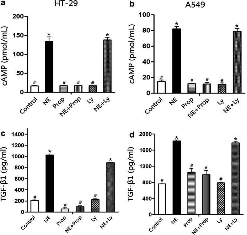
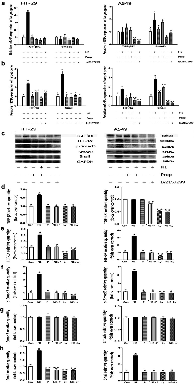
Results: NE stimulated TGF-β1 secretion and intracellular cAMP synthesis, induced morphological alterations in HT-29 and A549 cells, and enhanced their ability of migration and invasion. EMT markers induction was observed in NE-treated cancer cells. The effect of NE could be inhibited by propranolol or Ly2157299. β-AR/TGF-β1 signaling/p-Smad3/Snail and β-AR/TGF-β1 signaling/HIF-1α/Snail were two signaling pathways.

Conclusion: These findings demonstrated that TGF-β1 signaling pathway was a significant factor of NE-induced cancer cells EMT. The data also suggested that psychological stress might be a risk factor which enhances the ability of migration or invasion of cancer cells.

Keywords: Adrenergic receptor; Chronic stress; Epithelial–mesenchymal transition; Norepinephrine; Transforming growth factor-β1.


Conflict of interest statement

We declare that no conflict of interest exists in this manuscript.

Figures

Fig. 1
Fig. 1
Norepinephrine induced EMT morphological alterations in HT-29 and A549 cells. Cells were cultured in serum-free medium (control) and serum-free medium containing 10 μM NE (NE) for 4 days. Cellular morphology was observed using optical microscope (×100 and ×200) and scanning electron microscope (×5000–10,000). Cells treated with 10 μM NE showed a spindle-shaped, fibroblast-like phenotype (a, b), increased microvilli (c, d white triangle), and pseudopodium (d white arrows), while the control cells maintained the classic epithelial morphology
Fig. 2
Fig. 2
Propranolol and Ly2157299 attenuated norepinephrine-stimulated cancer cells migration and invasion. Serum-starved HT-29 (a, c, d) and A549 (b, e, f) cells were treated with 10 μM NE, 10 μM Prop, or 10 μM Ly2157299 as the figure labeled. Representative photographs are shown as cells that migrated to the lower membrane through filter pores (a, b ×200). The number of migratory (c, e) or invasive cells (d, f) was counted in five random views and is presented in columns as mean ± SD. NE increased cells migration and invasion. Prop and Ly2157299 blocked NE effect (cf), and they not affected cells viability (g, h). *P < 0.05 versus control group; # P < 0.05 versus NE group
Fig. 3
Fig. 3
Immunofluorescence staining of E-cadherin and vimentin in HT-29 and A549 cells. HT-29 (a, b) and A549 (c, d) cells were treated with 10 μM NE, 10 μM Prop, or 10 μM Ly2157299 for 4 days as the figure labeled. After then, cells were incubated with rabbit anti-human E-cadherin and vimentin and then observed under confocal laser scanning microscope. DAPI was used to counterstain cell nuclei. In the NE-treated group, E-cadherin fluorescence signal (a, c red) was weaker than in other groups, while vimentin fluorescence signal (b, d green) was stronger compared with others
Fig. 4
Fig. 4
Propranolol and Ly2157299 attenuated norepinephrine-induced EMT makers’ changes in HT-29 and A549 cells. After 4-day incubation with 10 μM NE, 10 μM Prop, or 10 μM Ly2157299, mRNA (a) and protein (b) expressions of E-cadherin and vimentin in HT-29 and A549 cells were examined by RT-PCR and western blot, respectively. Quantification of E-cadherin (c) and vimentin (d) protein is presented in columns as mean ± SD. NE decreased E-cadherin expression and increased vimentin expression, while Prop and Ly2157299 reversed this effect. *P < 0.05 versus control group; # P < 0.05 versus NE group
Fig. 5
Fig. 5
Norepinephrine stimulated cAMP synthesis and TGF-β1 secretion. Intracellular cAMP and TGF-β1 in culture supernatant were determined. Data are presented in columns as mean ± SD. NE triggered HT-29 (a) and A549 (b) cells cAMP synthesis. TGF-β1 in supernatant from HT-29 (c) and A549 cells (d) was increased after incubating with NE for 72 and 48 h, respectively. β-Adrenoceptor antagonist Prop could inhibit this effect, but Ly2157299 could not. *P < 0.05 versus control group; # P < 0.05 versus NE group
Fig. 6
Fig. 6
Propranolol and Ly2157299 blocked norepinephrine-induced HIF-1α, p-Smad3, and Snail expressions. NE induced TGF-βRI mRNA and protein expression in HT-29 cells (a, c, d) and Smad3 mRNA expression in A549 cells (a). NE stimulated transcripts and protein expressions of HIF-1α (b, c, e) and Snail (b, c, h) expressions, and increased p-Smad3 protein expressions (c, f) in HT-29 and A549 cells. Prop and Ly2157299 could block NE effect. Quantification of proteins is presented in columns as mean ± SD (dg). *P < 0.05 versus control group; # P < 0.05 versus NE group

Similar articles

Cited by

References

    1. Akhurst RJ, Derynck R (2001) TGF-β signaling in cancer—a double-edged sword. Trends Cell Biol 11(11):S44–S51. doi:10.1016/S0962-8924(01)02130-4 - PubMed
    1. Barbieri A, Bimonte S, Palma G, Luciano A, Rea D, Giudice A, Scognamiglio G, La Mantia E, Franco R, Perdona S, De Cobelli O, Ferro M, Zappavigna S, Stiuso P, Caraglia M, Arra C (2015) The stress hormone norepinephrine increases migration of prostate cancer cells in vitro and in vivo. Int J Oncol. doi:10.3892/ijo.2015.3038 - PubMed
    1. Barron TI, Connolly RM, Sharp L, Bennett K, Visvanathan K (2011) Beta blockers and breast cancer mortality: a population-based study. J Clin Oncol 29(19):2635–2644. doi:10.1200/JCO.2010.33.5422 - PubMed
    1. Brabletz T, Hlubek F, Spaderna S, Schmalhofer O, Hiendlmeyer E, Jung A, Kirchner T (2005) Invasion and metastasis in colorectal cancer: epithelial–mesenchymal transition, mesenchymal–epithelial transition, stem cells and β-catenin. Cells Tissues Organs 179(1–2):56–65. doi:10.1159/000084509 - PubMed
    1. Calon A, Espinet E, Palomo-Ponce S, Tauriello DV, Iglesias M, Cespedes MV, Sevillano M, Nadal C, Jung P, Zhang XH, Byrom D, Riera A, Rossell D, Mangues R, Massague J, Sancho E, Batlle E (2012) Dependency of colorectal cancer on a TGF-β-driven program in stromal cells for metastasis initiation. Cancer Cell 22(5):571–584. doi:10.1016/j.ccr.2012.08.013 - PMC - PubMed

Publication types

MeSH terms

LinkOut - more resources