. 2015 Oct;25(10):1137-51.
doi: 10.1038/cr.2015.110. Epub 2015 Sep 11.

Acute inflammation stimulates a regenerative response in the neonatal mouse heart

Affiliations

Acute inflammation stimulates a regenerative response in the neonatal mouse heart

Chunyong Han et al. Cell Res. 2015 Oct.

Abstract

Cardiac injury in neonatal 1-day-old mice stimulates a regenerative response characterized by reactive cardiomyocyte proliferation, which is distinguished from the fibrotic repair process in adults. Acute inflammation occurs immediately after heart injury and has generally been believed to exert a negative effect on heart regeneration by promoting scar formation in adults; however, little is known about the role of acute inflammation in the cardiac regenerative response in neonatal mice. Here, we show that acute inflammation induced cardiomyocyte proliferation after apical intramyocardial microinjection of immunogenic zymosan A particles into the neonatal mouse heart. We also found that cardiac injury-induced regenerative response was suspended after immunosuppression in neonatal mice, and that cardiomyocytes could not be reactivated to proliferate after neonatal heart injury in the absence of interleukin-6 (IL-6). Furthermore, cardiomyocyte-specific deletion of signal transducer and activator of transcription 3 (STAT3), the major downstream effector of IL-6 signaling, decreased reactive cardiomyocyte proliferation after apical resection. Our results indicate that acute inflammation stimulates the regenerative response in neonatal mouse heart, and suggest that modulation of inflammatory signals might have important implications in cardiac regenerative medicine.


Figures

Figure 1
Figure 1
The acute inflammatory response occurs immediately after AR and ZA microinjection in neonatal mouse hearts. (A) Immunostaining for Ly-6G (1A8) at 1 day post-resection (dpr) in the neonatal mouse heart. The high-magnification views of the boxed areas are presented on the right. AR, apical resection; scale bars, 100 μm. (B) qRT-PCR assays of the expression of inflammatory markers (il6, il1b and ccl3) in sham and AR hearts at 1, 4 and 7 dpr. Gene expression in AR hearts is presented as the fold change compared to sham hearts. n = 3 per group. (C, D) Apical intramyocardial microinjection of ZA into the neonatal 1-day-old mouse heart. ZA was conjugated with Alexa Fluor® 488 to track its distribution in the cardiac apex. The high-magnification views of the boxed areas are presented on the right. dpm, days post-microinjection. Scale bars, 100 μm. (E) Immunostaining for Ly-6G (1A8) in the PBS and ZA micro-injected apical myocardium at 1 dpm in neonatal mice. The high-magnification views of the boxed areas are presented on the right. Arrows indicate Ly-6G (1A8)+ leukocytes. Scale bars, 100 μm. (F) qRT-PCR assays of the expression of inflammatory markers (il6, il1b and ccl3) at 1, 4 and 7 dpm. Gene expression in the ZA micro-injected hearts is presented as the fold change compared with PBS micro-injected hearts. n = 3 per group; values are presented as the mean ± SEM; *P < 0.05.
Figure 2
Figure 2
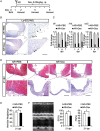
Injured hearts cannot regenerate after immunosuppression in neonatal mice. (A) Schematic showing dexamethasone (Dex) treatment after AR in neonatal 1-day-old mice. (B) Immunostaining for Ly-6G (1A8) in PBS- and Dex-treated hearts at 1 dpr. The high-magnification views of the boxed areas are presented on the right. Arrows indicate Ly-6G (1A8)+ leukocytes. Scale bars, 100 μm. (C) qRT-PCR assays of the expression of il6, il1b and ccl3. Gene expression data in the Dex-treated hearts are presented as a fold change compared to the PBS-treated hearts. n = 3 per group. (D) Masson's staining of PBS- and Dex-treated hearts at 21 dpr. The high-magnification views of the boxed areas are presented below. n = 3 per group; scale bars, 100 μm. (E) Quantification of ventricle weights of the PBS- and Dex-treated hearts at 21 dpr. n = 6 per group. (F) Representative images of M-mode echocardiography of hearts at 21 dpr between PBS- and Dex- treated mice. Systolic function of the left ventricular was measured by quantifying ejection fraction (EF) and fractional shortening (FS). n = 3 per group. Values are presented as the mean ± SEM; *P < 0.05; NS, not statistically significant.
Figure 3
Figure 3
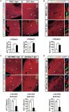
The acute inflammatory response is necessary and sufficient for reactive cardiomyocyte proliferation in neonatal mouse hearts. (A) Cardiomyocyte proliferation was identified by immunostaining for pH3 (green) and α-actinin (red) co-localization and Ki67 (green) and α-actinin (red) co-localization in PBS and ZA micro-injected hearts at 7 dpm. (B) The newly formed cardiomyocytes were shown as Brdu and α-actinin double-positive cells following a pulse-chase experiment in PBS and ZA micro-injected hearts at 21 dpm. (C) Immunostaining for pH3 (green) and Ki67 (green) with cardiac troponin T (cTnT) (red) co-localization in PBS- and Dex- treated hearts at 7 dpr. (D) The newly formed cardiomyocytes were shown as Brdu and α-actinin double-positive cells in PBS- and Dex-treated hearts at 21 dpr. In A and C, arrows indicate cardiomyocytes positive for pH3 or Ki67 staining. The inset is a high-magnification image of a pH3- or Ki67-positive cardiomyocyte which is denoted by an arrowhead. Scale bars, 75 μm. In B and D, magnification images of a Brdu+ cardiomyocyte (arrow) are shown on the right. Scale bars, 25 μm. CMs, cardiomyocytes; n = 3 per group; values are presented as the mean ± SEM; *P < 0.05.
Figure 4
Figure 4
IL-6 signaling is required for the cardiac regenerative response in neonatal mice. (A) Cardiomyocyte proliferation was identified by immunostaining for pH3 (green) and Ki67 (green) together with α-actinin (red) co-localization in wild-type (WT) and IL-6 knockout (IL-6−/−) hearts at 7 dpr in neonatal mice. Arrows indicate cardiomyocytes positive for pH3 or Ki67 staining. The inset is a high-magnification image of a pH3- or Ki67-positive cardiomyocyte which is denoted by an arrowhead. Scale bars, 75 μm. (B) The newly formed cardiomyocytes were shown as Brdu and α-actinin double-positive cells in WT and IL-6−/− hearts at 21 dpr. Magnification images of the Brdu+ cardiomyocyte (arrow) are shown on the right. Scale bars, 25 μm. (C) Masson staining of WT and IL-6−/− hearts at 21 dpr. The high-magnification views of the boxed areas are presented on the right. Scale bars, 200 μm. (D) Representative images of M-mode echocardiography of hearts at 21 dpr between WT and IL-6−/− mice. Left ventricular systolic function was quantified by EF and FS. (E) Quantification of cardiomyocyte proliferation in the PBS and IL-6 micro-injected hearts at 1, 4 and 7 dpm in neonatal mice. (F) Quantification of newly formed cardiomyocytes in the PBS and IL-6 micro-injected hearts at 21 dpm. CMs, cardiomyocytes; n = 3 per group; values are presented as the mean ± SEM; *P < 0.05; NS, not statistically significant.
Figure 5
Figure 5
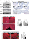
Stat3 plays a key role in the regenerative response in the neonatal mouse heart. (A) Western blotting analyses of phospho-AKT and AKT, phospho-Erk1/2 and Erk1/2, and phospho-STAT3 and STAT3 between the sham and AR hearts at 1, 4 and 7 dpr. (B) Immunostaining for phospho-STAT3 (Tyr705) in the resected, ZA micro-injected and IL-6 micro-injected hearts at 1 day post-surgery in neonatal mouse hearts. The high-magnification views of the boxed areas are presented below. Scale bars, 200 μm. (C) Cardiomyocyte proliferation was identified by immunostaining for pH3 (green) and Ki67 (green) together with α-actinin (red) co-localization in the STAT3+/+ and STAT3−/− mice at 7 dpr in neonatal mice. Arrows indicate cardiomyocytes positive for pH3 or Ki67 staining. The inset is a high-magnification image of a pH3- or Ki67-positive cardiomyocyte which is denoted by an arrowhead. Scale bars, 75 μm. (D) The newly formed cardiomyocytes were shown as Brdu and α-actinin double-positive cells following a pulse-chase experiment in STAT3+/+ and STAT3−/− hearts at 21 dpr. Magnification images of the Brdu+ cardiomyocyte (arrow) are shown on the right. Scale bars, 25 μm. CMs, cardiomyocytes; n = 3 per group; values are presented as the mean ± SEM; *P < 0.05.
Figure 6
Figure 6
The pro-proliferative activity of IL-6 on neonatal cardiomyocytes is mediated by cardiomyocyte STAT3 signaling. (A) Western blotting analyses of phospho-STAT3 (Tyr705) (p-STAT3) and STAT3 after in vitro treatment of neonatal rat ventricular cardiomyocytes with DMSO, IL-6 or both IL-6 and S3I-201 (STAT3 inhibitor). (B, C) Immunofluorescence analysis of pH3 (green) and α-actinin (red) co-localization, and Ki67 (green) and α-actinin (red) co-localization in response to IL-6 or both IL-6 and S3I-201 administration in vitro in neonatal ventricular cardiomyocytes of rats. CMs, cardiomyocytes. Scale bars, 75 μm. Values are presented as the mean ± SEM; *P < 0.05.

References

    1. 1Ptaszek LM, Mansour M, Ruskin JN, Chien KR. Towards regenerative therapy for cardiac disease. Lancet 2012; 379:933–942. - PubMed
    1. 2Xin M, Olson EN, Bassel-Duby R. Mending broken hearts: cardiac development as a basis for adult heart regeneration and repair. Nat Rev Mol Cell Biol 2013; 14:529–541. - PMC - PubMed
    1. 3Bergmann O, Bhardwaj RD, Bernard S, et al. Evidence for cardiomyocyte renewal in humans. Science 2009; 324:98–102. - PMC - PubMed
    1. 4Senyo SE, Steinhauser ML, Pizzimenti CL, et al. Mammalian heart renewal by pre-existing cardiomyocytes. Nature 2013; 493:433–436. - PMC - PubMed
    1. 5Ali SR, Hippenmeyer S, Saadat LV, Luo L, Weissman IL, Ardehali R. Existing cardiomyocytes generate cardiomyocytes at a low rate after birth in mice. Proc Natl Acad Sci USA 2014; 111:8850–8855. - PMC - PubMed

Publication types

MeSH terms