. 2015 Oct;130(4):511-23.
doi: 10.1007/s00401-015-1475-3. Epub 2015 Sep 10.

Frontotemporal dementia caused by CHMP2B mutation is characterised by neuronal lysosomal storage pathology

Collaborators, Affiliations

Frontotemporal dementia caused by CHMP2B mutation is characterised by neuronal lysosomal storage pathology

Emma L Clayton et al. Acta Neuropathol. 2015 Oct.

Abstract

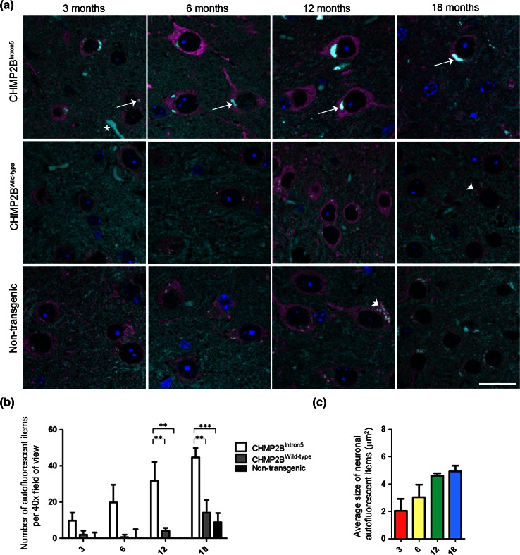
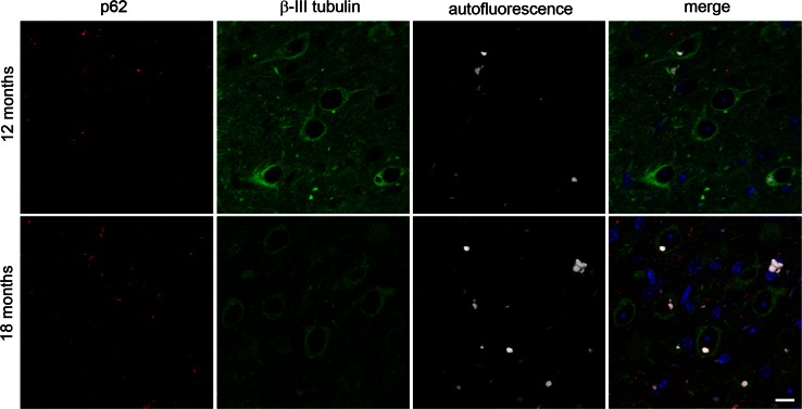
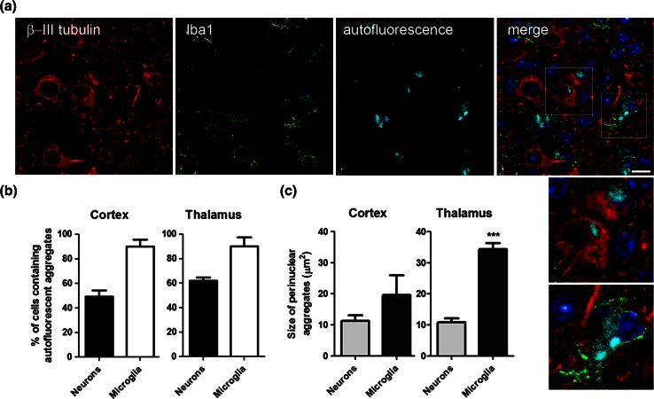
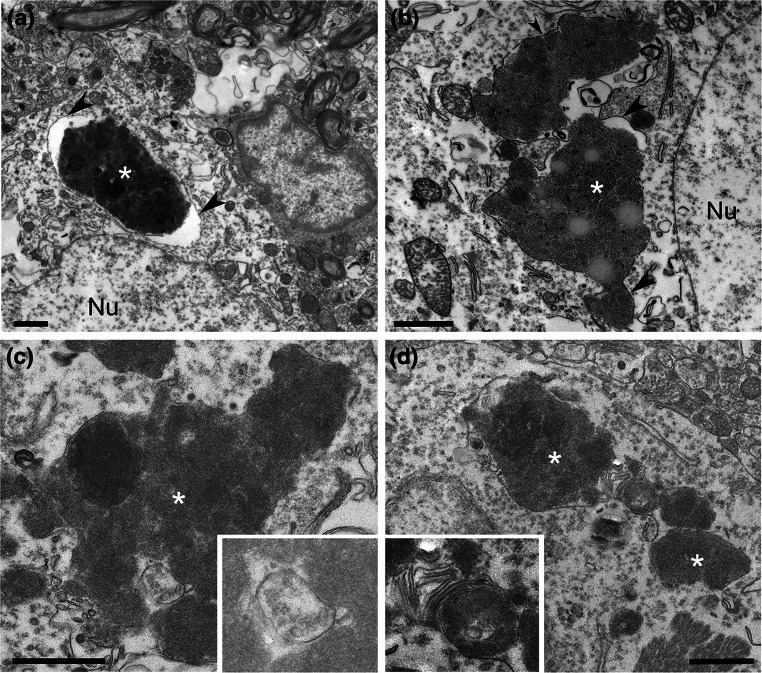
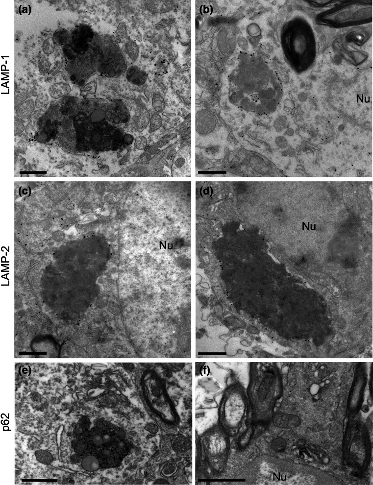
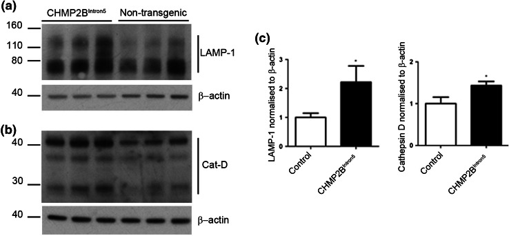
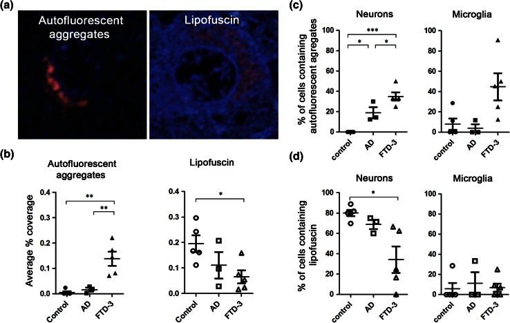
Mutations in the charged multivesicular body protein 2B (CHMP2B) cause frontotemporal dementia (FTD). We report that mice which express FTD-causative mutant CHMP2B at physiological levels develop a novel lysosomal storage pathology characterised by large neuronal autofluorescent aggregates. The aggregates are an early and progressive pathology that occur at 3 months of age and increase in both size and number over time. These autofluorescent aggregates are not observed in mice expressing wild-type CHMP2B, or in non-transgenic controls, indicating that they are a specific pathology caused by mutant CHMP2B. Ultrastructural analysis and immuno- gold labelling confirmed that they are derived from the endolysosomal system. Consistent with these findings, CHMP2B mutation patient brains contain morphologically similar autofluorescent aggregates. These aggregates occur significantly more frequently in human CHMP2B mutation brain than in neurodegenerative disease or age-matched control brains. These data suggest that lysosomal storage pathology is the major neuronal pathology in FTD caused by CHMP2B mutation. Recent evidence suggests that two other genes associated with FTD, GRN and TMEM106B are important for lysosomal function. Our identification of lysosomal storage pathology in FTD caused by CHMP2B mutation now provides evidence that endolysosomal dysfunction is a major degenerative pathway in FTD.

Keywords: CHMP2B; ESCRT; FTD; Lysosomal storage disorder; Lysosome.


Figures

Fig. 1
Fig. 1
Progressive accumulation of autofluorescent aggregates in CHMP2BIntron5 mice. a Spectrally unmixed images showing autofluorescent aggregates in the thalamus of CHMP2BIntron5 mice, but not in CHMP2BWild-type or non-transgenic control samples at 3, 6, 12 or 18 months of age. Nuclei are stained with DAPI (blue), neurons with β-III tubulin (magenta) and autofluorescence is shown in cyan. Arrows indicate autofluorescent aggregates, arrowheads indicate lipofuscin and an asterisk indicates a blood vessel. Scale bar 20 μm. b Quantification of the number of autofluorescent items in the thalamus of CHMP2BIntron5, CHMP2BWild-type and non-transgenic control mice. c Average size of neuronal autofluorescent items at 3, 6, 12 and 18 months in the thalamus of CHMP2BIntron5 mice. Data are shown as mean ± SEM. Significance was determined using a two-way ANOVA, with post hoc Bonferroni test. *p < 0.05, **p < 0.01, ***p < 0.001. All statistically significant differences are shown
Fig. 2
Fig. 2
Autofluorescent aggregates and p62-positive inclusions are distinct pathologies. Spectrally unmixed images of p62 staining (red) and autofluorescence (white) in the thalamus of 12- and 18-month-old CHMP2BIntron5 mice shows little overlap between p62 and autofluorescence. Neurons are stained with β-III tubulin (green), and nuclei with DAPI (blue). Scale bar 10 μm
Fig. 3
Fig. 3
Autofluorescent aggregates are found in neurons and microglia in CHMP2BIntron5 mice. a Cell-type specific staining in the thalamus of 18-month-old CHMP2BIntron5 brain shows that autofluorescent aggregates (cyan) can be localised to neurons (β-III tubulin—red) or microglia (Iba1—green). Nuclei are stained with DAPI (blue). The dotted boxes in the merged image are enlarged in the panel below. Scale bar 10 μm. b Quantification of the percentage of neurons or microglia containing autofluorescent aggregates in the cortex and the thalamus of 18-month CHMP2BIntron5 mice. c Quantification of the size of perinuclear autofluorescent aggregates in neurons or microglia from the cortex and the thalamus of 18-month CHMP2BIntron5 mice. Data are shown as mean ± SEM. Significance was determined using a Student’s t test. ***p < 0.001. All statistically significant differences are shown
Fig. 4
Fig. 4
CHMP2BIntron5 mouse brains display large intraneuronal deposits. 18-month CHMP2BIntron5 mouse brains were prepared for conventional EM. Four examples of large deposits are shown (asterisks ad), located next to large neuronal nuclei (Nu). These deposits were often single membrane-bound (arrowheads) and occasionally contained features suggestive of endocytic origin, such as multivesicular bodies (c inset) or lamellar membrane whorls typical of late endosome/lysosomes (d inset). Scale bars 1 μm
Fig. 5
Fig. 5
Intraneuronal deposits are of endolysosomal origin. 18-month CHMP2BIntron5 mouse brains were prepared for pre-embedding immuno-EM. Neuronal deposits stained positively for LAMP-1 (a, b) or LAMP-2 (c, d), but not for p62 (e), although p62-positive structures could be observed in oligodendrocytes (f). Nu nucleus. Scale bars 1 μm
Fig. 6
Fig. 6
Accumulation of lysosomal proteins in CHMP2BIntron5 mouse brain. 18-month-old CHMP2BIntron5 mouse brain homogenates were immunoblotted for components of the lysosome known to accumulate in lysosomal storage disorders. a LAMP-1 and b cathepsin D were both found to be elevated in aged CHMP2BIntron5 mouse brains compared to non-transgenic controls (c). Data are shown as mean ± SEM. Significance was determined using a Student’s t test. *p < 0.05. Actin blots confirm equal loading
Fig. 7
Fig. 7
Autofluorescent aggregates are a characteristic pathology of FTD-3 patient brain. Autofluorescent pathology in frontal cortex tissue from FTD-3, AD and neurologically normal control brains was investigated. a Examples of an autofluorescent aggregate in FTD-3 patient frontal cortex, and the morphologically distinct granular lipofuscin in control cortex. DAPI labels the nuclei (blue). b Blinded quantification of the coverage of autofluorescent aggregates or lipofuscin per field of view in control, AD or FTD-3 patient frontal cortex. Percentage of either neurons or microglia containing autofluorescence (c) or lipofuscin (d). Data are shown as individual symbols per case with bars representing mean ± SEM. Closed symbols autofluorescent aggregates, open symbols lipofuscin. Significance was determined using a one-way ANOVA, with post hoc Bonferroni test. *p < 0.05, **p < 0.01, ***p < 0.001. All statistically significant differences are shown

References

    1. Ahmed Z, Sheng H, Xu YF, Lin WL, Innes AE, Gass J, Yu X, Hou H, Chiba S, Yamanouchi K, Leissring M, Petrucelli L, Nishihara M, Hutton ML, McGowan E, et al. Accelerated lipofuscinosis and ubiquitination in granulin knockout mice suggest a role for progranulin in successful aging. Am J Pathol. 2010;177:311–324. doi: 10.2353/ajpath.2010.090915. - DOI - PMC - PubMed
    1. Belly A, Bodon G, Blot B, Bouron A, Sadoul R, Goldberg Y. CHMP2B mutants linked to frontotemporal dementia impair maturation of dendritic spines. J Cell Sci. 2010;123:2943–2954. doi: 10.1242/jcs.068817. - DOI - PMC - PubMed
    1. Bennion CJ, Pickering-Brown SM. Pathogenesis/genetics of frontotemporal dementia and how it relates to ALS. Exp Neurol. 2014;262 Pt B:84–90. doi: 10.1016/j.expneurol.2014.06.001. - DOI - PMC - PubMed
    1. Benussi L, Binetti G, Sina E, Gigola L, Bettecken T, Meitinger T, Ghidoni R. A novel deletion in progranulin gene is associated with FTDP-17 and CBS. Neurobiol Aging. 2008;29:427–435. doi: 10.1016/j.neurobiolaging.2006.10.028. - DOI - PubMed
    1. Berg TO, Fengsrud M, Stromhaug PE, Berg T, Seglen PO. Isolation and characterization of rat liver amphisomes. Evidence for fusion of autophagosomes with both early and late endosomes. J Biol Chem. 1998;273:21883–21892. doi: 10.1074/jbc.273.34.21883. - DOI - PubMed

Publication types

MeSH terms

Substances

LinkOut - more resources