. 2015 Sep 11:5:13886.
doi: 10.1038/srep13886.

Non-Classical monocytes display inflammatory features: Validation in Sepsis and Systemic Lupus Erythematous

Affiliations

Non-Classical monocytes display inflammatory features: Validation in Sepsis and Systemic Lupus Erythematous

Ratnadeep Mukherjee et al. Sci Rep. .

Abstract

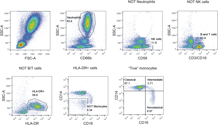
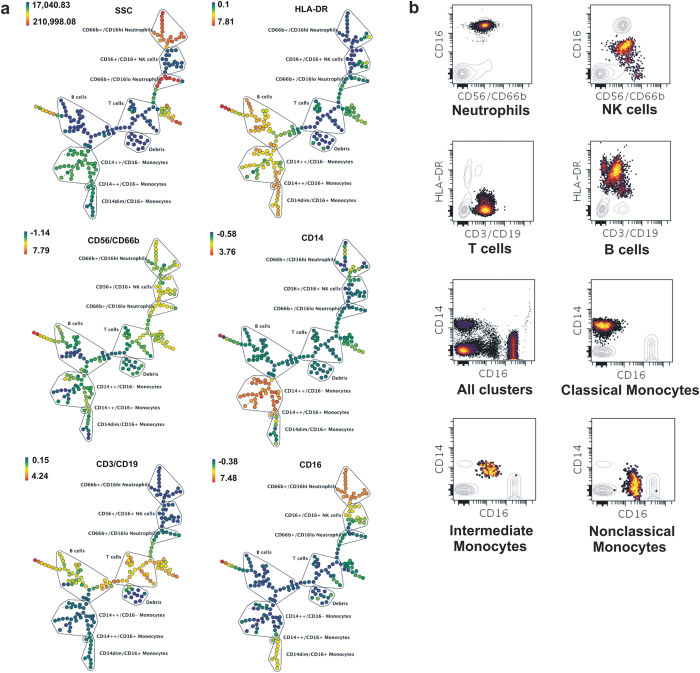
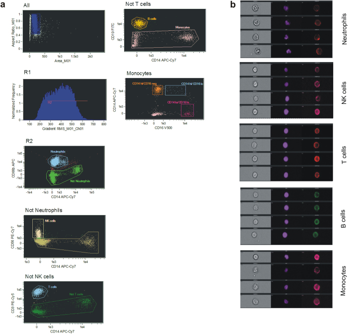
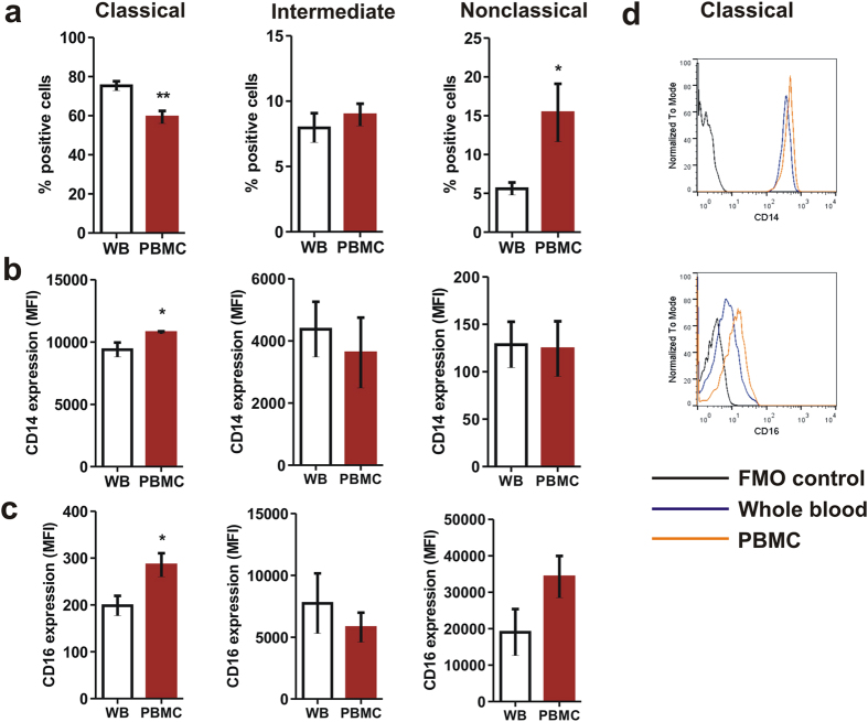
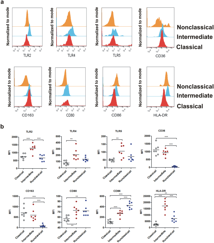
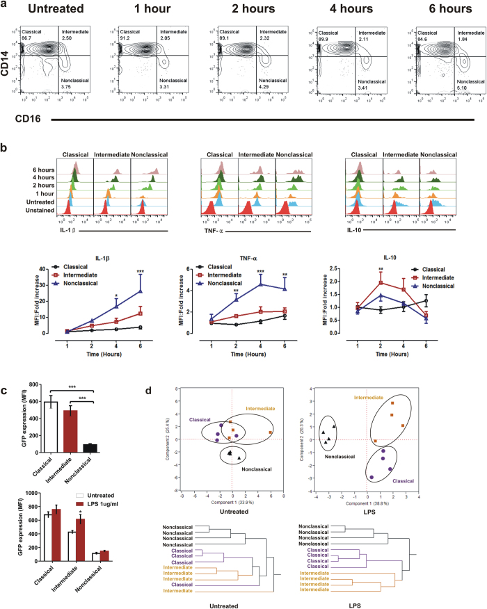
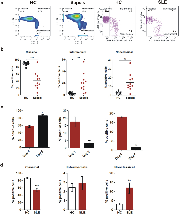
Given the importance of monocytes in pathogenesis of infectious and other inflammatory disorders, delineating functional and phenotypic characterization of monocyte subsets has emerged as a critical requirement. Although human monocytes have been subdivided into three different populations based on surface expression of CD14 and CD16, published reports suffer from contradictions with respect to subset phenotypes and function. This has been attributed to discrepancies in reliable gating strategies for flow cytometric characterization and purification protocols contributing to significant changes in receptor expression. By using a combination of multicolour flow cytometry and a high-dimensional automated clustering algorithm to confirm robustness of gating strategy and analysis of ex-vivo activation of whole blood with LPS we demonstrate the following: a. 'Classical' monocytes are phagocytic with no inflammatory attributes, b. 'Non-classical' subtype display 'inflammatory' characteristics on activation and display properties for antigen presentation and c. 'Intermediate' subtype that constitutes a very small percentage in circulation (under physiological conditions) appear to be transitional monocytes that display both phagocytic and inflammatory function. Analysis of blood from patients with Sepsis, a pathogen driven acute inflammatory disease and Systemic Lupus Erythmatosus (SLE), a chronic inflammatory disorder validated the broad conclusions drawn in the study.


Figures

Figure 1
Figure 1. Identification of blood monocyte subsets by polychromatic flow cytometry.
Gating strategy for identification of monocyte subsets showing successive exclusion of neutrophils, NK cells, B and T cells on conventional bivariate scatterplots of side scatter signal vs. exclusion marker specific for cell type. Remaining population was further selected for HLA-DR which was discriminated on a CD14 vs. CD16 scatterplot to give three monocyte subsets.
Figure 2
Figure 2. SPADE analysis of circulating immune cells showing validity of manual gating.
Confirmation of manual gating strategy by SPADE implemented as a web-based extension of Cytobank. Number of nodes was set at 200 with 10 per cent downsampling of events. (a) Each “bubble” on a given tree corresponds to a particular cell-type that was selected according to expression of its lineage marker as indicated by the colour code (expression decreasing from red to blue). (b) Associated bivariate scatterplots confirm the gating strategy by highlighting only a selected bubble at a time while greying out other populations.
Figure 3
Figure 3. Multispectral imaging cytometry based visualization and identification of circulating immune cells.
(a) Single cells were selected first (R1) followed by gating of cells in focus (R2). Thereafter, stepwise negative selection of nonmonocyte populations was done to finally include “true” monocytes. (b) Representative brightfield (left), nuclear (middle) and surface antigen (right) images of each different cell type as gated in (a) confirming individual cell types based on nuclear morphology, brightfield image and lineage marker expression.
Figure 4
Figure 4. Gradient purification of PBMCs from whole blood leads to changes in monocyte phenotype.
Comparison of monocyte subsets between whole blood monocytes and ficoll purified monocytes showing (a) significant decrease in proportion of ‘classical’ monocytes and increase in ‘nonclassical’ monocytes as a consequence of gradient purification that also led to increase in (b) CD14 and (c) CD16 expression on classical monocytes. Statistical significance was ascertained using a paired t-test. (d) Representative overlaid histograms showing increased expression of CD14 and CD16 on classical monocyte surface as a consequence of Ficoll purification. Data are mean ± SEM of five normal subjects. *P < 0.05, **P < 0.01. MFI: Median Fluorescence Intensity.
Figure 5
Figure 5. Differential expression of surface receptors on monocyte subsets.
(a) Representative overlaid histograms of eight different surface markers showing differential expression between monocyte subsets. Histograms were created using FlowJo software. (b) Different monocyte subtypes showing expression of a specific repertoire of surface antigens. Assessment of statistical significance was performed by one-way ANOVA followed by Bonferroni’s post-test. Data are mean ± S.E.M. of seven normal subjects. *P < 0.05, **P < 0.01, ***P < 0.001. MFI: Median Fluorescence Intensity.
Figure 6
Figure 6. Functional analysis of human monocyte subsets.
(a) Representative contour plots of monocyte subsets showing unchanged relative percentages even after 6 hours of LPS stimulation. (b) Analysis of cytokine production by monocyte subsets. Whole blood was left untreated or treated with LPS for the indicated time points along with Brefeldin A at a final concentration of 3 μg/ml to prevent secretion and stained for intracellular cytokines along with surface markers for exclusion gating as described previously. Brefeldin A was added one hour prior to termination of culture at each time point. Top panel shows representative overlaid histograms whereas bottom panel data are represented as mean ± SEM of normalized fold change over untreated samples obtained from five donors. *P < 0.05, **P < 0.01, ***P < 0.001 from one-way ANOVA followed by Bonferroni’s post-test. MFI: Median Fluorescence Intensity. (c) Differential phagocytic function of monocyte subsets as demonstrated by uptake of GFP-expressing E.coli under normal conditions as well as post activation with LPS. Whole blood was incubated with or without live bacteria for 30 minutes followed by staining for surface markers. Statistical significance was assessed either by one-way (left panel) or two-way (right panel) ANOVA followed by Bonferroni’s post-test. *P < 0.05, **P < 0.01, ***P < 0.001. (d) Principal components analysis and hierarchical clustering of untreated and LPS stimulated monocytes showing relationship between three monocyte subsets. Median fluorescence intensities obtained from CD14, CD16, TLR2, TLR4, TLR5, CD80, CD86, CD36, CD163, HLA-DR, IL-1β, TNF-α and IL-10 staining were used as parameters for principal components analysis and clustering.
Figure 7
Figure 7. Expansion of CD16 positive monocytes during inflammatory diseases.
(a) Representative bivariate scatterplots of monocyte subset percentage showing comparison of sepsis and SLE patients with their respective age and sex matched healthy controls. (b) Comparison of monocyte subsets (percentage) between healthy subjects (n = 9) and Sepsis patients (n = 11). (b) Analysis of monocyte subsets on Day 1 and Day 5 (post treatment) of Sepsis patients (n = 3). (c) Comparison of monocyte subsets (percentage) between healthy subjects (n = 13) and SLE patients (n = 10). *P < 0.05, **P < 0.01, ***P < 0.001 assessed by unpaired t-test.

References

    1. Auffray C., Sieweke M. H. & Geissmann F. Blood monocytes: development, heterogeneity, and relationship with dendritic cells. Annu Rev Immunol 27, 669–692 (2009). - PubMed
    1. Gordon S. & Taylor P. R. Monocyte and macrophage heterogeneity. Nat Rev Immunol 5, 953–964 (2005). - PubMed
    1. Chow A., Brown B. D. & Merad M. Studying the mononuclear phagocyte system in the molecular age. Nat Rev Immunol 11, 788–798 (2011). - PubMed
    1. Hume D. A. The mononuclear phagocyte system. Curr Opin Immunol 18, 49–53 (2006). - PubMed
    1. Hume D. A. et al. The mononuclear phagocyte system revisited. J Leukoc Biol 72, 621–627 (2002). - PubMed

Publication types