Modulation of cyclins and p53 in mesangial cell proliferation and apoptosis during Habu nephritis
- PMID: 26359229
- PMCID: PMC4819602
- DOI: 10.1007/s10157-015-1163-6
Modulation of cyclins and p53 in mesangial cell proliferation and apoptosis during Habu nephritis
Abstract
Background: Mesangial cell (MC) proliferation and apoptosis are the main pathological changes observed in mesangial proliferative nephritis. In this study, we explored the role of cyclins and p53 in modulating MC proliferation and apoptosis in a mouse model of Habu nephritis.
Methods: The Habu nephritis group was prepared by injection of Habu toxin. Mesangiolysis and mesangial expansion were determined by periodic acid-Schiff (PAS) reagent staining. Immunohistochemical analysis of PCNA and KI67, and TUNEL staining were used to detect cell proliferation and apoptosis, respectively. Expression levels of cyclins and p53 were examined by Western blotting.
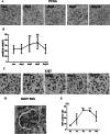
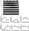
Results: PAS staining showed that mesangial dissolution appeared on days 1 and 3, and mesangial proliferation with extracellular matrix accumulation was apparent by days 7 and 14. Both PCNA and KI67 immunohistochemical analysis showed that MC proliferation began on day 3, peaked on day 3 and 7, and recovered by day 14. TUNEL staining results showed that MC apoptosis began to increase on day 1, continued to rise on day 7, and peaked on day 14. Western blot analysis showed that cyclin D1 was upregulated on day 1, cyclins A2 and E were upregulated on days 3 and 7, and p53 was upregulated on days 3, 7 and 14. There was no change in the expression levels of Bax or p21.
Conclusion: We explored the tendency for MC proliferation and apoptosis during the process of Habu nephritis and found that cyclins and p53 may modulate the disease pathology. This will help us determine the molecular pathogenesis of MC proliferation and provide new targets for disease intervention.
Keywords: Cell cycle; Cyclins; Mesangial proliferative nephritis; p53.
Figures


Similar articles
-
CXCL10 expression induced by Mxi1 inactivation induces mesangial cell apoptosis in mouse Habu nephritis.Cell Signal. 2015 May;27(5):943-50. doi: 10.1016/j.cellsig.2015.01.019. Epub 2015 Feb 12. Cell Signal. 2015. PMID: 25683914
-
Hydrogen peroxide-inducible clone-5 regulates mesangial cell proliferation in proliferative glomerulonephritis in mice.PLoS One. 2015 Apr 2;10(4):e0122773. doi: 10.1371/journal.pone.0122773. eCollection 2015. PLoS One. 2015. PMID: 25835392 Free PMC article.
-
Knockdown of Cxcl10 Inhibits Mesangial Cell Proliferation in Murine Habu Nephritis Via ERK Signaling.Cell Physiol Biochem. 2017;42(5):2118-2129. doi: 10.1159/000479914. Epub 2017 Aug 15. Cell Physiol Biochem. 2017. PMID: 28810249
-
Effect of simvastatin on proliferative nephritis and cell-cycle protein expression.Kidney Int Suppl. 1999 Jul;71:S84-7. doi: 10.1046/j.1523-1755.1999.07121.x. Kidney Int Suppl. 1999. PMID: 10412745 Review.
-
Cell biology of mesangial cells: the third cell that maintains the glomerular capillary.Anat Sci Int. 2017 Mar;92(2):173-186. doi: 10.1007/s12565-016-0334-1. Epub 2016 Feb 24. Anat Sci Int. 2017. PMID: 26910209 Review.
Cited by
-
The Xanthine Oxidase Inhibitor Febuxostat Suppresses the Progression of IgA Nephropathy, Possibly via Its Anti-Inflammatory and Anti-Fibrotic Effects in the gddY Mouse Model.Int J Mol Sci. 2018 Dec 10;19(12):3967. doi: 10.3390/ijms19123967. Int J Mol Sci. 2018. PMID: 30544662 Free PMC article.
-
Gene expression profiling analysis reveals that the long non‑coding RNA uc.412 is involved in mesangial cell proliferation.Mol Med Rep. 2019 Dec;20(6):5297-5303. doi: 10.3892/mmr.2019.10753. Epub 2019 Oct 16. Mol Med Rep. 2019. PMID: 31638227 Free PMC article.
-
Application of herbal traditional Chinese medicine in the treatment of lupus nephritis.Front Pharmacol. 2022 Nov 24;13:981063. doi: 10.3389/fphar.2022.981063. eCollection 2022. Front Pharmacol. 2022. PMID: 36506523 Free PMC article. Review.
-
Paeoniflorin Inhibits Mesangial Cell Proliferation and Inflammatory Response in Rats With Mesangial Proliferative Glomerulonephritis Through PI3K/AKT/GSK-3β Pathway.Front Pharmacol. 2019 Sep 9;10:978. doi: 10.3389/fphar.2019.00978. eCollection 2019. Front Pharmacol. 2019. PMID: 31551783 Free PMC article.
-
TUNEL Assay: A Powerful Tool for Kidney Injury Evaluation.Int J Mol Sci. 2021 Jan 2;22(1):412. doi: 10.3390/ijms22010412. Int J Mol Sci. 2021. PMID: 33401733 Free PMC article. Review.
References
-
- Ah E. Mesangial cell biology. Exp Cell Res. 2012;318(9):978–985. - PubMed
-
- Hartner A, Marek I, Cordasic N, Haas C, Schocklmann H, Hulsmann-Volkert G, et al. Glomerular regeneration is delayed in nephritic alpha 8-integrin-deficient mice: contribution of alpha 8-integrin to the regulation of mesangial cell apoptosis. Am J Nephrol. 2008;28(1):168–178. doi: 10.1159/000110022. - DOI - PubMed
Publication types
MeSH terms
Substances
LinkOut - more resources
Full Text Sources
Other Literature Sources
Research Materials
Miscellaneous

