Comparative Study
. 2015 Oct 13;6(31):31263-71.
doi: 10.18632/oncotarget.5075.

Acceleration of atherogenesis in ApoE-/- mice exposed to acute or low-dose-rate ionizing radiation

Affiliations
Comparative Study

Acceleration of atherogenesis in ApoE-/- mice exposed to acute or low-dose-rate ionizing radiation

Mariateresa Mancuso et al. Oncotarget. .

Abstract

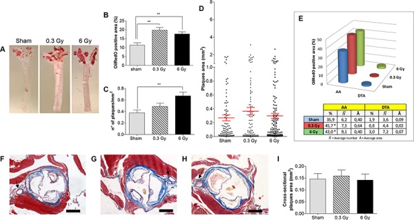
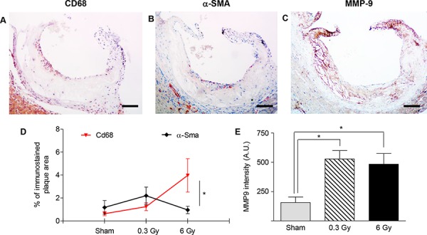
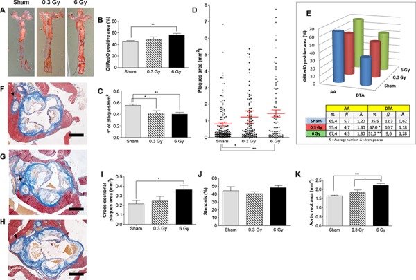
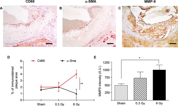
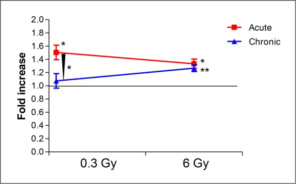
There is epidemiological evidence for increased non-cancer mortality, primarily due to circulatory diseases after radiation exposure above 0.5 Sv. We evaluated the effects of chronic low-dose rate versus acute exposures in a murine model of spontaneous atherogenesis. Female ApoE-/- mice (60 days) were chronically irradiated for 300 days with gamma rays at two different dose rates (1 mGy/day; 20 mGy/day), with total accumulated doses of 0.3 or 6 Gy. For comparison, age-matched ApoE-/- females were acutely exposed to the same doses and sacrificed 300 days post-irradiation. Mice acutely exposed to 0.3 or 6 Gy showed increased atherogenesis compared to age-matched controls, and this effect was persistent. When the same doses were delivered at low dose rate over 300 days, we again observed a significant impact on global development of atherosclerosis, although at 0.3 Gy effects were limited to the descending thoracic aorta. Our data suggest that a moderate dose of 0.3 Gy can have persistent detrimental effects on the cardiovascular system, and that a high dose of 6 Gy poses high risks at both high and low dose rates. Our results were clearly nonlinear with dose, suggesting that lower doses may be more damaging than predicted by a linear dose response.

Keywords: ApoE mice; aorta; atherosclerosis; radiation.


Conflict of interest statement

CONFLICTS OF INTEREST

No, there is no conflict of interest that I should disclose, having read the above statement.

Figures

Figure 1
Figure 1. Acute irradiation
A. Representative en face preparations of aortas from female ApoE−/− mice 300 days after acute irradiation with 0.3 Gy, 6 Gy and from age-matched controls. B–D. Morphometric analyses performed on digital images from en face preparations of aortas (n = 8) representing percentage of ORO-stained area (B), plaques density (C) and plaques size (D). E. Regional distribution of plaques. F–H. Representative aortic root sections from sham- (F), 0.3 Gy- (G) and 6 Gy-irradiated mice (H) (Masson's trichrome stain). I. Graphic representation of plaques area (n = 18) measured on aortic root cross-sections. Data are shown as mean ± SEM. Differences were tested with Student's t-test. **P < 0.001. Arrows: coronary artery. Bars: 500 μm.
Figure 2
Figure 2. Plaques vulnerability after acute irradiation
A–C. Representative sections of atherosclerotic plaques immunostained with antibodies against CD68 (A), α-SMA (B) and MMP-9 (C). Images refer to ApoE−/− mice 300 days after acute irradiation with 6 Gy. D. Mean percentage of total plaque area occupied by CD68- or α-SMA-positive cells. E. Intensity measurement of anti-MMP9 immunohistochemical staining by HistoQUEST software. Quantitative analysis involved 18 plaques/group. Data are shown as mean ± SEM. Differences were tested with Student's t-test. *P < 0.05. Bars: 100 μm.
Figure 3
Figure 3. Chronic irradiation
A. Representative ORO-stained aortas from female ApoE−/− mice after chronic irradiation with 0.3 Gy or 6 Gy over 300 days, and age-matched controls. Graphic representation of quantitative analyses performed on digital images from en face preparations of aortas (n = 8) showing: B. Percentage of ORO-stained aortic area. C. Plaque density. D. Plaques size. E. Regional distribution. F–H. Masson's trichrome staining of aortic root cross-sections from each experimental group. I–K. Dimensional analyses of plaques area (I), aortic stenosis (J) and aortic total area (K). Data are shown as mean ± SEM. Differences were tested with Student's t-test. *P < 0.05; **P < 0.001; ***P < 0.0001. Arrows: coronary artery. Bars: 500 μm.
Figure 4
Figure 4. Plaques vulnerability after chronic irradiation
A–C. Representative sections of atherosclerotic plaques after chronic irradiation with 6 Gy using antibodies anti-CD68 (A), α-SMA (B) and MMP-9 (C) D. Mean percentage of total plaque area occupied by CD68- or α-SMA-positive cells. E. Intensity measurement of immunohistochemical staining with anti-MMP9 by HistoQUEST software. Quantitative analysis involved 18 plaques/group. Data are shown as mean ± SEM. Differences were tested with Student's t-test. *P < 0.05; ***P < 0.0001. Bars: 100 μm.
Figure 5
Figure 5. Comparison of fold increases of percent ORO-stained aortic area over sham-irradiated controls between high (x rays) and low dose-rate (gamma rays) groups
Assuming a quality factor of 1 for x rays and gamma rays, a high radiation dose (6 Gy) would be equally efficient in enhancement of atherogenesis at both high and low dose rate. In contrast, a moderate dose (0.3 Gy) could significantly enhance atherogenesis only when delivered acutely (P = 0.0143, acute versus sham irradiated). When comparing fold increases, mice receiving 0.3 Gy acutely have enhanced atherogenesis compared with mice receiving the same dose as a chronic exposure (P = 0.0298).

References

    1. Little MP, Gola A, Tzoulaki I. A model of cardiovascular disease giving a plausible mechanism for the effect of fractionated low-dose ionizing radiation exposure. PLoS Comput Biol. 2009;5:e1000539. - PMC - PubMed
    1. Adams MJ, Hardenbergh PH, Constine LS, Lipshultz SE. Radiation-associated cardiovascular disease. Crit Rev Oncol Hematol. 2003;45:55–75. - PubMed
    1. Annals of the ICRP. Early and late effects of radiation in normal tissues and organs: threshold doses for tissue reactions and other non-cancer effects of radiation in a radiation protection context. 2012 public draft. http://www.icrp.org published at ICRP website. - PubMed
    1. Schultz-Hector S, Trott KR. Radiation-induced cardiovascular diseases: is the epidemiologic evidence compatible with the radiobiologic data? Int J Radiat Oncol Biol Phys. 2007;67:10–18. - PubMed
    1. Hendry JH, Akahoshi M, Wang LS, Lipshultz SE, Stewart FA, Trott KR. Radiation-induced cardiovascular injury. Radiat Environ Biophys. 2008;47:189–193. - PubMed

Publication types

MeSH terms

Substances