. 2015 Oct 30;290(44):26856-65.
doi: 10.1074/jbc.M115.683334. Epub 2015 Sep 10.

Structural and functional studies of the Pseudomonas aeruginosa minor pilin, PilE

Affiliations

Structural and functional studies of the Pseudomonas aeruginosa minor pilin, PilE

Ylan Nguyen et al. J Biol Chem. .

Abstract

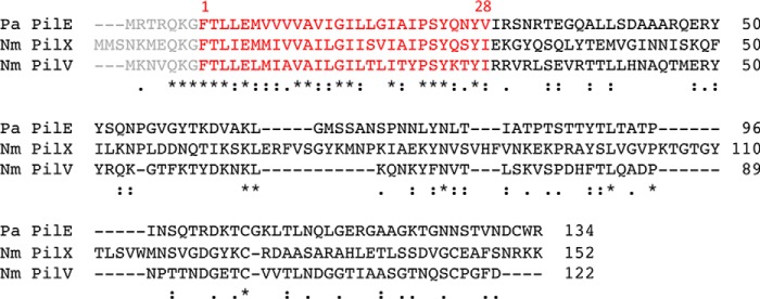
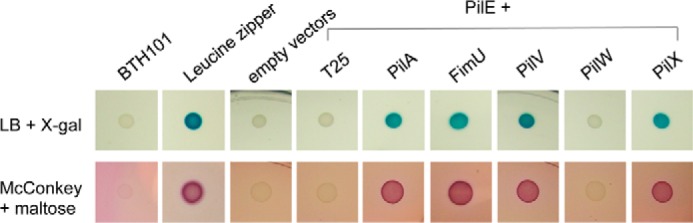
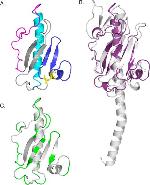
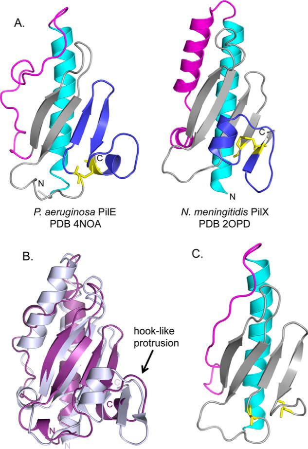
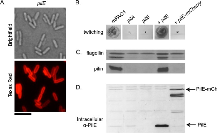
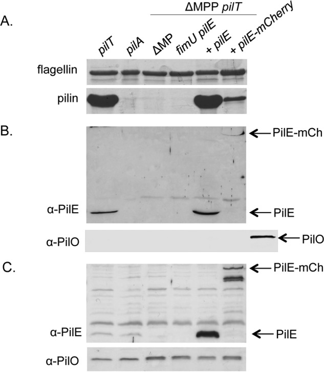
Many bacterial pathogens, including Pseudomonas aeruginosa, use type IVa pili (T4aP) for attachment and twitching motility. T4aP are composed primarily of major pilin subunits, which are repeatedly assembled and disassembled to mediate function. A group of pilin-like proteins, the minor pilins FimU and PilVWXE, prime pilus assembly and are incorporated into the pilus. We showed previously that minor pilin PilE depends on the putative priming subcomplex PilVWX and the non-pilin protein PilY1 for incorporation into pili, and that with FimU, PilE may couple the priming subcomplex to the major pilin PilA, allowing for efficient pilus assembly. Here we provide further support for this model, showing interaction of PilE with other minor pilins and the major pilin. A 1.25 Å crystal structure of PilEΔ1-28 shows a typical type IV pilin fold, demonstrating how it may be incorporated into the pilus. Despite limited sequence identity, PilE is structurally similar to Neisseria meningitidis minor pilins PilXNm and PilVNm, recently suggested via characterization of mCherry fusions to modulate pilus assembly from within the periplasm. A P. aeruginosa PilE-mCherry fusion failed to complement twitching motility or piliation of a pilE mutant. However, in a retraction-deficient strain where surface piliation depends solely on PilE, the fusion construct restored some surface piliation. PilE-mCherry was present in sheared surface fractions, suggesting that it was incorporated into pili. Together, these data provide evidence that PilE, the sole P. aeruginosa equivalent of PilXNm and PilVNm, likely connects a priming subcomplex to the major pilin, promoting efficient assembly of T4aP.

Keywords: Neisseria; Neisseria meningitidis; PilC1; PilY1; Pseudomonas aeruginosa (P. aeruginosa); X-ray crystallography; bacterial adhesion; bacterial pathogenesis; bacterial two-hybrid; minor pilins; pilus assembly; protein-protein interaction; twitching motility; type IV pili.


Figures

FIGURE 1.
FIGURE 1.
Sequence alignment of PilE with PilXNm and PilVNm. Sequence alignment of P. aeruginosa PilE from strain PAO1 and N. meningitidis PilX and PilV from strain 8013 using MUSCLE (49) is shown. Numbering is according to the mature pilin, with the leader sequence shown in gray. The first 28 residues that were removed for structural studies are colored red. Pa, P. aeruginosa.
FIGURE 2.
FIGURE 2.
Interactions of PilE with minor pilins and PilA. Protein-protein interactions were tested using a bacterial adenylate cyclase two-hybrid system (BACTH). Mature PilE was N-terminally tagged with T18, whereas PilA, FimU, PilV, PilW, and PilX were N-terminally tagged with T25. Interactions were tested in E. coli cya mutant strain BTH101 on LB agar + X-gal and MacConkey + maltose indicator plates, which result in blue or red colonies if there is an interaction. A leucine zipper was used as a positive control.
FIGURE 3.
FIGURE 3.
X-ray crystal structure of PilEΔ1–28. A, the x-ray crystal structure of selenomethionine-labeled PilEΔ1–28 was solved to 1.25 Å (PDB code: 4NOA). The N-terminal α-helix is colored cyan, αβ-loop is in magenta, β-sheet is in gray, and D-region is in blue. Cys residues are represented as sticks and colored yellow. B, structural alignment between PilEΔ1–28 (purple) and PilAPAK (gray, PDB code: 1OQW). 96 residues aligned with an RMSD of 3.8 Å. The arrow indicates the hook-like protrusion in both structures. C, mapping of residues differing between PilEPAO1 and PilEPA14. Non-conservative residues are colored green. Structural illustrations and alignments were generated with PyMOL (version 1.3, Schrödinger, LLC).
FIGURE 4.
FIGURE 4.
Comparison of PilEΔ1–28 with N. meningitidis PilXNm. A, side-by-side comparison of PilEΔ1–28 and PilXNm,Δ1–28 with the N-terminal α-helices colored in cyan, αβ-loops in magenta, β-sheets in gray, and D-regions in blue with the cysteines represented as sticks in yellow. B, structural alignment of PilEΔ1–28 (purple) and PilXNm,Δ1–28 (light blue). 104 residues are aligned with an RMSD of 4.3 Å. C, Phyre2-generated model of PilVNm based on PilE. Structural illustrations and alignments were generated with PyMOL (version 1.3, Schrödinger, LLC.).
FIGURE 5.
FIGURE 5.
Complementation of pilE with PilE mCherry. mCherry was fused to the C-terminal end of PilE, and the level of complementation of a pilE mutant was assessed. A, fluorescence microscopy analysis of PilE mCherry localization. Scale bar represents 5 μm. B, twitching motility was tested by stab-inoculating to the bottom of an LB 1% agar plate and staining with 1% crystal violet after a 24-h incubation at 37 °C. C, pili were sheared from the surface of cells of interest and separated on a 15% SDS-PAGE gel. The flagellin band is used as a loading control. D, intracellular levels of PilE were probed by Western blot analysis with a α-PilE peptide antibody (1:1000 dilution). Arrows indicate the bands of interest.
FIGURE 6.
FIGURE 6.
Pilus assembly by PilE mCherry in a retraction-deficient background. The ability of PilE mCherry to support pilus assembly in a retraction-deficient strain was tested in the ΔfimU pilE ΔMPP pilT mutant that lacks surface piliation in the absence of fimU and pilE. A, pilus assembly was probed by shearing proteins from the surface of the cells and analyzing the surface fractions by SDS-PAGE. The flagellin band is used as a loading control. B, incorporation of PilE mCherry into pili was examined by Western blot analysis of the surface fractions above and probing for PilE using an α-PilE peptide antibody (1:1000). Arrows indicate the bands of interest. The samples were probed with an antibody to inner membrane protein PilO (1:5000) as a control for cell lysis; the last lane is PAO1 lysate as a positive control for PilO. C, intracellular levels of PilE and PilO were probed by Western blot analysis with α-PilE peptide antibody (1:1000 dilution) and α-PilO antibody (1:5000), respectively.

References

    1. Pelicic V. (2008) Type IV pili: e pluribus unum? Mol. Microbiol. 68, 827–837 - PubMed
    1. Albers S. V., and Pohlschröder M. (2009) Diversity of archaeal type IV pilin-like structures. Extremophiles 13, 403–410 - PubMed
    1. Craig L., Pique M. E., and Tainer J. A. (2004) Type IV pilus structure and bacterial pathogenicity. Nat. Rev. Microbiol. 2, 363–378 - PubMed
    1. Ayers M., Howell P. L., and Burrows L. L. (2010) Architecture of the type II secretion and type IV pilus machineries. Future Microbiol. 5, 1203–1218 - PubMed
    1. Mattick J. S. (2002) Type IV pili and twitching motility. Annu. Rev. Microbiol. 56, 289–314 - PubMed

Publication types

MeSH terms

LinkOut - more resources