Review
. 2015:31:699-720.
doi: 10.1146/annurev-cellbio-100814-125353. Epub 2015 Sep 11.

Generating neuronal diversity in the mammalian cerebral cortex

Affiliations
Review

Generating neuronal diversity in the mammalian cerebral cortex

Simona Lodato et al. Annu Rev Cell Dev Biol. 2015.

Abstract

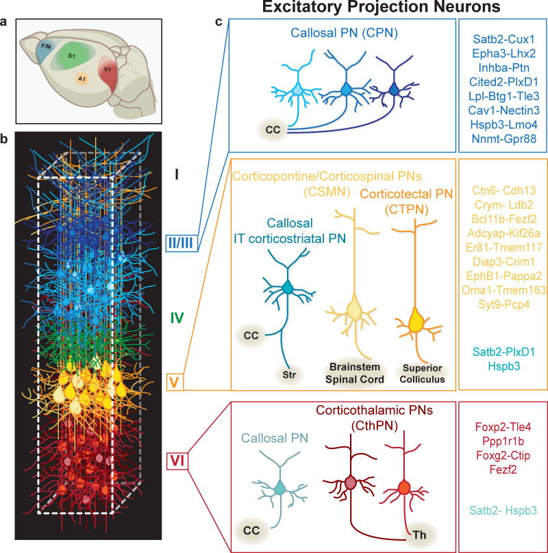
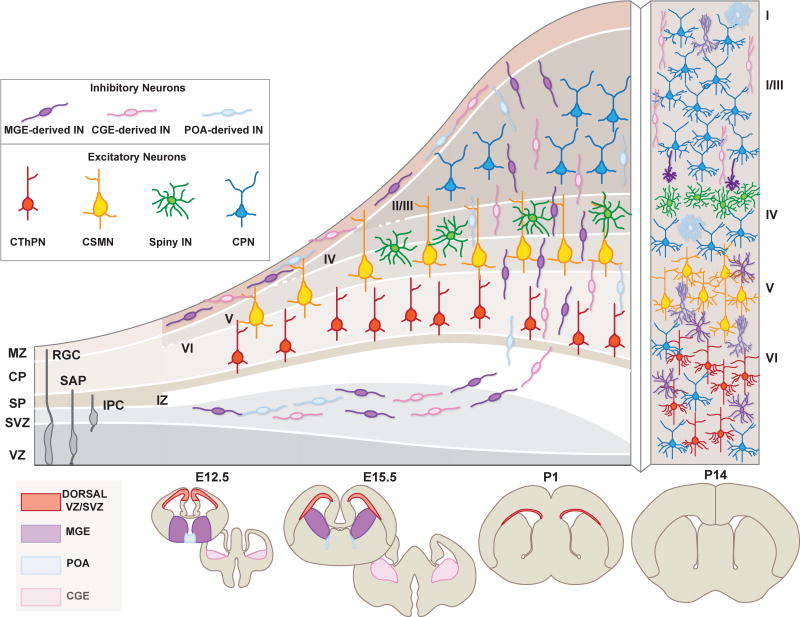
The neocortex is the part of the brain responsible for execution of higher-order brain functions, including cognition, sensory perception, and sophisticated motor control. During evolution, the neocortex has developed an unparalleled neuronal diversity, which still remains partly unclassified and unmapped at the functional level. Here, we broadly review the structural blueprint of the neocortex and discuss the current classification of its neuronal diversity. We then cover the principles and mechanisms that build neuronal diversity during cortical development and consider the impact of neuronal class-specific identity in shaping cortical connectivity and function.

Keywords: cortical networks; identity; interneurons; myelination profile; neocortex; projection neurons.


Figures

Figure 1
Figure 1
The neocortex is organized into areas, layers, and columns populated by a great diversity of excitatory and inhibitory neuronal subtypes. (a) Schematic representation of primary neocortical areas dedicated to processing distinct sensory modalities and governing fine motor control. F/M, frontal/motor cortex; S1, somatosensory cortex; A1, auditory cortex; V1, visual cortex. (b) Cortical columns contain horizontally-arranged layers with very diverse neuronal compositions. Only select examples are depicted here. (c) Layer II/III contains different classes of commissural neurons, primarily of distinct CPN identities. Layer V contains CPNs, often maintaining distinctive collaterals to the striatum (IT type of corticostriatal PNs), and different classes of subcerebral PNs that connect to the brainstem, spinal cord, and superior colliculus. Layer VI has different classes of CThPNs, connecting to separate thalamic nuclei and CPNs that connect through the CC. Cortical PN subtypes express unique gene signatures that in specific combinations identify each class (listed on right). Abbreviations: CC, corpus callosum; Th, thalamus; CPN, callosal projection neuron; CTPN, corticotectal projection neuron; CThPN, corticothalamic; IT, intratelencephalic; PN, projection neuron, IN, interneurons. Roman numerals refer to the six cortical layers.
Figure 2
Figure 2
Neocortical interneurons are characterized by their short-range projections, and can be broadly classified into excitatory and inhibitory interneurons. Here, we depict a schematic representation of the distinct excitatory and inhibitory interneuron classes within the six cortical. Excitatory spiny interneurons display mainly stellate and pyramidal morphology and are primarily located in the intragranular layer IV of the somatosenosry cortex (barrel cortex, shown in yellow boxes). In contrast, each cortical layer contains different types of inhibitory interneurons which display a wide array of morphologies and molecular identities. Both classes can be furher classified into subtypes which express distinctive combinations of molecular markers (listed on right).
Figure 3
Figure 3
Developmental origin and distribution in the neocortex of projection neuron (PN) and interneuron (IN) subtypes. Excitatory neurons originate from progenitors in the dorsal telencephalon, and cortical inhibitory INs derive from progenitors in the ventral telencephalon [mainly the medial and ganglionic eminences (MGE), the caudal ganglionic eminence (CGE), and the preoptic area (POA)]. Over time, these germinal zones give rise to a diversity of neuronal subtypes, both PNs and INs, that acquire distinct laminar addresses in the neocortex. After reaching the cortex, INs migrate tangentially in streams located above [marginal zone (MZ)] and below [subventricular zone (SVZ)] the cortical plate (CP), before switching to a mode of radial migration to invade the CP. By the end of neurogenesis, PN and IN classes coexist at specific locations in the cortical layers and begin to wire into the local cortical microcircuit. Abbreviations: CPN, callosal projection neuron; CThPN, corticothalamic; CSMN, corticospinal motor neuron; RGC, radial glial cell; SAP, subapical progenitor; IPC, intermediate precursor cell; VZ, ventricular zone; SVZ, subventricular zone; SP, subplate; IZ, intermediate zone; CP, cortical plate; MZ, marginal zone. E, embryonic; P, postnatal. Roman numerals refer to the six cortical layers.

References

    1. Aboitiz F, Morales D, Montiel J. The evolutionary origin of the mammalian isocortex: towards an integrated developmental and functional approach. Behav Brain Sci. 2003;26(5):535–52. discussion 552–85. - PubMed
    1. Alcamo EA, Chirivella L, Dautzenberg M, Dobreva G, Farinas I, et al. Satb2 regulates callosal projection neuron identity in the developing cerebral cortex. Neuron. 2008;57(3):364–77. - PubMed
    1. Anderson SA, Eisenstat DD, Shi L, Rubenstein JL. Interneuron migration from basal forebrain to neocortex: dependence on Dlx genes. Science. 1997;278(5337):474–76. - PubMed
    1. Angevine JBJ, Sidman RL. Autoradiographic study of cell migration during histogenesis of cerebral cortex in mouse. Nature. 1961;192:766–68. - PubMed
    1. Arlotta P, Molyneaux BJ, Chen J, Inoue J, Kominami R, Macklis JD. Neuronal subtype-specific genes that control corticospinal motor neuron development in vivo. Neuron. 2005;45(2):207–21. - PubMed

Publication types