Review
. 2015:31:575-91.
doi: 10.1146/annurev-cellbio-100814-125323. Epub 2015 Sep 10.

Polarized protein transport and lumen formation during epithelial tissue morphogenesis

Affiliations
Review

Polarized protein transport and lumen formation during epithelial tissue morphogenesis

Alex J Blasky et al. Annu Rev Cell Dev Biol. 2015.

Abstract

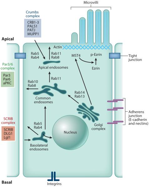
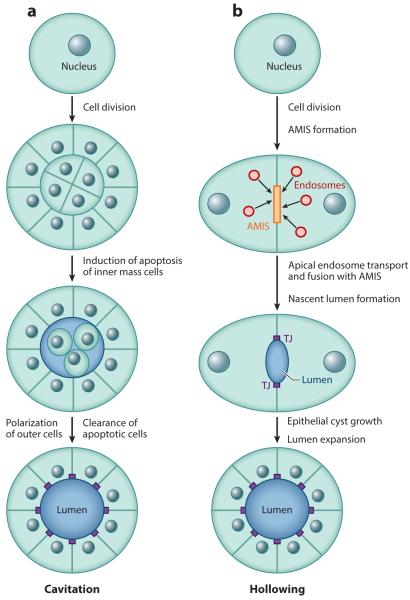
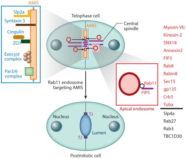
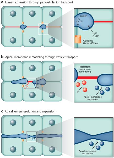
One of the major challenges in biology is to explain how complex tissues and organs arise from the collective action of individual polarized cells. The best-studied model of this process is the cross talk between individual epithelial cells during their polarization to form the multicellular epithelial lumen during tissue morphogenesis. Multiple mechanisms of apical lumen formation have been proposed. Some epithelial lumens form from preexisting polarized epithelial structures. However, de novo lumen formation from nonpolarized cells has recently emerged as an important driver of epithelial tissue morphogenesis, especially during the formation of small epithelial tubule networks. In this review, we discuss the latest findings regarding the mechanisms and regulation of de novo lumen formation in vitro and in vivo.

Keywords: coalescence; epithelial; lumenogenesis.


Figures

Figure 1
Figure 1
The proteins that establish and maintain epithelial cell polarity.
Figure 2
Figure 2
The (a) cavitation and (b) hollowing models of apical lumen formation in vitro. Red circles represent apical endosomes. Abbreviations: AMIS, apical membrane initiation site; TJ, tight junction.
Figure 3
Figure 3
The role of the midbody during apical membrane initiation site (AMIS) formation and apical endosome recruitment. Red indicates apical endosomes and all proteins known to be associated with them. Blue indicates AMIS-associated proteins. Proteins listed in black are known to be required for lumen formation but have unclear subcellular localization.
Figure 4
Figure 4
Model depicting apical lumen coalescence during the formation of the zebrafish intestinal lumen. (a) Definition of the apical lumen initiation site and initial formation of the zebrafish gut lumen require localization of apical and tight junction proteins. Both Claudin 15 and Na+/K+-ATPase are required for the paracellular ion transport necessary for lumen fluid accumulation and initial gut lumen expansion. (b) Gut lumen expansion occurs along adjacent basolateral membranes (red line) through Rab11-mediated apical trafficking (blue) and concurrent with basolateral membrane recycling (red ). (c) The mechanisms regulating the coalescence of multiple expanding apical lumens into a single continuous lumen remain unresolved. The Hedgehog pathway protein Smoothened is required for lumen resolution, but not lumen expansion, suggesting lumen expansion and resolution are two distinct processes.

References

    1. Alvers AL, Ryan S, Scherz PJ, Huisken J, Bagnat M. Single continuous lumen formation in the zebrafish gut is mediated by smoothened-dependent tissue remodeling. Development. 2014;141:1110–19. - PMC - PubMed
    1. Ameen NA, Salas PJ. Microvillus inclusion disease: a genetic defect affecting apical membrane protein traffic in intestinal epithelium. Traffic. 2000;1:76–83. - PubMed
    1. Andrew DJ, Ewald AJ. Morphogenesis of epithelial tubes: insights into tube formation, elongation, and elaboration. Dev. Biol. 2010;341:34–55. - PMC - PubMed
    1. Apodaca G, Gallo LI, Bryant DM. Role of membrane traffic in the generation of epithelial cell asymmetry. Nat. Cell Biol. 2012;14:1235–43. - PMC - PubMed
    1. Apodaca G, Katz LA, Mostov KE. Receptor-mediated transcytosis of IgA in MDCK cells is via apical recycling endosomes. J. Cell Biol. 1994;125:67–86. - PMC - PubMed

Publication types

LinkOut - more resources