Review
. 2015 Nov-Dec;21(6):498-502.
doi: 10.5152/dir.2015.15068.

Split-bolus MR urography: synchronous visualization of obstructing vessels and collecting system in children

Affiliations
Review

Split-bolus MR urography: synchronous visualization of obstructing vessels and collecting system in children

Bilal Battal et al. Diagn Interv Radiol. 2015 Nov-Dec.

Abstract

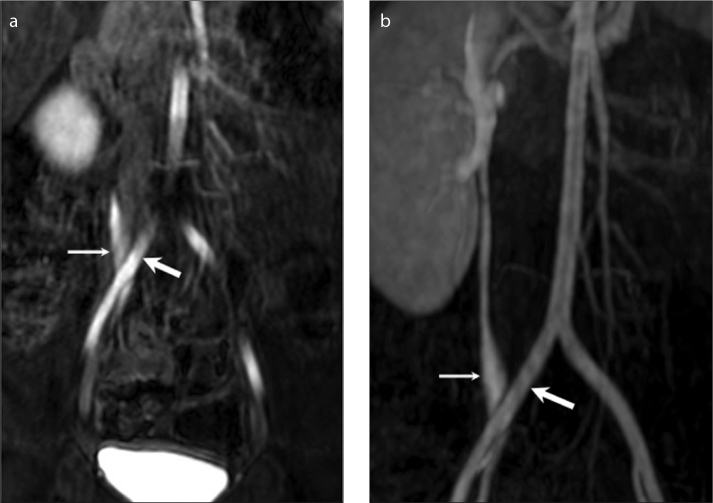
Several vascular abnormalities related with urinary system such as crossing accessory renal vessels, retroiliac ureters, retrocaval ureters, posterior nutcracker syndrome, and ovarian vein syndrome may be responsible for urinary collecting system obstruction. Split-bolus magnetic resonance urography (MRU) using contrast material as two separate bolus injections provides superior demonstration of the collecting system and obstructing vascular anomalies simultaneously and enables accurate preoperative radiologic diagnosis. In this pictorial review we aimed to outline the split-bolus MRU technique in children, list the coexisting congenital collecting system and vascular abnormalities, and exhibit the split-bolus MRU appearances of concurrent urinary collecting system and vascular abnormalities.


Figures

Figure 1.
Figure 1.
Coronal MIP image of T1-weighted excretory MRU shows left ureteropelvic junction stenosis and mild hydronephrosis.
Figure 2.
Figure 2.
Coronal thick-slab T2-weighted MRU reveals right ureteropelvic junction stenosis and severe hydronephrosis.
Figure 3.
Figure 3.
Scheme of the split-bolus MRU technique. 2D, two-dimensional; TSE, turbo spin-echo; 3D, three-dimensional; MIP, maximum intensity projection; MPR, multiplanar reconstruction.
Figure 4. a–d.
Figure 4. a–d.
A 13-year-old girl with right ureteropelvic junction (UPJ) stenosis. A coronal MIP image (a) obtained from second bolus excretory urographic data demonstrates simultaneous enhancement of the collecting system and renal arteries including a right accessory renal artery (thick arrow) without close relation with right UPJ. Three consecutive coronal images (b–d) obtained during the arterial phase of the second bolus of contrast medium show concurrent enhancement of the collecting system (asterisks) and renal arteries. A right accessory renal artery (thick arrow) is not the cause or the exacerbating factor for UPJ stenosis (thin arrows indicate renal arteries).
Figure 5. a–d.
Figure 5. a–d.
A 17-year-old male with surgically proven left UPJ stenosis with crossing renal vein. A coronal MIP image (a) obtained from the first bolus excretory urographic data shows the left UPJ stenosis. Coronal dynamic series (b) obtained during the late arterial/venous phase of the first bolus of contrast reveal a pair of left renal veins, the lower one of which (arrow) passes close to UPJ. Two consecutive coronal images (c, d) obtained during the arterial phase of the second bolus of contrast medium show concurrent enhancement of the collecting system (asterisk) and renal veins. A crossing renal vein (arrow) is in close contact with the UPJ and may complicate the surgical intervention.
Figure 6. a–d.
Figure 6. a–d.
A two-year-old boy with left UPJ stenosis related with an accessory renal artery. Coronal dynamic series (a) obtained during the arterial phase of the second bolus show simultaneous enhancement of the collecting system (asterisk) and the crossing accessory renal artery (thick arrow). A coronal MIP image (b) generated from the second bolus excretory urographic data reveals crossing accessory renal artery (thick arrow). Two consecutive sagittal reformatted images (c, d) obtained from the second bolus show that the accessory crossing renal artery (thick arrow) is not the cause of UPJ (thin arrow) stenosis. But awareness of its location is essential during endoureterotomy.
Figure 7. a, b.
Figure 7. a, b.
Retroiliac ureter in a two-year-old boy with left renal agenesis. Coronal dynamic series (a) obtained from the second bolus of intravenous contrast medium and corresponding coronal MIP images (b) show a right retroiliac ureter (thin arrow) with mild dilatation (thick arrow indicates common iliac artery).
Figure 8. a–d.
Figure 8. a–d.
Posterior nutcracker syndrome in a seven-year-old girl with asymptomatic microhematuria. Three consecutive axial reformatted images obtained from the late arterial/venous phases (a, b, c) and their corresponding coronal MIP image (d) demonstrate a retroaortic left renal vein (arrow) joining the inferior vena cava (asterisk) at a lower level than normal. The retroaortic segment of the left renal vein is prominently narrowed due to aortic compression; the proximal part is prominently enlarged.

References

    1. Levendecker JR, Barnes CE, Zagoria RJ. MR urography: techniques and clinical applications. Radiographics. 2008;28:23–46. http://dx.doi.org/10.1148/rg.281075077. - DOI - PubMed
    1. Battal B, Kocaoglu M, Akgun V, Aydur E, Dayanc M, Ilica T. Feasibility of MR urography in patients with urinary diversion. J Med Imaging Radiat Oncol. 2011;55:542–550. http://dx.doi.org/10.1111/j.1754-9485.2011.02318.x. - DOI - PubMed
    1. Koçyiğit A, Yüksel S, Bayram R, Yılmaz İ, Karabulut N. Efficacy of magnetic resonance urography in detecting renal scars in children with vesicoureteral reflux. Pediatr Nephrol. 2014;29:1215–1220. http://dx.doi.org/10.1007/s00467-014-2766-y. - DOI - PubMed
    1. Kocyigit A, Serinken M, Ceven Z, et al. A strategy to optimize CT use in children with mild blunt head trauma utilizing clinical risk stratification; could we improve CT use in children with mild head injury? Clin Imaging. 2014;38:236–240. http://dx.doi.org/10.1016/j.clinimag.2013.12.004. - DOI - PubMed
    1. Kocaoglu M, Bulakbasi N, Ilica AT, Gok F, Tayfun C, Somuncu I. Intravenous contrast-enhanced dynamic MR urography: diagnosis of vesicoureteral reflux during bladder filling with time-signal intensity curves. J Magn Reson Imaging. 2006;24:349–355. http://dx.doi.org/10.1002/jmri.20637. - DOI - PubMed