. 2015 Sep 10;162(6):1379-90.
doi: 10.1016/j.cell.2015.08.035.

Broadly Neutralizing Antibody 8ANC195 Recognizes Closed and Open States of HIV-1 Env

Affiliations

Broadly Neutralizing Antibody 8ANC195 Recognizes Closed and Open States of HIV-1 Env

Louise Scharf et al. Cell. .

Abstract

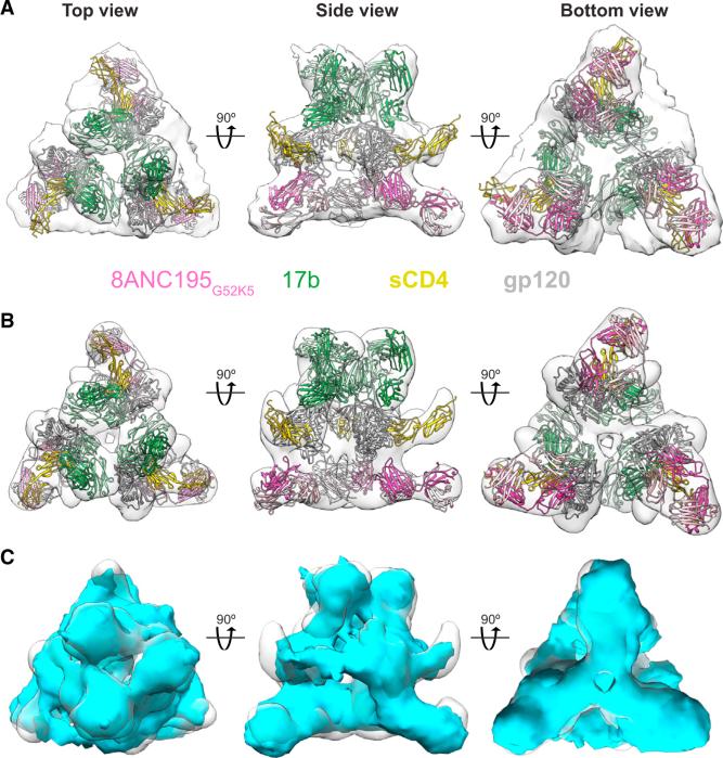
The HIV-1 envelope (Env) spike contains limited epitopes for broadly neutralizing antibodies (bNAbs); thus, most neutralizing antibodies are strain specific. The 8ANC195 epitope, defined by crystal and electron microscopy (EM) structures of bNAb 8ANC195 complexed with monomeric gp120 and trimeric Env, respectively, spans the gp120 and gp41 Env subunits. To investigate 8ANC195's gp41 epitope at higher resolution, we solved a 3.58 Å crystal structure of 8ANC195 complexed with fully glycosylated Env trimer, revealing 8ANC195 insertion into a glycan shield gap to contact gp120 and gp41 glycans and protein residues. To determine whether 8ANC195 recognizes the CD4-bound open Env conformation that leads to co-receptor binding and fusion, one of several known conformations of virion-associated Env, we solved EM structures of an Env/CD4/CD4-induced antibody/8ANC195 complex. 8ANC195 binding partially closed the CD4-bound trimer, confirming structural plasticity of Env by revealing a previously unseen conformation. 8ANC195's ability to bind different Env conformations suggests advantages for potential therapeutic applications.


Figures

Figure 1
Figure 1. Overview of 8ANC195G52K5-BG505 SOSIP Structure
8ANC195G52K5 Fabs are dark pink (HC) and light pink (LC), gp120 subunits are different shades of light gray, and gp41 subunits are different shades of dark gray. N-linked glycans at the Fab-trimer interface are yellow. (A) 8ANC195G52K5-Env structure seen from the top in space-filling (left) and ribbon diagram (right) representations. (B) 8ANC195G52K5-Env structure seen from the side in space-filling (left) and ribbon diagram (right) representations. (C) Conformations of HIV-1 Env trimers shown schematically (adapted from figures in Liu et al., 2008) as seen from above (top) and the side (bottom) (CD4-binding site, yellow; remainder of gp120, light gray; gp41, dark gray; membrane, blue-gray). The closed structure was observed for unliganded virion-bound trimers (Liu et al., 2008) and structures involving liganded BG505 SOSIP trimers (Julien et al., 2013a; Lyumkis et al., 2013; Pancera et al., 2014). The partially open structure was observed for virion-bound trimers associated with b12 or A12 (Liu et al., 2008). Open structures were observed for trimers associated with CD4 or the Fab from the CD4-induced Ab 17b (Merk and Subramaniam, 2013). See also Figure S1 and Table S1.
Figure 2
Figure 2. Interfaces in 8ANC195G52K5-BG505 SOSIP Structure
(A) 8ANC195G52K5 epitope. BG505 SOSIP is shown in space-filling representation with the gp120 and gp41 portions of one protomer in white and medium gray, respectively (other two protomers are light gray). Glycan and protein portions of the 8ANC195G52K5 epitope are highlighted in the indicated colors on one protomer. (B) Close-up of contact areas. Relevant portions of 8ANC195G52K5 are shown in wire representation with atoms within buried surface areas on BG505 SOSIP shown as colored surfaces. (C) Details of interactions with glycans on gp120 and gp41. Buried surface areas on 8ANC195G52K5 are shown as colored surfaces. See also Figure S2 and Table S2.
Figure 3
Figure 3. Contacts between 8ANC195G52K5 Fab and BG505 SOSIP Protein Residues and Glycans
Proteins are shown as Ca traces with interface residues as sticks (oxygen, red; nitrogen, blue). CDRs and framework regions (FWRs) of 8ANC195G52K5 at the interface are highlighted and labeled. Putative H bonds are shown as yellow dots. Env glycans at the interface are yellow (Asn234gp120), orange (Asn276gp120), and tan (Asn637gp41) and are enclosed in electron density from a 2Fo-Fc simulated annealing omit map contoured at 1.0 σ (gray mesh) in the lower structure panels. Schematic structures of the ordered portions of these N-linked glycans are shown at the bottom. See also Tables S3 and S4.
Figure 4
Figure 4. Effects of Env Trimer Conformational State on Binding Affinity of 8ANC195
(A) Schematic representation of coupled surfaces and injected analytes. A control IgG (mG053), an IgG recognizing closed trimers (PGT121 or PGT145), CD4-Fc (recognizing open trimers), and a CD4-induced Ab (17b or 21c) recognizing open CD4-bound trimers were immobilized on separate flow cells of a biosensor chip. BG505 SOSIP trimers were injected, resulting in binding to surfaces except for the control flow cell. 8ANC195, 8ANC195G52K5, or a G52HC/8ANC195LC chimera Fab was then injected over the IgG-BG505 SOSIP complex. (B) Summary of affinities (KD values; reported as mean ± SD for three independent experiments) for combinations of capture proteins and analytes. Since PGT145 indirectly inhibited binding of 8ANC195 to BG505 SOSIP trimers and PGT121 enhanced 8ANC195 binding (Derking et al., 2015), our affinity measurements likely underestimated (PGT145-captured trimers) or overestimated (PGT121-captured trimers) the affinity of 8ANC195 for closed trimer. N.D., not determined. See also Figure S3.
Figure 5
Figure 5. EM Reconstructions of BG505 SOSIP-sCD4-17b-8ANC195G52K5 Complex
The X-ray structures of a gp120-scD4-17b complex (PDB: 1RZK) and 8ANC195 Fab (PDB: 4P9M) were fit to EM densities and are shown from the top, side, and bottom. (A) ~23Å resolution EM density derived from cryo-ET and sub-tomogram averaging. (B) ~17Å resolution EM density derived from negative-stain single-particle reconstruction. (C) Superposition of densities from cryo-ET/sub-tomogram averaging (cyan) and negative-stain single-particle (light gray) reconstructions. See also Figures S4 and S5.
Figure 6
Figure 6. Comparison of Different Conformations of Env Trimers in BG505 SOSIP-sCD4-17b-8ANC195G52K5 and Other Env Trimer Complexes Illustrated by 17b and gp120 Positions
(A) Overlay of 17b Fabs from BG505 SOSIP-sCD4-17b-8ANC195G52K5 complex (gray space-filling representations) with 17b Fabs complexed with Env trimers in different conformational states (thick Cα traces in shades of blue). 17b Fabs shown after superimposition of gp120s from trimers (trimers and other ligands not shown). (Left) 17b modeled onto 3DNN coordinates of a closed trimer. (Middle) 17b modeled onto 3DNL coordinates of a partly open trimer. (Right) 17b taken from 3DNO structure of a CD4- and 17b-bound open trimer. Note that three 17b Fabs cannot be accommodated without clashes when modeled onto the gp120s of closed and partly open trimer structures. (B) Cα trace of the three 17b Fabs from the BG505 SOSIP-sCD4-17b-8ANC195G52K5 complex. (C) Positions of gp120s (gray space-filling representation with locations of CD4-binding site in yellow, V3 loop in green, and base of V1V2 domain in red) in Env trimers adopting the indicated conformations. The locations of Asp368gp120 are shown as a black dot for each trimer conformation, with the distance between the Cα of this residue in adjacent protomers indicated. Distances are presented as the mean and SD for the analogous distance in representative structures in each conformation. See also Figure S6 and Table S5.

References

    1. Abela IA, Berlinger L, Schanz M, Reynell L, Günthard HF, Rusert P, Trkola A. Cell-cell transmission enables HIV-1 to evade inhibition by potent CD4bs directed antibodies. PLoS Pathog. 2012;8:e1002634. - PMC - PubMed
    1. Adams PD, Afonine PV, Bunkóczi G, Chen VB, Davis IW, Echols N, Headd JJ, Hung LW, Kapral GJ, Grosse-Kunstleve RW, et al. PHENIX: a comprehensive Python-based system for macromolecular structure solution. Acta Crystallogr. D Biol. Crystallogr. 2010;66:213–221. - PMC - PubMed
    1. Binley JM, Ban YE, Crooks ET, Eggink D, Osawa K, Schief WR, Sanders RW. Role of complex carbohydrates in human immunodeficiency virus type 1 infection and resistance to antibody neutralization. J. Virol. 2010;84:5637–5655. - PMC - PubMed
    1. Blattner C, Lee JH, Sliepen K, Derking R, Falkowska E, de la Peña AT, Cupo A, Julien JP, van Gils M, Lee PS, et al. Structural delineation of a quaternary, cleavage-dependent epitope at the gp41-gp120 interface on intact HIV-1 Env trimers. Immunity. 2014;40:669–680. - PMC - PubMed
    1. Capon DJ, Chamow SM, Mordenti J, Marsters SA, Gregory T, Mitsuya H, Byrn RA, Lucas C, Wurm FM, Groopman JE, et al. Designing CD4 immunoadhesins for AIDS therapy. Nature. 1989;337:525–531. - PubMed

Publication types

MeSH terms

Associated data