. 2015 Sep 11;10(9):e0137652.
doi: 10.1371/journal.pone.0137652. eCollection 2015.

AQUA Cloning: A Versatile and Simple Enzyme-Free Cloning Approach

Affiliations

AQUA Cloning: A Versatile and Simple Enzyme-Free Cloning Approach

Hannes M Beyer et al. PLoS One. .

Abstract

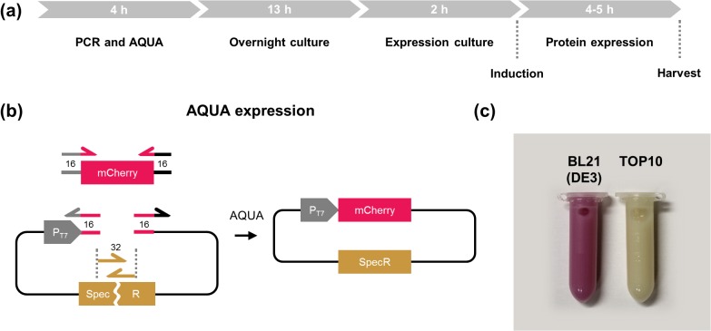
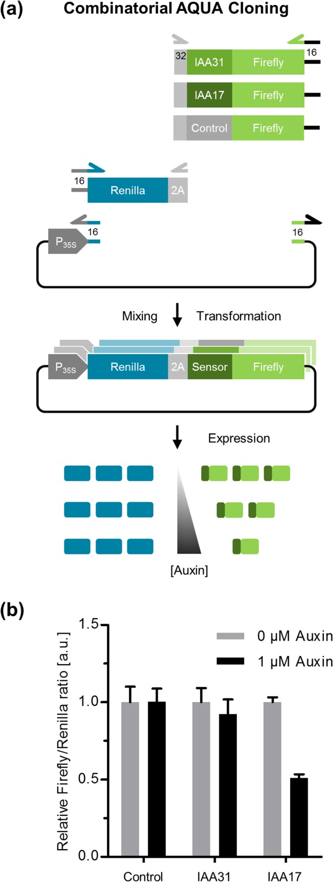
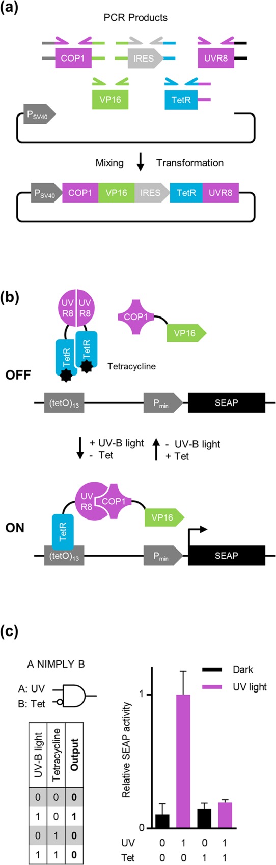
Assembly cloning is increasingly replacing conventional restriction enzyme and DNA-ligase-dependent cloning methods for reasons of efficiency and performance. Here, we describe AQUA (advanced quick assembly), a simple and versatile seamless assembly cloning approach. We demonstrate the applicability and versatility of AQUA Cloning in selected proof-of-principle applications including targeted insertion-, deletion- and site-directed point-mutagenesis, and combinatorial cloning. Furthermore, we show the one pot de novo assembly of multiple DNA fragments into a single circular plasmid encoding a complex light- and chemically-regulated Boolean A NIMPLY B logic operation. AQUA Cloning harnesses intrinsic in vivo processing of linear DNA fragments with short regions of homology of 16 to 32 bp mediated by Escherichia coli. It does not require any kits, enzymes or preparations of reagents and is the simplest assembly cloning protocol to date.


Conflict of interest statement

Competing Interests: The authors have declared that no competing interests exist.

Figures

Fig 1
Fig 1. AQUA Cloning: advanced quick assembly cloning.
(a) DNA parts are produced by PCR, or restriction digest (or both). Oligonucleotides are designed to contribute flanking homologous regions to adjacent DNA fragments of optimally 32 bp in length. DNA parts are assembled into a circular plasmid by sequence-determined directionality. (b) AQUA Cloning work-flow. (1) DNA parts are generated by PCR amplification, or derived from an enzymatic digestion. (2) Next, DNA parts are purified by gel-electrophoresis and (3) mixed and simply incubated in water prior to transformation into chemically competent E. coli Top10 cells for in vivo assembly. (4) Finally, obtained colonies are confirmed for correct assembly by standard methods such as analytical PCR, restriction digest, or comprehensive sequencing.
Fig 2
Fig 2. AQUA Cloning conditions.
(a) The DNA fragment encoding the red fluorescent protein mCherry was PCR amplified with flanking extensions of 16, 24, or 32 bp of homologous sequence overlaps to an SV40 promoter-driven mammalian expression vector which was digested, or PCR amplified. AQUA Cloning was performed with pre-incubations on ice, at room temperature (RT), or at 50°C. DNA mixtures were transformed into chemically competent E. coli Top10 cells and colonies were obtained the next day. (b) Typical numbers of colony forming units (CFU) obtained from each condition. The highest number of CFU was derived from a pre-incubation at room temperature and with 32 bp of shared homology with PCR originating DNA fragments (arrow). The accuracies for each condition were determined by analytical colony PCR from eight selected clones and were extrapolated to the total number of CFU (grey). Abbreviations: PSV40, simian virus 40 early promoter.
Fig 3
Fig 3. 4-fragment assembly.
(a) A quadruple de novo assembly was designed by generating four PCR amplified linear DNA fragments (i) mCherry with an NLS sequence, (ii) an IRES element for bicistronicity, (iii) EGFP with an NES sequence and (iv) the PSV40 promoter-driven mammalian expression vector. Each pair of adjacent fragments shared 32 bp of overlapping sequence. (b) Experimental verification of the correct assembly in (a) by transient transfection into HEK-293T cells followed by confocal imaging. Scale bar, 10 μm. Abbreviations: IRES, internal ribosomal entry site; NES, nuclear export signal; NLS, nuclear localization signal, PSV40, simian virus 40 early promoter.
Fig 4
Fig 4. Site-directed mutagenesis with AQUA Cloning: deletion-, insertion- and point-mutagenesis.
(a) For targeted deletion of the Lck membrane anchor from an Lck-fused EGFP, a primer pair was designed for whole-plasmid PCR with the forward primer annealing to EGFP and sharing overlaps to the PSV40 promoter region. The reverse primer anneals to the PSV40 promoter and shares overlaps to EGFP. The resulting PCR product hence excludes the Lck membrane anchor and the extremes are complementary by 32 bp allowing AQUA assembly into a circular plasmid for cytosolic expression of EGFP. The resulting EGFP expression plasmid was further used as a PCR template with a primer pair where the 5’-extensions were replaced with a mitochondrial target signal (MTS) which localized expressed EGFP into the mitochondria of the host cell. The flanking MTS sequence of the resulting PCR product serves as region of homology for AQUA Cloning to construct the expression plasmid with the MTS sequence inserted. (b) Confocal imaging after transient transfection into NIH/3T3 cells reveals successful AQUA Cloning: the deletion of the Lck-membrane anchor resulted in cytosolic expression of EGFP (middle) of previously membrane localized protein (left), and the insertion of a MTS in recruitment of EGFP into the mitochondria (right). Scale bars, 10 μm. Corresponding fluorescence intensity profiles are given below each figure. (c) For site-directed substitution of a single amino acid in EGFP, a primer pair was designed surrounding the target site. 5’-extensions were attached to encode the Y66H substitution (leading to blue fluorescence) and at the same time contributing a region of homology for AQUA Cloning of a single linear PCR product. (d) Green and blue fluorescence of recombinant purified EGFP and EGFP-Y66H, respectively upon excitation with 366 nm light. (e) Excitation and emission spectra of EGFP and EGFP-Y66H.
Fig 5
Fig 5. AQUA Expression—combined cloning and protein expression.
(a) Timeline for AQUA Expression. Cloning and production of recombinant protein in E. coli may be performed within 24 h starting with the PCR until the bacteria are harvested the next day. (b) A 3-DNA fragment cloning was performed by inserting the coding sequence for the red fluorescent protein mCherry into a bacterial T7 promoter-driven expression vector. The vector was split into two parts within the resistance gene for the antibiotic spectinomycin. Therefore, only correctly assembled fragments allow cell growth. (c) AQUA Expression in the expression strain BL21 (DE3) results in red colored bacteria due to mCherry protein production, while the TOP10 strain—lacking the required T7 RNA polymerase—remains colorless.
Fig 6
Fig 6. Combinatorial AQUA Cloning for plant cells.
(a) For a combinatorial cloning of 3 plasmids, each originating from 3 linear DNA parts (variants of a ratiometric auxin sensor) out of a pool of 5 parts, were constructed. The consensus sequences derived from the Aux/IAA 31 and Aux/IAA 17 proteins, or an auxin insensitive sequence, each fused to the firefly luciferase were prepared. The fragments were cloned together with the renilla luciferase, separated by a 2A peptide for equimolar expression, into a 35S promoter-driven plant expression vector. Expression of the sensor constructs in plant cells should enable the auxin-dependent degradation of firefly, while renilla remains unaffected for intrinsic signal normalization. (b) Relative firefly:renilla ratios obtained from protoplasts expressing the sensor variants after 1 h of auxin treatment. Error bars represent standard error of the mean, n = 6.
Fig 7
Fig 7. Multi-fragment assembly for the development of a light- and chemically-regulated Boolean logic operation.
(a) Each DNA fragment for the assembly was PCR-amplified with 32 bp of homology region at each fusion-site as indicated schematically. The first cistron is composed of a fusion of the tetracycline repressor protein TetR to the UVR8 ultraviolet B (UV-B) light photoreceptor. The second cistron, separated by an IRES element, comprises a fusion of the interaction factor COP1 to the transcriptional activation domain VP16. AQUA Cloning of the 6 DNA fragments yields the desired bicistronic mammalian expression vector. (b) Functional principle of the light- and chemically-regulated A NIMPLY B logic gate. In the absence of tetracycline, TetR binds to its cognate operator element (TRE) of an appropriate reporter plasmid. The UV-B light mediated activation of UVR8 provokes the dissociation of the otherwise homodimeric conformation and allows the interaction and hence the recruitment of COP1 which is fused to the VP16 activation domain. The close proximity of the transactivator VP16 to the minimal promoter (Pmin) mediates the transcription of the encoded SEAP reporter gene under UV-B light irradiation and without the addition of tetracycline. (c) Truth table (left) and experimental results (right) of the A NIMPLY B gate. SEAP activity is detected only in the presence of UV-B light and in the absence of tetracycline, validating a functional A NIMPLY B logic gate. Abbreviations: COP1, CONSTITUTIVELY PHOTOMORPHOGENIC 1; IRES, internal ribosome entry site; Pmin, minimal human cytomegalovirus immediate early promoter; PSV40, simian virus 40 early promoter; SEAP, human placental secreted alkaline phosphatase; TetR, tetracycline repressor protein; UVR8, UV resistance locus 8 protein; VP16, viral transactivation domain.

References

    1. Weber W, Fussenegger M. Emerging biomedical applications of synthetic biology. Nat Rev Genet. 2012;13: 21–35. - PMC - PubMed
    1. Church GM, Elowitz MB, Smolke CD, Voigt CA, Weiss R. Realizing the potential of synthetic biology. Nat Rev Mol Cell Biol. 2014;15: 289–294. 10.1038/nrm3767 - DOI - PMC - PubMed
    1. Gibson DG, Young L, Chuang R, Venter JC, Iii CAH, Smith HO, et al. Enzymatic assembly of DNA molecules up to several hundred kilobases. Nat Methods. 2009;6: 12–16. - PubMed
    1. De Kok S, Stanton LH, Slaby T, Durot M, Holmes VF, Patel KG, et al. Rapid and reliable DNA assembly via ligase cycling reaction. ACS Synth Biol. 2014;3: 97–106. 10.1021/sb4001992 - DOI - PubMed
    1. Van den Ent F, Löwe J. RF cloning: a restriction-free method for inserting target genes into plasmids. J Biochem Biophys Methods. 2006;67: 67–74. - PubMed

Publication types